
Main menu

  • Home
  • Content
    • Current
    • Ahead of print
    • Past Issues
    • JNM Supplement
    • SNMMI Annual Meeting Abstracts
    • Continuing Education
    • JNM Podcasts
  • Subscriptions
    • Subscribers
    • Institutional and Non-member
    • Rates
    • Journal Claims
    • Corporate & Special Sales
  • Authors
    • Submit to JNM
    • Information for Authors
    • Assignment of Copyright
    • AQARA requirements
  • Info
    • Reviewers
    • Permissions
    • Advertisers
  • About
    • About Us
    • Editorial Board
    • Contact Information
  • More
    • Alerts
    • Feedback
    • Help
    • SNMMI Journals
  • SNMMI
    • JNM
    • JNMT
    • SNMMI Journals
    • SNMMI

User menu

  • Subscribe
  • My alerts
  • Log in
  • My Cart

Search

  • Advanced search
Journal of Nuclear Medicine
  • SNMMI
    • JNM
    • JNMT
    • SNMMI Journals
    • SNMMI
  • Subscribe
  • My alerts
  • Log in
  • My Cart
Journal of Nuclear Medicine

Advanced Search

  • Home
  • Content
    • Current
    • Ahead of print
    • Past Issues
    • JNM Supplement
    • SNMMI Annual Meeting Abstracts
    • Continuing Education
    • JNM Podcasts
  • Subscriptions
    • Subscribers
    • Institutional and Non-member
    • Rates
    • Journal Claims
    • Corporate & Special Sales
  • Authors
    • Submit to JNM
    • Information for Authors
    • Assignment of Copyright
    • AQARA requirements
  • Info
    • Reviewers
    • Permissions
    • Advertisers
  • About
    • About Us
    • Editorial Board
    • Contact Information
  • More
    • Alerts
    • Feedback
    • Help
    • SNMMI Journals
  • View or Listen to JNM Podcast
  • Visit JNM on Facebook
  • Join JNM on LinkedIn
  • Follow JNM on Twitter
  • Subscribe to our RSS feeds
Research ArticleClinical Investigations

Toward Radioguided Surgery with β− Decays: Uptake of a Somatostatin Analogue, DOTATOC, in Meningioma and High-Grade Glioma

Francesco Collamati, Alessandra Pepe, Fabio Bellini, Valerio Bocci, Giacomo Chiodi, Marta Cremonesi, Erika De Lucia, Mahila E. Ferrari, Paola M. Frallicciardi, Chiara M. Grana, Michela Marafini, Ilaria Mattei, Silvio Morganti, Vincenzo Patera, Luca Piersanti, Luigi Recchia, Andrea Russomando, Alessio Sarti, Adalberto Sciubba, Martina Senzacqua, Elena Solfaroli Camillocci, Cecilia Voena, Davide Pinci and Riccardo Faccini
Journal of Nuclear Medicine January 2015, 56 (1) 3-8; DOI: https://doi.org/10.2967/jnumed.114.145995
Francesco Collamati
1Dipartimento di Fisica, Sapienza Università di Roma, Roma, Italy
2INFN Sezione di Roma, Roma, Italy
  • Find this author on Google Scholar
  • Find this author on PubMed
  • Search for this author on this site
Alessandra Pepe
3Dipartimento di Scienze di Base e Applicate per l’Ingegneria, Sapienza Università di Roma, Roma, Italy
  • Find this author on Google Scholar
  • Find this author on PubMed
  • Search for this author on this site
Fabio Bellini
1Dipartimento di Fisica, Sapienza Università di Roma, Roma, Italy
2INFN Sezione di Roma, Roma, Italy
  • Find this author on Google Scholar
  • Find this author on PubMed
  • Search for this author on this site
Valerio Bocci
2INFN Sezione di Roma, Roma, Italy
  • Find this author on Google Scholar
  • Find this author on PubMed
  • Search for this author on this site
Giacomo Chiodi
2INFN Sezione di Roma, Roma, Italy
  • Find this author on Google Scholar
  • Find this author on PubMed
  • Search for this author on this site
Marta Cremonesi
4Istituto Europeo di Oncologia, Milano, Italy
  • Find this author on Google Scholar
  • Find this author on PubMed
  • Search for this author on this site
Erika De Lucia
5Laboratori Nazionali di Frascati dell’INFN, Frascati, Italy
  • Find this author on Google Scholar
  • Find this author on PubMed
  • Search for this author on this site
Mahila E. Ferrari
4Istituto Europeo di Oncologia, Milano, Italy
  • Find this author on Google Scholar
  • Find this author on PubMed
  • Search for this author on this site
Paola M. Frallicciardi
2INFN Sezione di Roma, Roma, Italy
6Museo Storico della Fisica e Centro Studi e Ricerche “E. Fermi,” Roma, Italy
  • Find this author on Google Scholar
  • Find this author on PubMed
  • Search for this author on this site
Chiara M. Grana
4Istituto Europeo di Oncologia, Milano, Italy
  • Find this author on Google Scholar
  • Find this author on PubMed
  • Search for this author on this site
Michela Marafini
2INFN Sezione di Roma, Roma, Italy
6Museo Storico della Fisica e Centro Studi e Ricerche “E. Fermi,” Roma, Italy
  • Find this author on Google Scholar
  • Find this author on PubMed
  • Search for this author on this site
Ilaria Mattei
5Laboratori Nazionali di Frascati dell’INFN, Frascati, Italy
7Dipartimento di Fisica, Università RomaTre, Roma, Italy; and
  • Find this author on Google Scholar
  • Find this author on PubMed
  • Search for this author on this site
Silvio Morganti
2INFN Sezione di Roma, Roma, Italy
  • Find this author on Google Scholar
  • Find this author on PubMed
  • Search for this author on this site
Vincenzo Patera
2INFN Sezione di Roma, Roma, Italy
3Dipartimento di Scienze di Base e Applicate per l’Ingegneria, Sapienza Università di Roma, Roma, Italy
  • Find this author on Google Scholar
  • Find this author on PubMed
  • Search for this author on this site
Luca Piersanti
2INFN Sezione di Roma, Roma, Italy
3Dipartimento di Scienze di Base e Applicate per l’Ingegneria, Sapienza Università di Roma, Roma, Italy
  • Find this author on Google Scholar
  • Find this author on PubMed
  • Search for this author on this site
Luigi Recchia
2INFN Sezione di Roma, Roma, Italy
  • Find this author on Google Scholar
  • Find this author on PubMed
  • Search for this author on this site
Andrea Russomando
1Dipartimento di Fisica, Sapienza Università di Roma, Roma, Italy
2INFN Sezione di Roma, Roma, Italy
8Center for Life Nano Science@Sapienza, Istituto Italiano di Tecnologia, Roma, Italy
  • Find this author on Google Scholar
  • Find this author on PubMed
  • Search for this author on this site
Alessio Sarti
5Laboratori Nazionali di Frascati dell’INFN, Frascati, Italy
3Dipartimento di Scienze di Base e Applicate per l’Ingegneria, Sapienza Università di Roma, Roma, Italy
  • Find this author on Google Scholar
  • Find this author on PubMed
  • Search for this author on this site
Adalberto Sciubba
2INFN Sezione di Roma, Roma, Italy
3Dipartimento di Scienze di Base e Applicate per l’Ingegneria, Sapienza Università di Roma, Roma, Italy
  • Find this author on Google Scholar
  • Find this author on PubMed
  • Search for this author on this site
Martina Senzacqua
1Dipartimento di Fisica, Sapienza Università di Roma, Roma, Italy
2INFN Sezione di Roma, Roma, Italy
  • Find this author on Google Scholar
  • Find this author on PubMed
  • Search for this author on this site
Elena Solfaroli Camillocci
8Center for Life Nano Science@Sapienza, Istituto Italiano di Tecnologia, Roma, Italy
  • Find this author on Google Scholar
  • Find this author on PubMed
  • Search for this author on this site
Cecilia Voena
2INFN Sezione di Roma, Roma, Italy
  • Find this author on Google Scholar
  • Find this author on PubMed
  • Search for this author on this site
Davide Pinci
2INFN Sezione di Roma, Roma, Italy
  • Find this author on Google Scholar
  • Find this author on PubMed
  • Search for this author on this site
Riccardo Faccini
1Dipartimento di Fisica, Sapienza Università di Roma, Roma, Italy
2INFN Sezione di Roma, Roma, Italy
  • Find this author on Google Scholar
  • Find this author on PubMed
  • Search for this author on this site
  • Article
  • Figures & Data
  • Info & Metrics
  • PDF
Loading

Article Figures & Data

Figures

  • Tables
  • FIGURE 1.
    • Download figure
    • Open in new tab
    • Download powerpoint
    FIGURE 1.

    Example of ROI definition for meningioma (left), HGG (center), and nontumor (right).
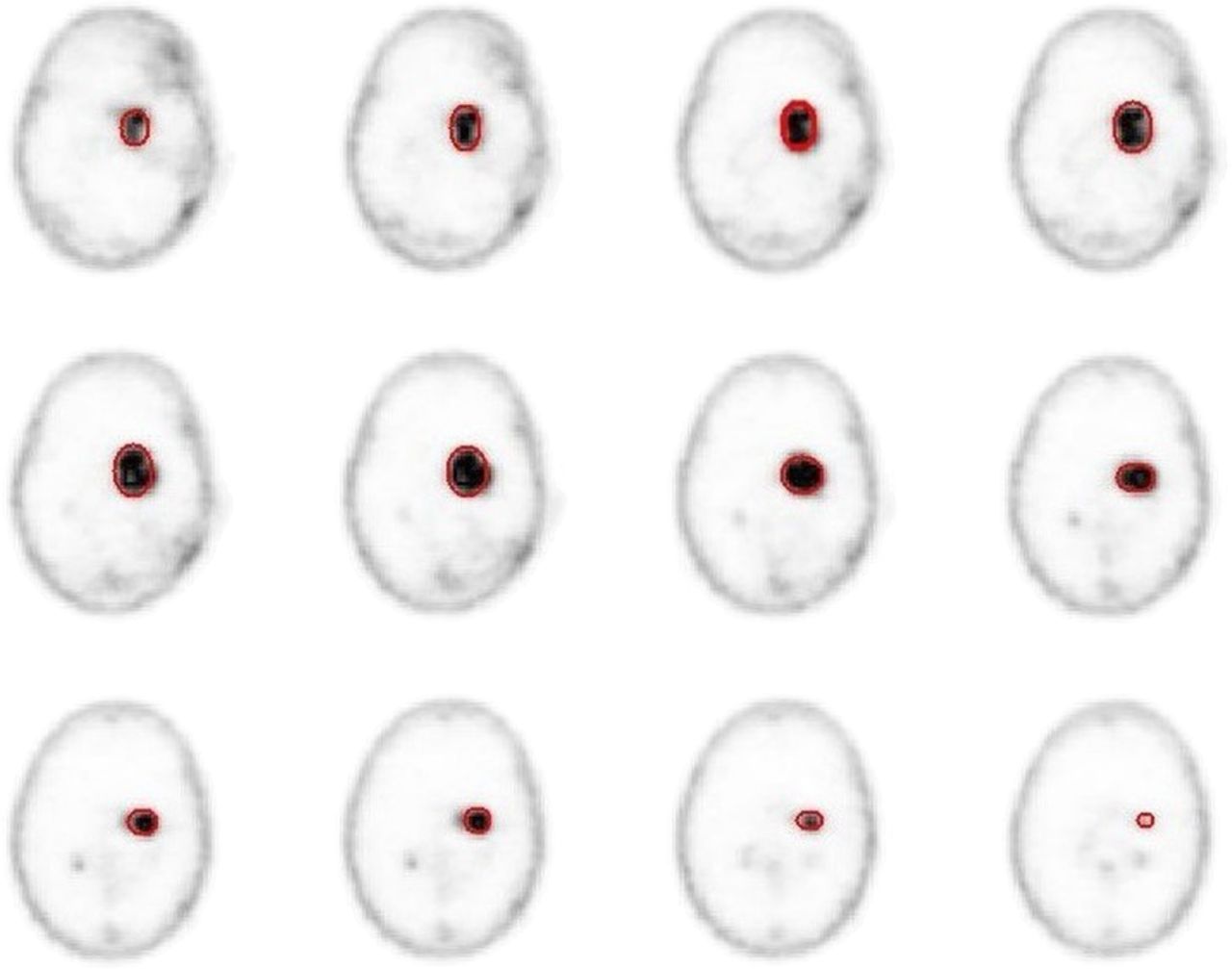
  • FIGURE 2.
    • Download figure
    • Open in new tab
    • Download powerpoint
    FIGURE 2.

    Example of ROI definition in several slices of PET scan of meningioma patient.
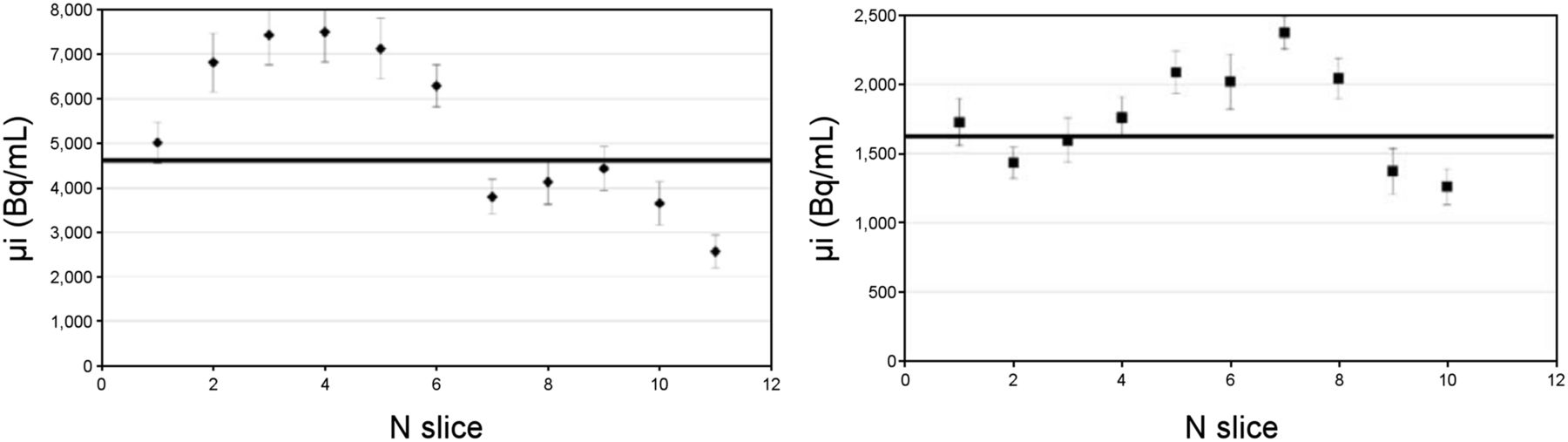
  • FIGURE 3.
    • Download figure
    • Open in new tab
    • Download powerpoint
    FIGURE 3.

    Example of μi estimated in different slices in case of meningioma (left) and glioma (right).
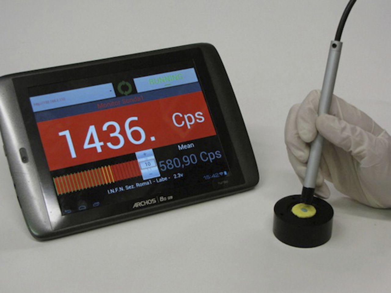
  • FIGURE 4.
    • Download figure
    • Open in new tab
    • Download powerpoint
    FIGURE 4.

    Photograph of probe prototype.
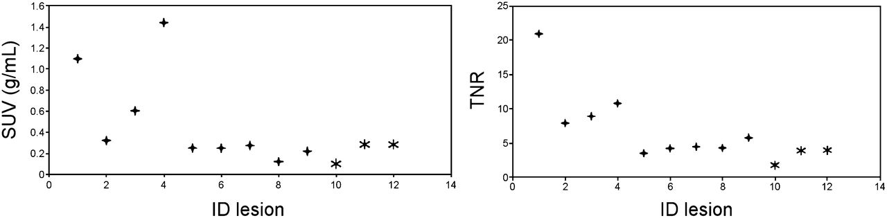
  • FIGURE 5.
    • Download figure
    • Open in new tab
    • Download powerpoint
    FIGURE 5.

    Measured SUV (left) and TNR (right) on all studied meningioma lesions. Lesions from same patient share same symbols. ID = sequential number identifying a lesion.

  • FIGURE 6.
    • Download figure
    • Open in new tab
    • Download powerpoint
    FIGURE 6.

    Measured SUV (left) and TNR (right) on all studied HGG lesions. ID = sequential number identifying a lesion.

Tables

  • Figures
    • View popup
    TABLE 1

    Results for Meningioma Patients

    Patient no.Nles.Weight (kg)Aadm (MBq)ν*† (Hz)νNT*† (Hz)Embedded Image*† (s)Embedded Image* (MBq/kg)DiagnosisPrevious treatment
    M0116322032.21.90.20.7AtypicalS
    M0218016017.62.60.61.9AtypicalS/RT/PRRT
    M0339530533.73.50.30.9Likely atypicalS/RT
    50.33.50.30.5
    76.83.50.10.3
    M0414820089.44.50.10.2AtypicalS/RT/CT
    M0535713066.74.40.20.3RelapseS/RT/CT/PRRT
    53.24.40.20.5
    57.64.40.20.4
    M06290145107.61.80.10.1UnknownPRRT
    56.11.80.20.4
    M0717423750.23.90.20.5AnaplasticS/RT
    M08310522355.73.60.20.5AtypicalS/RT
    31.23.60.20.9
    29.63.60.40.9
    M0924814513.42.40.92.7AtypicalS/RT
    15.12.40.72.5
    M1017024014.61.20.61.8AtypicalS/RT
    12.61.20.81.9
    M1117522012.73.81.65.0AtypicalUnknown
    • ↵* Data assume 12 h between administration of 90Y and surgery.

    • ↵† Estimations assume that reference activity Aref = 3 MBq/kg is administered.

    • Nles. = number of lesions; Aadm = administered activity; ν = signal rate expected on probe in each lesion; νNT = nontumor rate expected on probe in each lesion; Embedded Image = time needed to identify a 0.1-mL residual; Embedded Image = the minimum activity that needs to be administered to have Embedded Image; S = surgery; RT = radiotherapy; PRRT = peptide receptor radionuclide therapy; CT = chemotherapy.

    • View popup
    TABLE 2

    Results for HGG Patients

    Patient no.Weight (kg)Aadm (MBq)ν*† (Hz)νNT*† (Hz)Embedded Image*† (s)Embedded Image* (MBq/kg)DiagnosisPrevious treatment
    G019724616.51.40.51.5HGGS/RT/CT/PRRT
    G02682235.21.12.68.5HGGRT/CT/B
    G03801529.61.91.44.3HGGS/RT/CT
    G049319822.43.70.61.8HGGS/RT/CT/PRRT
    G05901924.62.07.423.6HGGS/RT/CT/PRRT
    G06601854.41.65.820.0HGGS/RT/CT
    G07631944.81.75.117.6HGGS/RT/CT
    G08702662.10.8—40.0HGGRT/CT
    G09852553.71.15.317.6HGGS/RT/CT
    G10802242.21.6——OligodendrogliomaS/RT/CT/I
    G11702345.12.05.518.8HGGRT/CT
    G1215385.02.05.918.8Pontine gliomaRT/CT/PRRT
    • ↵* Data assume 12 h between administration of 90Y and surgery.

    • ↵† Estimations assume that reference activity Aref = 3 MBq/kg is administered.

    • Nles. = number of lesions; Aadm = administered activity; ν = signal rate expected on probe in each lesion; νNT = nontumor rate expected on probe in each lesion; Embedded Image = time needed to identify a 0.1-mL residual; Embedded Image = the minimum activity that needs to be administered to have Embedded Image; S = surgery; RT = radiotherapy; CT = chemotherapy; PRRT = peptide receptor radionuclide therapy; B = bevacizumab; I = immunotherapy.

PreviousNext
Back to top

In this issue

Journal of Nuclear Medicine: 56 (1)
Journal of Nuclear Medicine
Vol. 56, Issue 1
January 1, 2015
  • Table of Contents
  • Table of Contents (PDF)
  • About the Cover
  • Index by author
Print
Download PDF
Article Alerts
Sign In to Email Alerts with your Email Address
Email Article

Thank you for your interest in spreading the word on Journal of Nuclear Medicine.

NOTE: We only request your email address so that the person you are recommending the page to knows that you wanted them to see it, and that it is not junk mail. We do not capture any email address.

Enter multiple addresses on separate lines or separate them with commas.
Toward Radioguided Surgery with β− Decays: Uptake of a Somatostatin Analogue, DOTATOC, in Meningioma and High-Grade Glioma
(Your Name) has sent you a message from Journal of Nuclear Medicine
(Your Name) thought you would like to see the Journal of Nuclear Medicine web site.
Citation Tools
Toward Radioguided Surgery with β− Decays: Uptake of a Somatostatin Analogue, DOTATOC, in Meningioma and High-Grade Glioma
Francesco Collamati, Alessandra Pepe, Fabio Bellini, Valerio Bocci, Giacomo Chiodi, Marta Cremonesi, Erika De Lucia, Mahila E. Ferrari, Paola M. Frallicciardi, Chiara M. Grana, Michela Marafini, Ilaria Mattei, Silvio Morganti, Vincenzo Patera, Luca Piersanti, Luigi Recchia, Andrea Russomando, Alessio Sarti, Adalberto Sciubba, Martina Senzacqua, Elena Solfaroli Camillocci, Cecilia Voena, Davide Pinci, Riccardo Faccini
Journal of Nuclear Medicine Jan 2015, 56 (1) 3-8; DOI: 10.2967/jnumed.114.145995

Citation Manager Formats

  • BibTeX
  • Bookends
  • EasyBib
  • EndNote (tagged)
  • EndNote 8 (xml)
  • Medlars
  • Mendeley
  • Papers
  • RefWorks Tagged
  • Ref Manager
  • RIS
  • Zotero
Share
Toward Radioguided Surgery with β− Decays: Uptake of a Somatostatin Analogue, DOTATOC, in Meningioma and High-Grade Glioma
Francesco Collamati, Alessandra Pepe, Fabio Bellini, Valerio Bocci, Giacomo Chiodi, Marta Cremonesi, Erika De Lucia, Mahila E. Ferrari, Paola M. Frallicciardi, Chiara M. Grana, Michela Marafini, Ilaria Mattei, Silvio Morganti, Vincenzo Patera, Luca Piersanti, Luigi Recchia, Andrea Russomando, Alessio Sarti, Adalberto Sciubba, Martina Senzacqua, Elena Solfaroli Camillocci, Cecilia Voena, Davide Pinci, Riccardo Faccini
Journal of Nuclear Medicine Jan 2015, 56 (1) 3-8; DOI: 10.2967/jnumed.114.145995
Twitter logo Facebook logo LinkedIn logo Mendeley logo
  • Tweet Widget
  • Facebook Like
  • Google Plus One
Bookmark this article

Jump to section

  • Article
    • Abstract
    • MATERIALS AND METHODS
    • RESULTS
    • DISCUSSION
    • CONCLUSION
    • DISCLOSURE
    • Footnotes
    • REFERENCES
  • Figures & Data
  • Info & Metrics
  • PDF

Related Articles

  • This Month in JNM
  • PubMed
  • Google Scholar

Cited By...

  • The Complementary Role of 68Ga-DOTATATE PET/CT in Diagnosis of Recurrent Meningioma
  • Improved Detection of Transosseous Meningiomas Using 68Ga-DOTATATE PET/CT Compared with Contrast-Enhanced MRI
  • Non-routine Tracers for PET Imaging of High-grade Glioma
  • Time Evolution of DOTATOC Uptake in Neuroendocrine Tumors in View of a Possible Application of Radioguided Surgery with {beta}- Decay
  • Google Scholar

More in this TOC Section

  • Cardiac Presynaptic Sympathetic Nervous Function Evaluated by Cardiac PET in Patients with Chronotropic Incompetence Without Heart Failure
  • Validation and Evaluation of a Vendor-Provided Head Motion Correction Algorithm on the uMI Panorama PET/CT System
  • Left Ventricular Strain from Myocardial Perfusion PET Imaging: Method Development and Comparison to 2-Dimensional Echocardiography
Show more Clinical Investigations

Similar Articles

Keywords

  • radioguided surgery
  • somatostatin analogue
  • meningioma
  • high-grade glioma
SNMMI

© 2025 SNMMI

Powered by HighWire