


Abstract
68Ga-DOTATATE PET/CT enables detection of meningioma tissue based on somatostatin receptor 2 expression. Transosseous extension of intracranial meningiomas is known to be an important risk factor for tumor recurrence and patient mortality. We analyzed the diagnostic performance of 68Ga-DOTATATE PET/CT and contrast-enhanced MRI (CE-MRI) for the detection of osseous infiltration using qualitative and quantitative imaging parameters. Methods: In this institutional review board–approved retrospective study, subjects were selected from 327 consecutive 68Ga-DOTATATE PET/CT examinations for evaluation of confirmed or suspected meningioma. Inclusion criteria were CE-MRI within 30 d and pathology-confirmed meningioma diagnosis with inclusion or exclusion of transosseous extension as the standard of reference. Imaging was analyzed by two readers. Tracer uptake values and meningioma volumes were determined. χ2, Mann–Whitney U, Wilcoxon signed rank, and McNemar tests, as well as receiver-operating-characteristic analyses, were performed to compare variables and diagnostic performance. Results: Eighty-two patients fulfilled the inclusion criteria. Patients with transosseous extension of meningioma (n = 67) showed significantly larger lesions (median, 12.8 vs. 3.3 mL; P < 0.001) and significantly higher tracer uptake values (median SUVmax, 14.2 vs. 7.6; P = 0.011) than patients with extraosseous meningiomas (n = 15). 68Ga-DOTATATE PET/CT in comparison to CE-MRI performed at a higher sensitivity (98.5% vs. 53.7%) while maintaining high specificity (86.7% vs. 93.3%) in the detection of osseous involvement (P < 0.001). In receiver-operating-characteristic analysis, PET/CT assessment performed better than CE-MRI (area under the curve, 0.932 vs. 0.773). PET/CT- and CE-MRI–based volume estimation yielded comparable results for extraosseous meningiomas (P = 0.132) and the extraosseous part of transosseous meningiomas (P = 0.636), whereas the volume of the intraosseous part was assessed as significantly larger by PET/CT (P < 0.001). Conclusion: 68Ga-DOTATATE PET/CT enables improved detection of the transosseous extension of intracranial meningiomas compared with CE-MRI.
Meningiomas are the most common intracranial neoplasms, representing approximately one third of all newly diagnosed primary central nervous system tumors in recent years (1). The recently published European Association of Neuro-Oncology guidelines for the diagnosis and treatment of meningiomas by Goldbrunner et al. recommend MRI as the primary radiologic modality of choice (2). The MRI diagnosis of meningioma relies on a morphologic analysis, which can be supplemented by CT. In recent decades, new molecular imaging strategies based on PET have evolved and allow the incorporation of functional information for diagnostic purposes (e.g., glucose metabolism using 18F-FDG (3,4) or somatostatin receptor 2 expression using 68Ga-DOTATATE (5–7)). In particular, 68Ga-DOTATATE PET/CT imaging has demonstrated benefits for noninvasive differential diagnosis (8), treatment planning (9–11), and growth prediction (12).
Transosseous extension of intracranial meningiomas has been identified as an important risk factor for tumor recurrence and patient mortality (13–16), as it frequently impedes gross total resection. The detection of osseous infiltration has relied on morphologic features such as hyperostosis or intraosseous contrast enhancement, which, however, provide only limited diagnostic value (17–20). A new approach for improved detection of osseous involvement was established for PET/CT imaging with the tracer 18F-fluoride (21,22), which indirectly demonstrates meningioma-induced bone changes. However, a systematic analysis for the detection of osseous involvement by the tracer 68Ga-DOTATATE, which depicts the meningioma-characteristic somatostatin receptor 2 expression, is still lacking.
The aim of our study was to evaluate the diagnostic performance of 68Ga-DOTATATE PET/CT imaging and contrast-enhanced MRI (CE-MRI) for the qualitative detection of osseous involvement in pre- and postoperative clinical time points using pathology-proven osseous infiltration as the standard of reference. In addition, we investigated quantitative 68Ga-DOTATATE PET/CT and CE-MRI parameters; tumor-to-background signals; and volume estimation of total, intraosseous, and extraosseous parts using both methods.
MATERIALS AND METHODS
Study Design and Population
The institutional review board of the Ludwig-Maximilians-University Hospital, Munich (Ethikkommission der Medizinischen Fakultät der Ludwig-Maximilians-Universität München), approved this retrospective study and waived the requirement for informed consent. This study was conducted according to the Helsinki Declaration of 1975 (and as revised in 2013). The study was based on a consecutive cohort of 327 patients evaluated with 68Ga-DOTATATE PET/CT for confirmed or suspected intracranial meningioma at our institution between August 2010 and January 2016.
Of this cohort, we included all subjects with CE-MRI within 30 d of either pre- or postoperative matching 68Ga-DOTATATE PET/CT imaging and a pathology-confirmed meningioma diagnosis with inclusion or exclusion of transosseous extension. We excluded all subjects who had a diagnosis other than meningioma, who had undergone repeated examinations, or whose PET/CT or MR images were not of diagnostic quality.
PET/CT and MR Image Acquisition
All patients underwent 68Ga-DOTATATE PET/CT (Biograph 64; Siemens Healthcare) 60 min after intravenous injection of a median 150 MBq (interquartile range, 129–187 MBq) of 68Ga-DOTATATE. First, contrast-enhanced CT scans (1.5 mL of iopromide [Ultravist-300; Bayer Healthcare] per kilogram of body weight) were obtained for anatomic localization. Subsequently, the PET scan was acquired by static emission data for 4 min per bed position. PET images were reconstructed using an iterative algorithm (ordered-subset expectation maximization: 4 iterations, 8 subsets). Contrast-enhanced CT data were reconstructed with a slice thickness of 2.0 mm (axial). The reconstructed PET, CT, and fused images were analyzed on the manufacturer’s imaging software (syngo.via; Siemens Healthcare). MRI was performed on 1.5- or 3.0-T scanners across different manufacturers. The protocol consisted of axial T2-weighted sequences (slice thickness of 2.0 mm), fluid-attenuated inversion recovery sequences (slice thickness of 5.0 mm), diffusion-weighted imaging (slice thickness of 5.0 mm), and 3-dimensional T1-weighted sequences (slice thickness of 1.0 mm) before and after administration of an intravenous contrast agent, gadobutrol (Gadovist; Bayer Healthcare) (0.1 mL of a 1.0 mmol/mL solution per kilogram of body weight).
Pathologic Analysis
All tumors were fixed in 4% neutral formalin for 24 h at room temperature, embedded in paraffin, and cut into consecutive 4-mm-thick sections. Pathologic diagnosis of meningioma was performed according to the current World Health Organization classification of tumors of the central nervous system (23). Only patients with clearly defined inclusion or exclusion of osseous involvement were considered.
Qualitative Imaging Analysis
PET/CT and corresponding CE-MRI studies were analyzed in consensus by two readers with several years of experience in hybrid imaging (one board-certified in nuclear medicine with 6 y of experience in PET/CT and MRI; the other board-certified in radiology and diagnostic nuclear medicine with 10 y of experience in PET/CT and MRI). The information from the CT, PET, and all MRI sequences was considered in establishing the diagnosis of osseous involvement. The readers were unaware of any clinical or pathologic information. Osseous involvement was assessed on a 3-point ordinal scale: 0 = no signs of; 1 = suggestive of; 2 = consistent with. Peritumoral edema was assessed on a 3-point ordinal scale: 0 = none; 1 = mild; 2 = extensive. Location and presence of meningiomatosis was noted.
Quantitative Imaging Analysis
Volumetric analyses of the total meningioma and the intraosseous part (when applicable) were performed in a masked and randomized fashion using OsiriX imaging software (version 4.0; Pixmeo). In PET/CT scans, the total volume of interest (VOI) was first determined by the tracer uptake using the software’s growing-region tool with an SUVmax cutoff of 2.3 as determined by a previous study (24). Subsequently, the total VOI was manually adjusted on each slice to fit the intraosseous tracer uptake using CT overlay representations to define the intraosseous versus extraosseous parts of the meningioma. SUVmax and SUVmean were determined for both VOIs (total and intraosseous) and the pituitary gland. In MRI scans, the total volume was determined by manually setting regions of interest on each axial slice using the software’s closed-polygon tool followed by calculation of the total VOI. Subsequently, the total VOI was adjusted to fit the intraosseous tracer uptake, resulting in calculation of the intraosseous VOI. The extraosseous VOI was calculated as [total VOI − intraosseous VOI]. Measurement of the tumor-to-background signal was based on SUVmean in PET/CT and signal in T1-weighted CE-MRI sequences. The contralateral nonaffected bone served as background for intraosseous parts; the contralateral subarachnoid space served as background for extraosseous parts of the meningioma.
Statistical Analysis
We performed all statistical analyses using SPSS Statistics 23 (IBM). Diagnostic accuracy was determined using 2 × 2 tables and is presented as sensitivity, specificity, and positive and negative likelihood ratios. As the prevalence of transosseous meningiomas in the study population does not reflect the real prevalence, we did not calculate the positive or negative predictive value, because these measures depend on the prevalence. The statistical difference between two diagnostic methods was evaluated using the McNemar test. Categoric variables were compared using the χ2 test. Nonparametric tests for nonnormally distributed continuous variables were performed using the Mann–Whitney U test for independent samples and the Wilcoxon signed rank test for related samples. Correlation between nonnormally distributed continuous variables was calculated using Spearman analysis. Receiver-operating-characteristic analyses were performed to compare the diagnostic performance using area under the curve. Binary logistic regression analysis was performed to test the association between predictors and the dependent variable pathology-confirmed transosseous extension of meningioma. All continuous and nonnormally distributed variables are presented as median followed by interquartile range. Normal distribution was assessed using the Kolmogorov–Smirnov test. Categoric variables are presented as frequency and percentage. P values below 0.05 were considered to indicate statistical significance.
RESULTS
Study Population
Among the initial cohort of 327 68Ga-DOTATATE PET/CT examinations, 42 patients had diagnoses other than meningioma. Of the remaining 285 examinations, 82 were repeated examinations of the same patient. Of 203 individual patients with an imaging-based diagnosis of meningioma, 86 did not have a corresponding MRI examination within 30 d of PET/CT and 12 had nondiagnostic MRI examinations. The remaining 105 patients were analyzed for morphologic and functional parameters. The final study cohort for the diagnostic accuracy analysis was formed by 82 patients for whom additional pathologic evaluation of osseous involvement was available. A detailed flow chart of patient selection is presented in Supplemental Figure 1 (supplemental materials are available at http://jnm.snmjournals.org).
Characteristics of Extraosseous and Transosseous Meningiomas
In the final study population for diagnostic accuracy evaluation, 15 patients (18.3%) had extraosseous meningiomas and 67 patients (81.7%) transosseous meningiomas. The transosseous meningioma patients had a significantly higher meningioma SUVmax (14.2 vs. 7.6; P = 0.011) and SUVmean (4.3 vs. 2.7; P = 0.001) than the extraosseous meningioma patients, whereas the pituitary gland values did not significantly differ (each with P > 0.05). Transosseous meningiomas had significantly larger volumes, whether assessed by PET/CT (12.8 vs. 3.3 mL; P < 0.001) or by CE-MRI (10.6 vs. 2.5 mL; P = 0.001). No significant differences were observed in patient age, sex, peritumoral edema, or meningiomatosis (each with P > 0.05). The detailed characteristics of extraosseous and transosseous meningiomas are shown in Table 1. A patient example is provided in Figure 1. Additional statistical analyses did not confirm statistically significant correlations between higher tracer uptake values and larger meningioma volumes (each with P > 0.05) and are provided in Supplemental Tables 1 and 2.
Characteristics of Extraosseous and Transosseous Meningiomas

Example of meningioma with transosseous extension. A 56-y-old woman presented with worsening headache. CE-MRI was performed and revealed large, homogeneously enhancing mass in left temporopolar region, consistent with meningioma (lower row). Before surgical resection, patient was examined using 68Ga-DOTATATE PET/CT, which additionally demonstrated transosseous extension as assessed by strong tracer uptake extending into sphenoid wing. This was not evident on nonenhanced T2- or T1-weighted images or on T1-weighted CE-MR images. Extension of meningioma into sphenoid wing was found during surgical resection and later confirmed by pathologic evaluation of transosseous secretory meningioma (World Health Organization grade I). w = weighted.
Diagnostic Performance of 68Ga-DOTATATE PET/CT and CE-MRI for Detection of Osseous Involvement in Intracranial Meningiomas
The diagnostic accuracy for the two diagnostic tests was determined by defining the readers’ interpretation of “consistent with” as positive. The overall sensitivity for osseous involvement of 68Ga-DOTATATE PET/CT was significantly higher than that of CE-MRI (98.5% vs. 53.7%; P < 0.001). The overall specificity for osseous involvement of 68Ga-DOTATATE PET/CT was high, although slightly lower than that of CE-MRI (86.7% vs. 93.3%; P = 0.200). The McNemar test showed significant differences in the diagnostic performance of the two methods (P < 0.001). The detailed diagnostic accuracy measures are presented in Table 2.
Diagnostic Performance of 68Ga-DOTATATE PET/CT and CE-MRI for Detection of Osseous Involvement in Intracranial Meningiomas
A receiver-operating-characteristic analysis for osseous involvement in the overall study population allows accounting for the 3-point scale of qualitative 68Ga-DOTATATE PET/CT and CE-MRI assessment. Qualitative assessment produced a larger area under the curve using PET/CT than using CE-MRI (0.932 vs. 0.773). The detailed results are shown in Table 3. A graphic depiction of the receiver-operating-characteristic curves is provided in Supplemental Figure 2.
Receiver-Operating-Characteristic Analysis of Imaging Parameters for Osseous Involvement
PET/CT Measurements in Transosseous Meningiomas
We compared the quantitative PET/CT measurements in the total tumor and in the segmented intraosseous tumor part to assess whether 68Ga-DOTATATE PET/CT provides adequate signal within the bone. We stratified the analysis for different meningioma locations. The intraosseous SUVmax equaled the total SUVmax in almost every location. Overall, the intraosseous SUVmean was significantly higher than the total SUVmean (4.6 vs. 4.3; P < 0.001). The detailed results are provided in Table 4. Signal measurements in transosseous meningiomas with consistent imaging diagnoses of osseous involvement on 68Ga-DOTATATE PET/CT and CE-MRI demonstrated significantly higher tumor-to-background signal for PET/CT than for CE-MRI for both the extraosseous part (13.4 vs. 3.7; P < 0.001) and the intraosseous part (7.5 vs. 1.9; P < 0.001). The results are shown in Supplemental Table 3.
PET/CT Measurements in Transosseous Meningiomas
Meningioma Volume Estimation Using 68Ga-DOTATATE PET/CT and CE-MRI
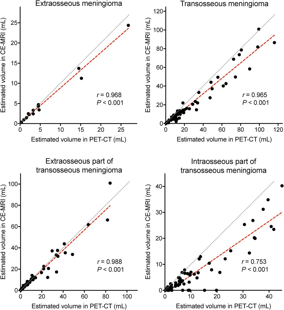
Separate volumetric assessments of the meningioma using 68Ga-DOTATATE PET/CT and CE-MRI yielded comparable results for extraosseous meningiomas (3.2 vs. 2.5 mL; P = 0.132) and the extraosseous part of transosseous meningiomas (6.6 vs. 6.7 mL; P = 0.636) based on the Wilcoxon signed rank test. In contrast, the intraosseous part of transosseous meningiomas was assessed as having significantly larger volumes by PET/CT (6.8 vs. 3.3 mL; P < 0.001). Consistently, analyses using Spearman correlation demonstrated lower correlation coefficients for the volume estimates of the intraosseous part of transosseous meningiomas (r = 0.753) than for the volume estimates of the extraosseous part (r = 0.988). The individual results are shown in a correlation scatterplot in Figure 2 and in a grouped volume plot in Supplemental Figure 3. Subgroup analyses for different meningioma locations returned similar relations in volume estimation; the results are presented in Figure 3. A representative patient example for the discrepancy in estimating the intraosseous meningioma volume is presented in Figure 4.

Comparison of estimated volumes in 68Ga-DOTATATE PET/CT and CE-MRI. Correlation scatterplots depict estimated volumes using 68Ga-DOTATATE PET/CT (x-axis) against estimated volumes using CE-MRI (y-axis). Spearman correlation coefficients (r) were calculated. Gray line represents perfect positive correlation. Red line is calculated using least-squares fit of data. Data are based on 82 meningioma patients with pathologic exclusion or inclusion of osseous involvement. In cases of imaging-based false-negative osseous involvement, intraosseous part was attributed no volume (0 mL). Wilcoxon signed rank test demonstrated significant differences between PET and CE-MRI for volume estimation of total and intraosseous part of transosseous meningiomas (each with P < 0.001).
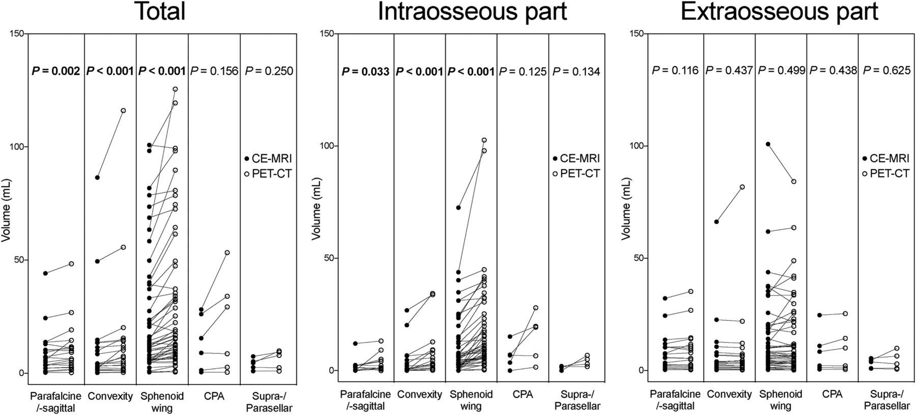
Comparison of estimated volumes stratified for meningioma location. Plots depict estimated volumes for total meningioma, intraosseous part, and extraosseous part using 68Ga-DOTATATE PET/CT (open circles) and CE-MRI (solid circles). Data are based on 105 meningioma patients, among whom 90 had imaging-based diagnosis of transosseous extension. Statistical comparison was performed using Wilcoxon signed rank test. Bold P values indicate statistical significance. CPA = cerebellopontine angle.

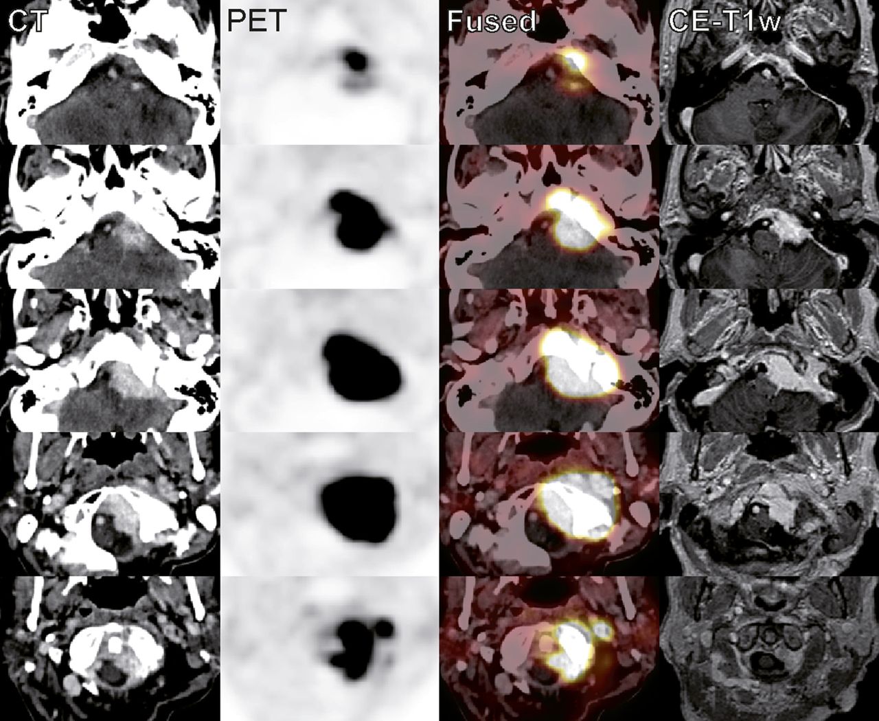
Example of estimated intraosseous volume discrepancy between 68Ga-DOTATATE PET/CT and CE-MRI in patient with skull base meningioma. A 75-y-old woman was followed up with 68Ga-DOTATATE PET/CT and CE-MRI after partial resection of transosseous transitional meningioma (World Health Organization grade I) in left cerebellopontine angle. Because of worsening and radiating pain in occipital region and hypoglossal nerve palsy, she was evaluated for stereotactic radiosurgery. Estimated volumes of transosseous extension were significantly larger on 68Ga-DOTATATE PET/CT than on CE-MRI (28.0 vs. 7.5 mL), whereas volumes of extraosseous part agreed well (25.3 vs. 22.6 mL). w = weighted.
DISCUSSION
The recently published European Association of Neuro-Oncology guidelines for the diagnosis and treatment of meningiomas by Goldbrunner et al. (2) reinforced the role of CE-MRI as the primary imaging modality of choice. However, the authors also underlined the considerable diagnostic challenges in the delineation of meningiomas, particularly in complex locations such as the skull base. The limited sensitivity of CE-MRI for the diagnosis of osseous involvement in our study is in line with a previous study comparing 18F-fluoride PET/CT with CE-MRI by Tateishi et al. (21). 68Ga-DOTATATE PET/CT provided an improved detection of osseous involvement similar to that of 18F-fluoride PET/CT based on the comparison of receiver-operating-characteristic analyses. In 18F-fluoride PET/CT, intraosseous meningioma extension is indirectly estimated by osseous 18F-fluoride tracer uptake, reflecting active bone formation and osseous turnover (22). In contrast, 68Ga-DOTATATE directly binds to meningioma tissue characterized by somatostatin-receptor overexpression.
The higher 68Ga-DOTATATE tracer uptake of transosseous meningiomas is in line with a previous report by Sommerauer et al. (12), which originally investigated the prognostic information of tracer uptake for growth prediction. Investigating a larger number of patients with transosseous meningiomas, we can confirm this observation. Notably, we did not detect a significant interrelation between meningioma volume and 68Ga-DOTATATE uptake values. Both the extraosseous and the intraosseous parts of transosseous meningiomas demonstrated strong tracer uptake. The slightly higher SUVmean of the intraosseous part than of the total meningioma is likely explained by the adaptation of the VOI, which may neglect areas of the tumor rim with lower tracer uptake. The difference in tracer uptake between extraosseous and transosseous meningiomas is interesting regarding its impact on growth prediction (12), as attempts to draw prognostic information from MRI parameters failed (25). As outlined by the recent European Association of Neuro-Oncology guidelines (2), evidence regarding surveillance and follow-up is scarce and recommendations were thus based on expert consensus opinion. In this respect, 68Ga-DOTATATE PET/CT has the potential to identify additional risk factors for tumor growth such as osseous involvement (13) or tracer uptake to guide the timing of follow-up imaging and improve patient surveillance.
Volumetric assessment of meningiomas using conventional imaging such as CE-MRI is cumbersome and often poorly approximated in daily clinical routine by the ABC/2 formula (26), thus introducing a considerable risk for misdiagnosis of tumor progression. Thereby, important prognostic information is neglected, as computer-aided volumetric analyses of meningiomas have proven to be more accurate in the assessment of tumor growth than the above-mentioned approximated volumes (27). In this light, 68Ga-DOTATATE PET/CT may facilitate automated volume measurements based on the PET tracer uptake, which provides improved tumor-to-background signal. A prior prospective study by Rachinger et al. (24) established an SUVmax cutoff of 2.3 for discrimination between meningioma and tumor-free tissue by spatially precise neuronavigated tissue-sampling during surgical resection. We applied this cutoff for the volume estimation by PET/CT and found good agreement with CE-MRI for extraosseous meningiomas and the extraosseous part of transosseous meningiomas. Given these features, 68Ga-DOTATATE PET/CT could make volume measurements and successive comparisons easily accessible in clinical routine and thereby improve therapy guidance. The intraosseous in contrast to the extraosseous part of transosseous meningiomas, however, showed considerable differences in volume estimation, as significantly larger intraosseous meningioma volumes were obtained using 68Ga-DOTATATE PET/CT than using CE-MRI. These differences might be explained by the considerably higher tumor-to-background signal that is provided by 68Ga-DOTATATE PET/CT. Visual comparison of the intraosseous PET and CE-MRI VOI shapes frequently revealed a very asymmetric distribution of the larger PET VOI around the CE-MRI VOI, exposing intraosseous meningioma extension undetected by CE-MRI. Because of this asymmetry between the PET and CE-MRI VOIs, a systematic overestimation of intraosseous meningioma extension by PET caused by spillover remains unlikely. On the basis of the differences in intraosseous as opposed to extraosseous volume estimation, we hypothesize, but cannot prove, that CE-MRI may underestimate the transosseous extension of intracranial meningiomas.
68Ga-DOTATATE PET adds important diagnostic molecular information to the primarily anatomic imaging modalities CT and MRI. Analogous to integrated PET/CT, hybrid PET/MRI scanners enable simultaneous acquisition of PET and MR images combining molecular PET information with high-resolution MR images yielding superior soft-tissue contrast. In addition, integrated acquisition of PET and MRI data provides optimized temporal and spatial data coregistration superior to software-based image fusion of separately acquired PET and MR images (28). In a comparison of 68Ga-DOTATOC PET/CT and PET/MRI for meningioma imaging, Afshar-Oromieh et al. demonstrated the feasibility of 68Ga-DOTATOC PET/MRI for the detection of intracranial meningiomas in a cohort of 15 patients (29). The authors reported that the acquired PET/MR images were free of artifacts and concluded that 68Ga-DOTATOC PET/MRI represents an ideal combination of high sensitivity and specificity with the best morphologic visualization. However, with regard to the comparison to 68Ga-DOTATOC PET/CT, the authors stated that no superiority of PET/MRI for treatment planning could be shown in this cohort and that larger studies are needed to compare the two techniques. In favor of PET/CT, important diagnostic CT information includes high-resolution bone imaging. Dedicated CT protocols aid in the localization of intraosseous meningioma manifestations and enable precise preoperative planning, particularly in difficult locations such as the skull base. In the case of calcified meningiomas, CT supports the assessment of gross tumor volume in radiation therapy planning, whereas calcifications cause extinction artifacts on MRI (5). Therefore, all 3 modalities—PET, MRI, and CT—contribute important information to diagnosis and treatment planning in meningioma. With 68Ga-DOTATATE PET as the most sensitive modality for the assessment of meningioma manifestations, the selection of either hybrid imaging modality, PET/CT or PET/MRI, is feasible. The third modality can be acquired separately, depending on clinical preference and availability.
There are limitations to our study that need to be considered when interpreting the data. First, pathologic confirmation of inclusion or exclusion of osseous involvement as the prerequisite for a study on diagnostic accuracy represents a selection bias toward symptomatic meningioma patient subgroups in which surgical resection was indicated. Second, the consecutive cohort of patients undergoing 68Ga-DOTATATE PET/CT examinations may cause a selection bias to patients with meningiomas in complex anatomic locations or with inconclusive MRI examinations. Third, the estimation of meningioma volumes could not be compared with a pathologic standard of reference because surgical resections were sometimes intended as partial resections or the resection material is too fragmented for ex vivo volume analysis. Therefore, no evidence on the pathologic meningioma volumes can be drawn. Yet, to our knowledge, this study represents the largest cohort of patients evaluated for osseous involvement by 68Ga-DOTATATE PET/CT and CE-MRI with pathologic confirmation as the standard of reference.
CONCLUSION
The present study evaluated the diagnostic performance of 68Ga-DOTATATE PET/CT and CE-MRI for the detection of osseous involvement by intracranial meningiomas, with pathology-proven bone infiltration as the standard of reference. 68Ga-DOTATATE PET/CT provided a higher sensitivity than CE-MRI while maintaining high specificity. Quantitative PET/CT analysis confirmed adequate tracer distribution in intraosseous meningioma parts and yielded significantly higher tumor-to-background signals than CE-MRI. Volumetric analysis using 68Ga-DOTATATE PET/CT returned significantly larger volume estimates for the intraosseous extension of transosseous meningiomas than CE-MRI, whereas the estimated volumes for extraosseous meningioma parts did not differ significantly between the two methods. 68Ga-DOTATATE PET/CT provides added diagnostic value in the assessment of transosseous meningiomas compared with CE-MRI.
DISCLOSURE
No potential conflict of interest relevant to this article was reported.
Footnotes
Published online Apr. 27, 2017.
- © 2017 by the Society of Nuclear Medicine and Molecular Imaging.
REFERENCES
- Received for publication February 17, 2017.
- Accepted for publication April 11, 2017.


Jump to section
Related Articles
Cited By...
- Meningioma: Molecular Updates from the 2021 World Health Organization Classification of CNS Tumors and Imaging Correlates
- Meningioma Revisited: Should Whole-Body Staging with [68Ga]Ga-DOTATOC PET/CT of High-Grade Meningiomas Become Standard Practice?
- DOTATATE PET/MR Imaging Differentiates Secondary-Progressive from de Novo World Health Organization Grade 3 Meningiomas
- Evaluation of [68Ga]-DOTATOC PET/MRI in Patients with Meningioma of the Subcranial and Intraorbital Space
- Cost-Effectiveness Analysis of 68Ga-DOTATATE PET/MRI in Radiotherapy Planning in Patients with Intermediate-Risk Meningioma
- The Complementary Role of 68Ga-DOTATATE PET/CT in Diagnosis of Recurrent Meningioma
- Improved Detection of Postoperative Residual Meningioma with [68Ga]Ga-DOTA-TOC PET Imaging Using a High-resolution Research Tomograph PET Scanner