


Article Figures & Data
Figures
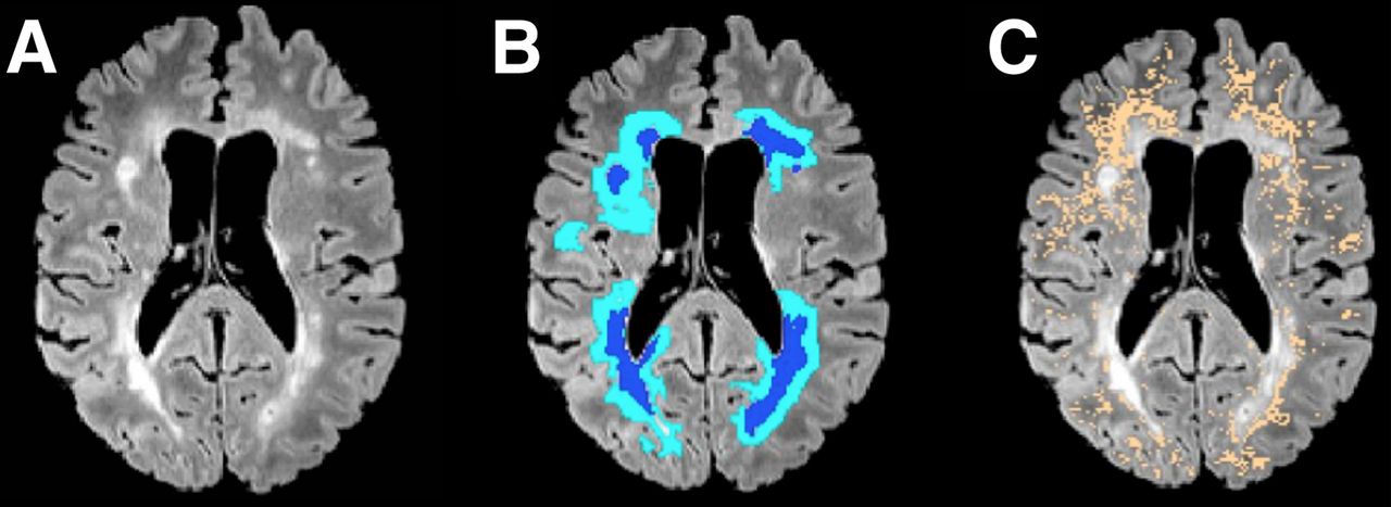
- FIGURE 1.
(A) T2 FLAIR image of representative MS patient (patient 1, Supplemental Table 1). Hyperintense areas correspond to demyelinating lesions. (B) Lesion (dark blue) and perilesional areas (light blue) corresponding to intersection of 6-mm-diameter sphere traced around lesions within image plane. (C) Nonlesional voxels with MTR values ranging between 90% and 98% of mean MTR of nonlesional WM tissue (NLLM) (copper).
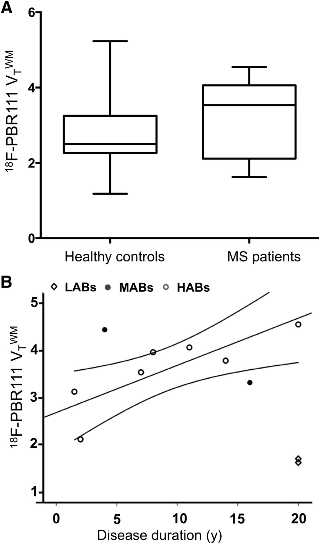
- FIGURE 2.
(A) 18F-PBR111 VT in whole WM of MS patients and genotype- and age-matched healthy control subjects. Lines in middle of boxes are median values, whereas hinges represent 25th and 75th percentiles, respectively. Whiskers represent maximum and minimum values. Contrast between MS patients and healthy controls showed trend for higher 18F-PBR111
 in MS patients (Wilcoxon rank P = 0.062). (B) Relationship between whole WM 18F-PBR111 VT and disease duration in MS patients for patients with different rs6971 genotypes (Spearman partial correlation in HABs: ρ = 0.86; P < 0.05, corrected for age).
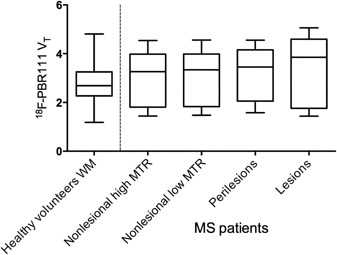
 - FIGURE 3.
18F-PBR111 uptake in healthy volunteers and across MS patient ROIs. Lines in middle of boxes are median values, whereas hinges represent 25th and 75th percentiles, respectively. Whiskers represent maximum and minimum values. Between-group contrasts showed that MS patients’ 18F-PBR111
and were greater than healthy volunteers’ (Wilcoxon rank P < 0.05). Within–MS patient contrasts showed was greater than and was greater than (Wilcoxon rank P < 0.05), and was greater than (Wilcoxon rank P < 0.005). - FIGURE 4.
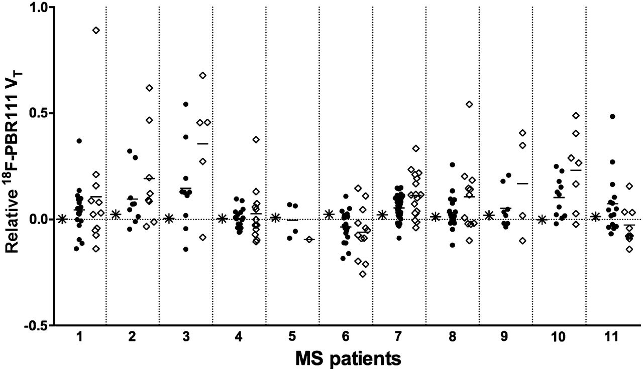
Relative 18F-PBR111 uptake (relative to NLHM WM) in NLLM WM, in individual T2 FLAIR lesions, and in perilesional volumes for MS patients studied. Ordinant represents relative difference in 18F-PBR111 VT in lesions (♢), in perilesional volumes (●), and in NLLM WM (*) relative to normal-appearing WM (NLHM WM). On abscissa, MS patients (Supplemental Table 1) are separately indicated.
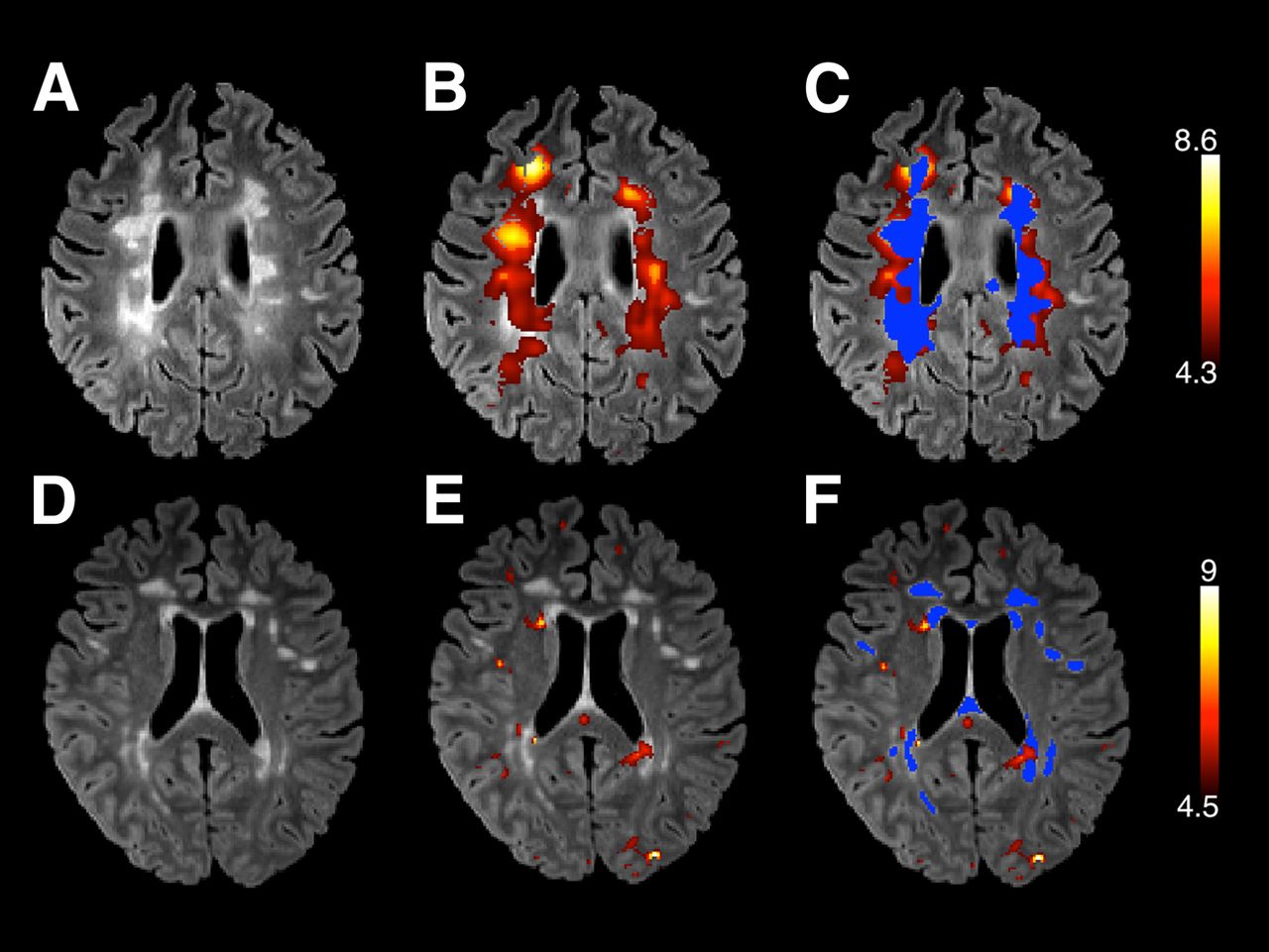
- FIGURE 5.
T2 FLAIR images (A and D), 18F-PBR111 VT parametric maps (B and E) overlaid (in warm colors) on T2 FLAIR images, and overlap between T2 FLAIR lesions (marked in blue) and 18F-PBR111 VT (in warm colors) (C and F) from 2 illustrative patients. Lower threshold for VT parametric maps corresponds to value of 18F-PBR111 VT in NLHM volume for each of 2 patients. Upper threshold is twice VT in NLHM. Upper row illustrates patient with recent active disease (Supplemental Table 1, patient 9). Here, T2 FLAIR hyperintense lesional areas correspond to areas of increased 18F-PBR111 signal. Lower row illustrates patient (Supplemental Table 1, patient 4) with relatively benign disease course showing focal regions of increased 18F-PBR111 VT that correspond poorly to T2 FLAIR hyperintense areas.
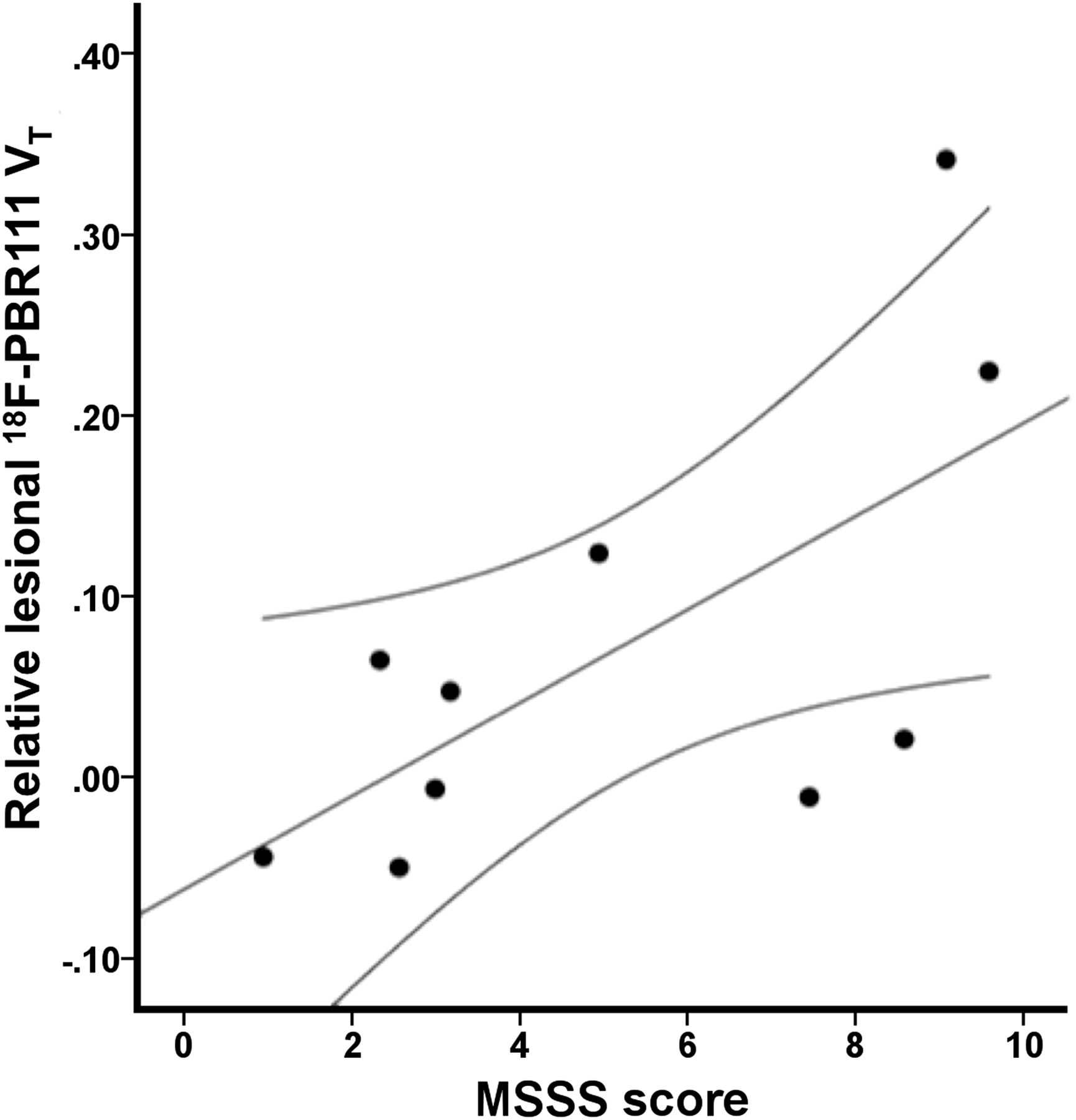
- FIGURE 6.
Positive relationship between MSSSs and 18F-PBR111 uptake in lesions (expressed relative to that in normal-appearing WM) (Spearman ρ = 0.62, P ≤ 0.05).


Additional Files
Supplemental Data
Files in this Data Supplement:


Jump to section
Related Articles
Cited By...
- Secondary Progressive Multiple Sclerosis: New Insights
- Quantitative magnetisation transfer imaging in relapsing-remitting multiple sclerosis: a systematic review and meta-analysis
- Natalizumab treatment reduces microglial activation in the white matter of the MS brain
- 11C-PBR28 and 18F-PBR111 Detect White Matter Inflammatory Heterogeneity in Multiple Sclerosis
- Imaging Microglial Activation with TSPO PET: Lighting Up Neurologic Diseases?
- Imaging robust microglial activation after lipopolysaccharide administration in humans with PET