
Main menu

  • Home
  • Content
    • Current
    • Ahead of print
    • Past Issues
    • JNM Supplement
    • SNMMI Annual Meeting Abstracts
    • Continuing Education
    • JNM Podcasts
  • Subscriptions
    • Subscribers
    • Institutional and Non-member
    • Rates
    • Journal Claims
    • Corporate & Special Sales
  • Authors
    • Submit to JNM
    • Information for Authors
    • Assignment of Copyright
    • AQARA requirements
  • Info
    • Reviewers
    • Permissions
    • Advertisers
  • About
    • About Us
    • Editorial Board
    • Contact Information
  • More
    • Alerts
    • Feedback
    • Help
    • SNMMI Journals
  • SNMMI
    • JNM
    • JNMT
    • SNMMI Journals
    • SNMMI

User menu

  • Subscribe
  • My alerts
  • Log in
  • Log out
  • My Cart

Search

  • Advanced search
Journal of Nuclear Medicine
  • SNMMI
    • JNM
    • JNMT
    • SNMMI Journals
    • SNMMI
  • Subscribe
  • My alerts
  • Log in
  • Log out
  • My Cart
Journal of Nuclear Medicine

Advanced Search

  • Home
  • Content
    • Current
    • Ahead of print
    • Past Issues
    • JNM Supplement
    • SNMMI Annual Meeting Abstracts
    • Continuing Education
    • JNM Podcasts
  • Subscriptions
    • Subscribers
    • Institutional and Non-member
    • Rates
    • Journal Claims
    • Corporate & Special Sales
  • Authors
    • Submit to JNM
    • Information for Authors
    • Assignment of Copyright
    • AQARA requirements
  • Info
    • Reviewers
    • Permissions
    • Advertisers
  • About
    • About Us
    • Editorial Board
    • Contact Information
  • More
    • Alerts
    • Feedback
    • Help
    • SNMMI Journals
  • View or Listen to JNM Podcast
  • Visit JNM on Facebook
  • Join JNM on LinkedIn
  • Follow JNM on Twitter
  • Subscribe to our RSS feeds
Research ArticleClinical Investigations

Systematic Comparison of the Performance of Integrated Whole-Body PET/MR Imaging to Conventional PET/CT for 18F-FDG Brain Imaging in Patients Examined for Suspected Dementia

Stefan Hitz, Cornelia Habekost, Sebastian Fürst, Gaspar Delso, Stefan Förster, Sibylle Ziegler, Stephan G. Nekolla, Michael Souvatzoglou, Ambros J. Beer, Timo Grimmer, Matthias Eiber, Markus Schwaiger and Alexander Drzezga
Journal of Nuclear Medicine June 2014, 55 (6) 923-931; DOI: https://doi.org/10.2967/jnumed.113.126813
Stefan Hitz
1Department of Nuclear Medicine, Technische Universität München, Munich, Germany
  • Find this author on Google Scholar
  • Find this author on PubMed
  • Search for this author on this site
Cornelia Habekost
1Department of Nuclear Medicine, Technische Universität München, Munich, Germany
  • Find this author on Google Scholar
  • Find this author on PubMed
  • Search for this author on this site
Sebastian Fürst
1Department of Nuclear Medicine, Technische Universität München, Munich, Germany
  • Find this author on Google Scholar
  • Find this author on PubMed
  • Search for this author on this site
Gaspar Delso
1Department of Nuclear Medicine, Technische Universität München, Munich, Germany
  • Find this author on Google Scholar
  • Find this author on PubMed
  • Search for this author on this site
Stefan Förster
1Department of Nuclear Medicine, Technische Universität München, Munich, Germany
2TUM Neuroimaging Center (TUM-NIC), Technische Universität München, Munich, Germany
  • Find this author on Google Scholar
  • Find this author on PubMed
  • Search for this author on this site
Sibylle Ziegler
1Department of Nuclear Medicine, Technische Universität München, Munich, Germany
  • Find this author on Google Scholar
  • Find this author on PubMed
  • Search for this author on this site
Stephan G. Nekolla
1Department of Nuclear Medicine, Technische Universität München, Munich, Germany
  • Find this author on Google Scholar
  • Find this author on PubMed
  • Search for this author on this site
Michael Souvatzoglou
1Department of Nuclear Medicine, Technische Universität München, Munich, Germany
  • Find this author on Google Scholar
  • Find this author on PubMed
  • Search for this author on this site
Ambros J. Beer
1Department of Nuclear Medicine, Technische Universität München, Munich, Germany
  • Find this author on Google Scholar
  • Find this author on PubMed
  • Search for this author on this site
Timo Grimmer
3Department of Psychiatry and Psychotherapy, Technische Universität München, Munich, Germany
  • Find this author on Google Scholar
  • Find this author on PubMed
  • Search for this author on this site
Matthias Eiber
4Department of Radiology, Technische Universität München, Munich, Germany; and
  • Find this author on Google Scholar
  • Find this author on PubMed
  • Search for this author on this site
Markus Schwaiger
1Department of Nuclear Medicine, Technische Universität München, Munich, Germany
  • Find this author on Google Scholar
  • Find this author on PubMed
  • Search for this author on this site
Alexander Drzezga
1Department of Nuclear Medicine, Technische Universität München, Munich, Germany
5Department of Nuclear Medicine, University Hospital of Cologne, Cologne, Germany
  • Find this author on Google Scholar
  • Find this author on PubMed
  • Search for this author on this site
  • Article
  • Figures & Data
  • Supplemental
  • Info & Metrics
  • PDF
Loading

Article Figures & Data

Figures

  • Tables
  • Additional Files
  • FIGURE 1.
    • Download figure
    • Open in new tab
    • Download powerpoint
    FIGURE 1.

    Voxel-based group comparison reveals statistically significant differences in measured regional activity in AC (left) and NAC (right) data between PET/MR and PET/CT. For display, green was used for relatively higher measured PET signal in PET/MR than PET/CT (extent threshold in AC, T = 2.50, P < 0.05, false discovery rate–corrected; in NAC, T = 2.05, P < 0.05, false discovery rate–corrected), and red represented relatively higher measured PET signal in PET/CT (extent threshold in AC, T = 2.12, P < 0.05, false discovery rate–corrected; in NAC, T = 2.77, P < 0.05, false discovery rate–corrected).
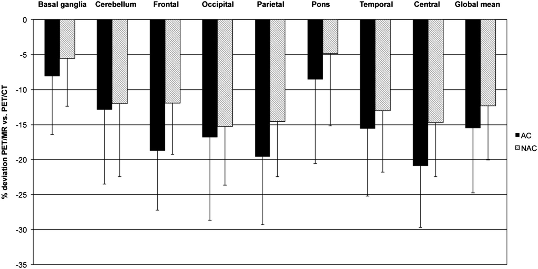
  • FIGURE 2.
    • Download figure
    • Open in new tab
    • Download powerpoint
    FIGURE 2.

    Percentage differences of values in different anatomic regions by ROI-based analysis. Error bars indicate SD.
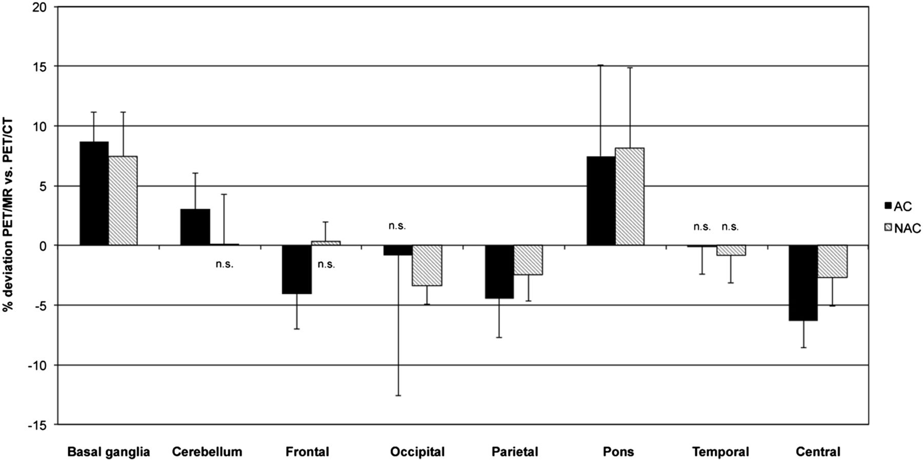
  • FIGURE 3.
    • Download figure
    • Open in new tab
    • Download powerpoint
    FIGURE 3.

    Percentage differences of values in different anatomic regions by ROI-based analysis in data corrected for global differences. Error bars indicate SD. n.s. = no significant difference between PET/MR and PET/CT.
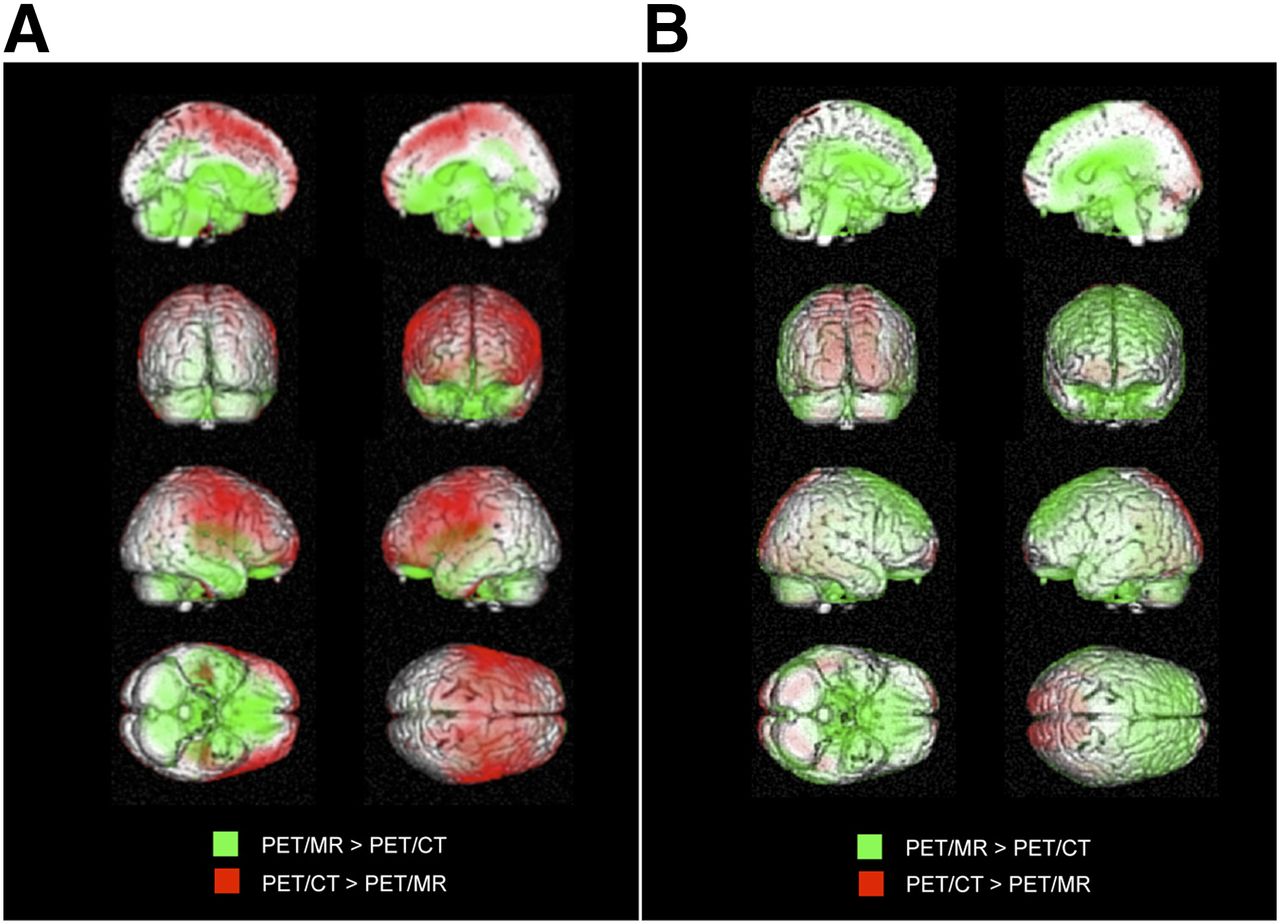
  • FIGURE 4.
    • Download figure
    • Open in new tab
    • Download powerpoint
    FIGURE 4.

    Patient 1 (PET/CT first): 3DSSPs generated by Neurostat after anatomic stereotactic normalization of same patient examined with PET/CT (A) and PET/MR (B). 3DSSPs of individual patient’s 18F-FDG PET data (upper) and pixelwise comparison of individual patient’s PET data to age-matched reference database, resulting in z score images (high z score = significantly reduced glucose metabolism; reference region: global mean) (lower). Compared with PET/CT, lower relative metabolic rate can be observed in frontoparietal portions of neocortex in PET/MR data on 3DSSP images (red arrows). On z score images, relatively stronger deviations from control are detected (green arrows) for PET/MR data.

Tables

  • Figures
  • Additional Files
    • View popup
    TABLE 1

    Patients’ Characteristics

    Patient no.SexAge (y)Examination sequenceTime interval (min) between PET/MR and PET/CT
    1M69PET/CT first34
    2F39PET/CT first36
    3M71PET/CT first46
    4F52PET/CT first35
    5M60PET/CT first33
    6M76PET/CT first70
    7F75PET/CT first33
    8F25PET/CT first48
    9F61PET/MR first40
    10F53PET/MR first40
    11F45PET/MR first37
    12M45PET/MR first29
    13F46PET/MR first41
    14F61PET/CT first29
    15F70PET/CT first26
    16M64PET/CT first37
    17F74PET/CT first37
    18M73PET/MR first25
    19F79PET/MR first50
    20M64PET/MR first42
    21F70PET/MR first22
    22F45PET/MR first22
    23F66PET/MR first34
    24F68PET/MR first44
    25F57PET/MR first32
    26F55PET/MR first40
    27F64PET/CT first44
    28M67PET/CT first30
    29F72PET/CT first40
    30M60PET/CT first25
    • View popup
    TABLE 2

    Study Characteristics

    CharacteristicPET/CT firstPET/MR firstP
    n1614Not significant
    Patient median age (y)65.559Not significant
    Time between PET/MR and PET/CT37.69 ± 10.8535.57 ± 8.55Not significant
    Male73Not significant
    Female911Not significant
    • View popup
    TABLE 3

    ROI Analysis

    Data typeBasal gangliaCerebellumFrontalOccipitalParietalPonsTemporalCentral
    AC data
     PET/MR AC1.04 ± 0.061.04 ± 0.050.98 ± 0.031.08 ± 0.051.03 ± 0.050.83 ± 0.050.95 ± 0.041.05 ± 0.04
     PET/CT AC0.96 ± 0.061.01 ± 0.051.02 ± 0.041.09 ± 0.091.08 ± 0.060.78 ± 0.080.95 ± 0.051.12 ± 0.04
     Difference between PET/CT and PET/MR−0.08−0.030.040.010.05−0.060.000.07
     % Difference PET/MR (PET/CT = 100%)8.663.01−4.08−0.86−4.497.48−0.11−6.33
     P (PET/CT vs. PET/MR)P < 0.001P < 0.001P < 0.001NSP < 0.001P < 0.001NSP < 0.001
    NAC data
     PET/MR NAC0.84 ± 0.090.97 ± 0.051.12 ± 0.051.16 ± 0.081.15 ± 0.070.62 ± 0.070.98 ± 0.041.16 ± 0.06
     PET/CT NAC0.78 ± 0.110.96 ± 0.051.12 ± 0.051.19 ± 0.091.18 ± 0.070.57 ± 0.070.99 ± 0.051.20 ± 0.07
     Difference between PET/CT and PET/MR−0.060.000.000.040.03−0.050.010.03
     % Difference PET/MR (PET/CT = 100%)7.410.130.36−3.34−2.488.13−0.85−2.68
     P (PET/CT vs. PET/MR)P < 0.001NSNSP < 0.001P < 0.001P < 0.001NSP < 0.001
    • Mean uptake values obtained by ROI analysis, normalized to individual global mean.

    • NS = no significant difference.

Additional Files

  • Figures
  • Tables
  • Supplemental Data

    Files in this Data Supplement:

    • Supplemental Data
PreviousNext
Back to top

In this issue

Journal of Nuclear Medicine: 55 (6)
Journal of Nuclear Medicine
Vol. 55, Issue 6
June 1, 2014
  • Table of Contents
  • Table of Contents (PDF)
  • About the Cover
  • Index by author
Print
Download PDF
Article Alerts
Sign In to Email Alerts with your Email Address
Email Article

Thank you for your interest in spreading the word on Journal of Nuclear Medicine.

NOTE: We only request your email address so that the person you are recommending the page to knows that you wanted them to see it, and that it is not junk mail. We do not capture any email address.

Enter multiple addresses on separate lines or separate them with commas.
Systematic Comparison of the Performance of Integrated Whole-Body PET/MR Imaging to Conventional PET/CT for 18F-FDG Brain Imaging in Patients Examined for Suspected Dementia
(Your Name) has sent you a message from Journal of Nuclear Medicine
(Your Name) thought you would like to see the Journal of Nuclear Medicine web site.
Citation Tools
Systematic Comparison of the Performance of Integrated Whole-Body PET/MR Imaging to Conventional PET/CT for 18F-FDG Brain Imaging in Patients Examined for Suspected Dementia
Stefan Hitz, Cornelia Habekost, Sebastian Fürst, Gaspar Delso, Stefan Förster, Sibylle Ziegler, Stephan G. Nekolla, Michael Souvatzoglou, Ambros J. Beer, Timo Grimmer, Matthias Eiber, Markus Schwaiger, Alexander Drzezga
Journal of Nuclear Medicine Jun 2014, 55 (6) 923-931; DOI: 10.2967/jnumed.113.126813

Citation Manager Formats

  • BibTeX
  • Bookends
  • EasyBib
  • EndNote (tagged)
  • EndNote 8 (xml)
  • Medlars
  • Mendeley
  • Papers
  • RefWorks Tagged
  • Ref Manager
  • RIS
  • Zotero
Share
Systematic Comparison of the Performance of Integrated Whole-Body PET/MR Imaging to Conventional PET/CT for 18F-FDG Brain Imaging in Patients Examined for Suspected Dementia
Stefan Hitz, Cornelia Habekost, Sebastian Fürst, Gaspar Delso, Stefan Förster, Sibylle Ziegler, Stephan G. Nekolla, Michael Souvatzoglou, Ambros J. Beer, Timo Grimmer, Matthias Eiber, Markus Schwaiger, Alexander Drzezga
Journal of Nuclear Medicine Jun 2014, 55 (6) 923-931; DOI: 10.2967/jnumed.113.126813
Twitter logo Facebook logo LinkedIn logo Mendeley logo
  • Tweet Widget
  • Facebook Like
  • Google Plus One
Bookmark this article

Jump to section

  • Article
    • Abstract
    • MATERIALS AND METHODS
    • RESULTS
    • DISCUSSION
    • CONCLUSION
    • DISCLOSURE
    • Acknowledgments
    • Footnotes
    • REFERENCES
  • Figures & Data
  • Supplemental
  • Info & Metrics
  • PDF

Related Articles

  • This Month in JNM
  • PubMed
  • Google Scholar

Cited By...

  • Hybrid PET/MR Imaging in Neurology: Present Applications and Prospects for the Future
  • Effect of Attenuation Correction on Regional Quantification Between PET/MR and PET/CT: A Multicenter Study Using a 3-Dimensional Brain Phantom
  • Google Scholar

More in this TOC Section

  • Feasibility of Ultra-Low-Activity 18F-FDG PET/CT Imaging Using a Long–Axial-Field-of-View PET/CT System
  • Cardiac Presynaptic Sympathetic Nervous Function Evaluated by Cardiac PET in Patients with Chronotropic Incompetence Without Heart Failure
  • Validation and Evaluation of a Vendor-Provided Head Motion Correction Algorithm on the uMI Panorama PET/CT System
Show more Clinical Investigations

Similar Articles

Keywords

  • PET/MR
  • PET/CT
  • brain imaging
  • Dixon MRI sequence
  • neurodegeneration
SNMMI

© 2025 SNMMI

Powered by HighWire