


Article Figures & Data
Figures
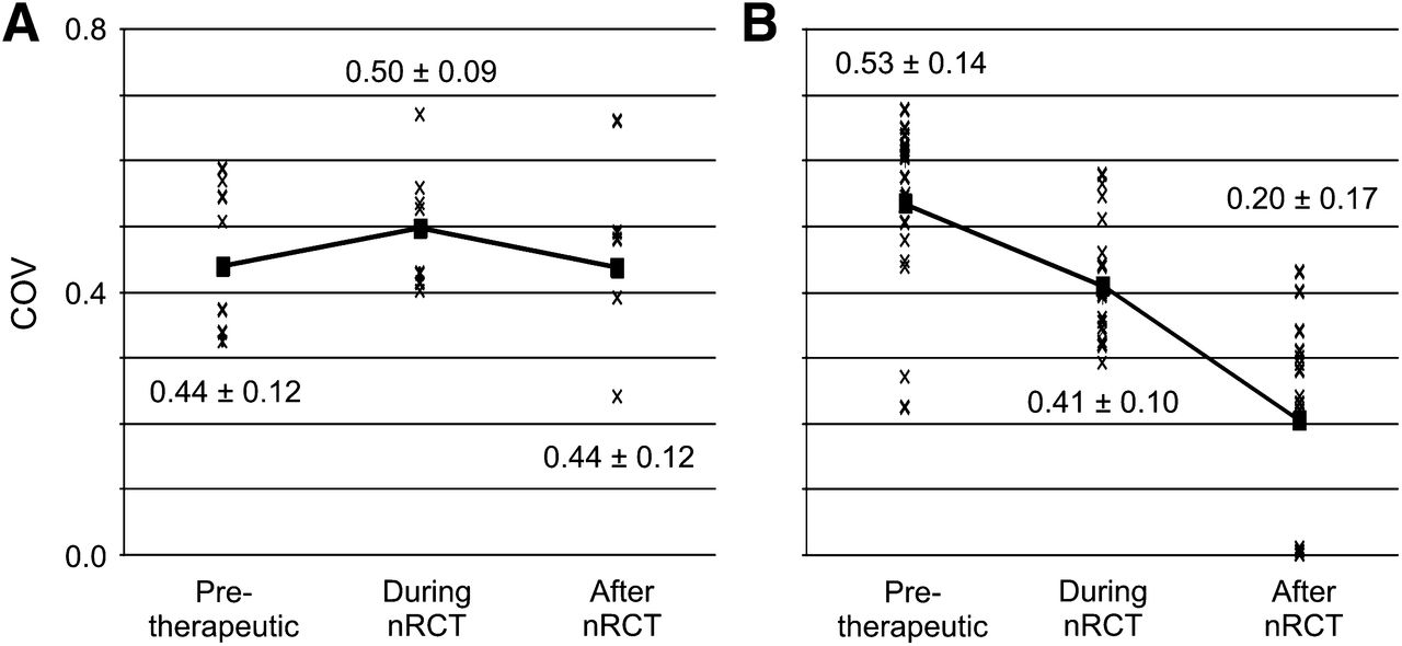
- FIGURE 1.
Individual COV values for histopathologic responders (A) and nonresponders (B) on PET/CT before, 2 wk after start, and after completion of nRCT. Squares indicate mean values over all patients in each group; solid lines indicate changes during therapy.
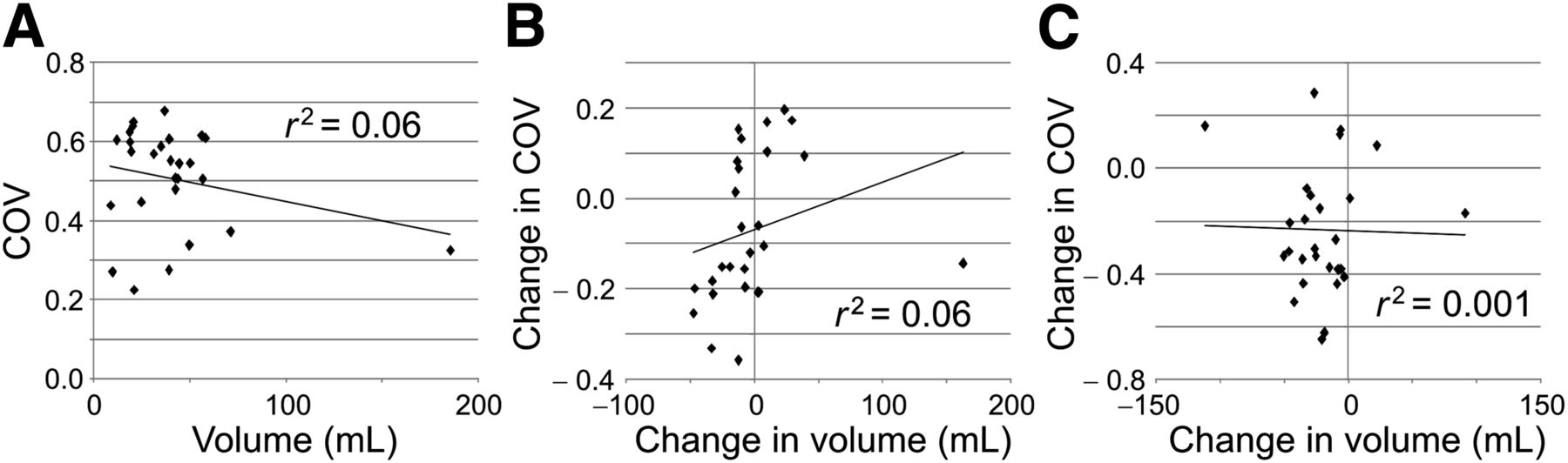
- FIGURE 2.
Correlation between COV and lesion volume at time point 1 (A), between changes in COV and volume early in therapy (time points 1 to 2) (B), and between changes in COV and volume after end of therapy (time points 1 to 3) (C).
- FIGURE 3.
Kaplan–Meier plots and number-at-risk tables for probability of progression-free survival. Low-risk group (solid lines) was identified by COV measured on 18F-FDG PET/CT before start of nRCT (A), by changes in COV (ΔCOV) between baseline and 2 wk after start of nRCT (early response assessment) (B), and by ΔCOV between baseline and 18F-FDG PET/CT 2 wk after completion of nRCT (late response assessment) (C). n = number of individuals in each group; e = number of events in each group. Time of censoring is marked by a dot.
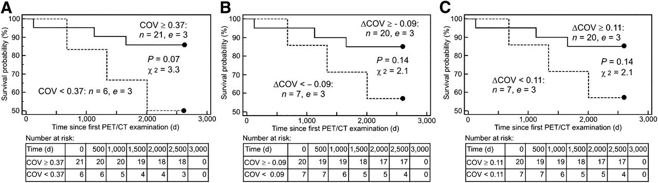
- FIGURE 4.
Kaplan–Meier plots and number-at-risk tables for probability of overall survival. Responders (solid lines) were identified by COV measured on 18F-FDG PET/CT before start of nRCT (A), by changes in COV (ΔCOV) between baseline and 2 wk after start of nRCT (early response assessment) (B), and by ΔCOV between baseline and 18F-FDG PET/CT 2 wk after completion of nRCT (late response assessment) (C). n = number of individuals in each group; e = number of events in each group. Time of censoring is marked by a dot.


Tables
Parameter AUC 95% confidence interval SUVmax 0.52 0.32–0.71 Skewness 0.55 0.33–0.75 Kurtosis 0.61 0.39–0.81 SUVmean 0.68 0.48–0.85 Diameter 0.68 0.48–0.85 COV 0.73 0.53–0.88 Volume 0.75 0.55–0.90 TLG 0.79 0.59–0.92 Parameter AUC 95% confidence interval Early response* Kurtosis 0.50 0.29–0.71 Skewness 0.50 0.29–0.71 SUVmean 0.57 0.36–0.75 TLG 0.60 0.39–0.78 Volume 0.66 0.45–0.83 Diameter 0.68 0.48–0.85 SUVmax 0.68 0.48–0.85 COV 0.83 0.63–0.94 Late response† Volume 0.51 0.31–0.70 SUVmean 0.51 0.32–0.71 Diameter 0.68 0.47–0.87 SUVmax 0.74 0.53–0.89 TLG 0.74 0.54–0.89 Skewness 0.74 0.52–0.90 Kurtosis 0.74 0.53–0.90 COV 0.89 0.71–0.98 Time point Mean progression-free survival (d) Mean overall survival (d) 1, COV ≥ 0.37 2,210 2,362 1, COV < 0.37 1,445 1,967 1→2, ΔCOV ≥ −0.09 2,192 2,351 1→2, ΔCOV < −0.09 1,518 2,056 1→3, ΔCOV ≥ 0.11 2,261 2,351 1→3, ΔCOV < 0.11 1,445 2,056 Overall patients 2,053 2,274


Jump to section
Related Articles
Cited By...
- Radiomics Features of 18F-fluorodeoxyglucose Positron-Emission Tomography as a Novel Prognostic Signature in Colorectal Cancer
- Immune Checkpoint Imaging in Oncology: A Game Changer Toward Personalized Immunotherapy?
- Investigating PSMA-Targeted Radioligand Therapy Efficacy as a Function of Cellular PSMA Levels and Intratumoral PSMA Heterogeneity
- Predictive Role of Temporal Changes in Intratumoral Metabolic Heterogeneity During Palliative Chemotherapy in Patients with Advanced Pancreatic Cancer: A Prospective Cohort Study
- Optimized Feature Extraction for Radiomics Analysis of 18F-FDG PET Imaging
- Glioma Survival Prediction with Combined Analysis of In Vivo 11C-MET PET Features, Ex Vivo Features, and Patient Features by Supervised Machine Learning
- Differential Prognostic Value of Metabolic Heterogeneity of Primary Tumor and Metastatic Lymph Nodes in Patients with Pharyngeal Cancer
- Impact of Image Reconstruction Settings on Texture Features in 18F-FDG PET
- Molecular Imaging to Plan Radiotherapy and Evaluate Its Efficacy