


Article Figures & Data
Figures
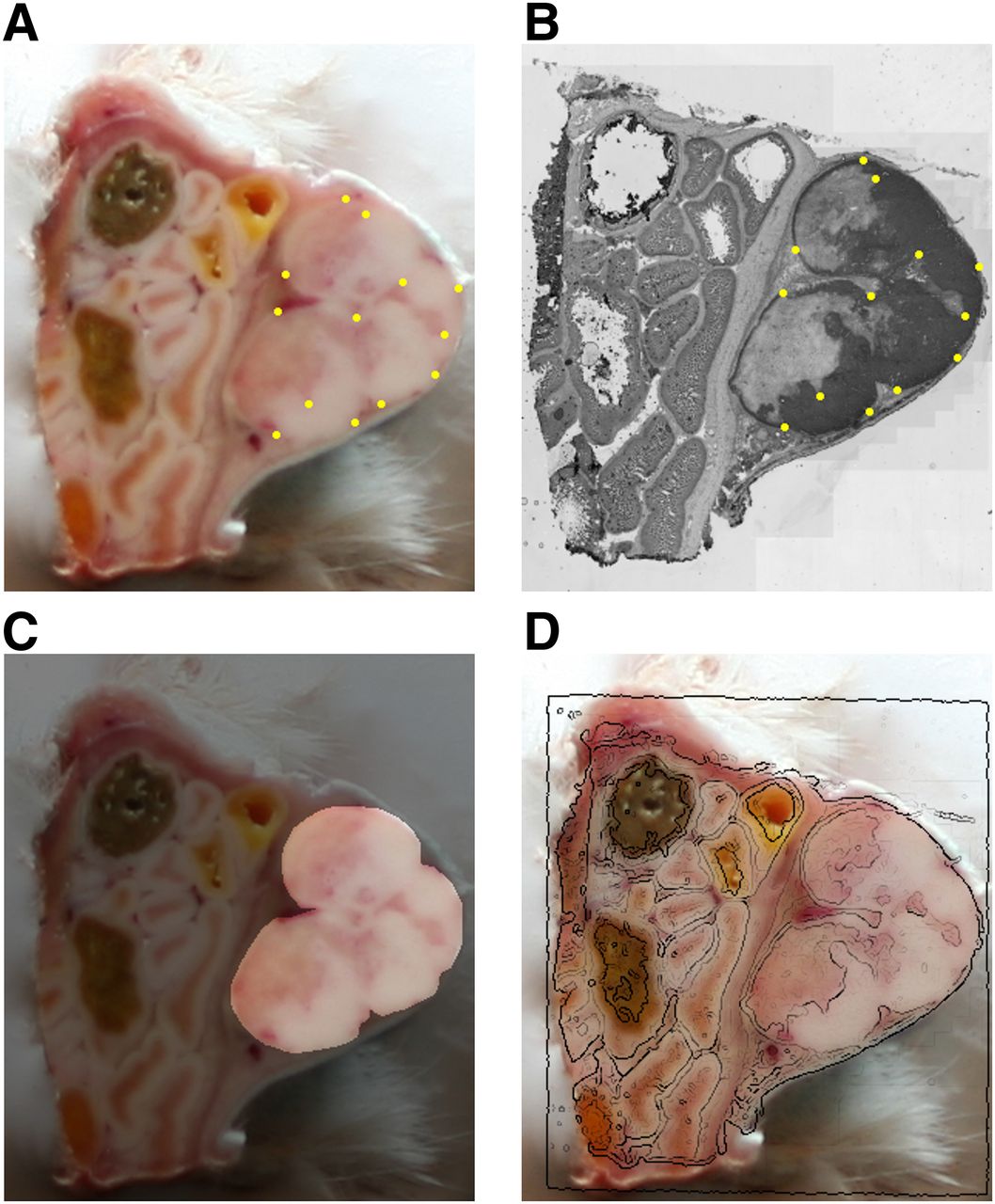
- FIGURE 1.
Example in-plane 2D registration of EGFr-stained histology image to corresponding tissue block photograph. (A) Block-face image with yellow dots denoting manually selected landmarks. (B) Blue color channel extracted from original RGB color histology image, with yellow dots denoting points corresponding to landmarks in A and EGFr target expression in dark gray. (C) Tumor mask used during registration and calculation of percentage necrosis. (D) Edges of registered histology image (detected using Canny edge detector) overlaid onto block-face photograph.
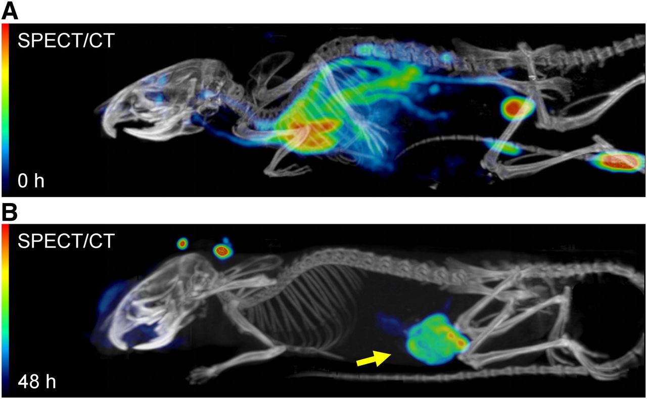
- FIGURE 2.
Total-body fused SPECT/CT maximum-intensity projections showing biodistribution of 111In-labeled zalutumumab directly after injection (A) and 2 d after injection (B). Arrow points to A431 xenograft.
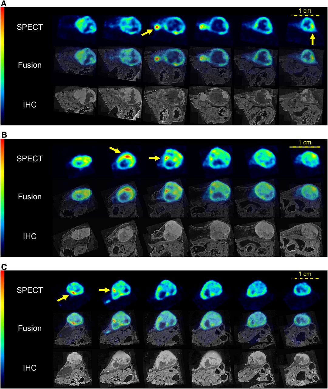
- FIGURE 3.
High-resolution in vivo SPECT slices of 111In distribution in A431 xenograft compared with cross-sections through registered 3D immunohistochemistry (IHC) stack. Top rows: equidistant consecutive SPECT slices. Middle rows: SPECT images fused with registered immunohistochemistry slices stained for EGFr expression. Bottom rows: nonfused immunohistochemistry slices. Arrows point to hot spots that seem to be located at outer rim of or outside EGFr regions. Panels A, B, and C show results from mice 1, 2, and 3, respectively.
- FIGURE 4.
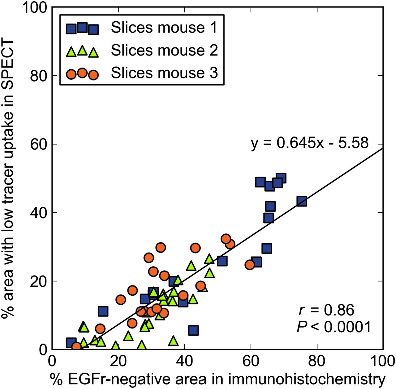
Scatterplot relating EGFr-negative (necrotic) tissue to regions with low SPECT uptake, both quantified as percentage of total slice area.
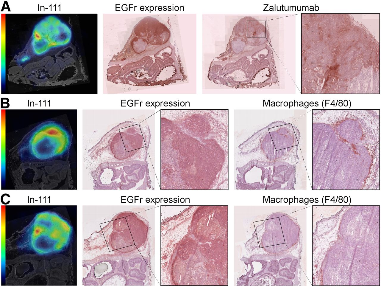
- FIGURE 5.
Expression and distribution of radiolabel (111In), EGFr, antibody (zalutumumab), and macrophages (F4/80). (A) SPECT slice from mouse 3 compared with 2 registered adjacent sections stained for EGFr expression (ex vivo incubation with zalutumumab) and presence of in vivo administered zalutumumab. Arrows point to 111In hot spot in region with neither EGFr uptake nor zalutumumab expression. Magnification shows heterogeneous distribution of zalutumumab. (B) Similar example from mouse 2 in which 111In hot spot was found to coincide with EGFr-positive tumor tissue and macrophages. (C) Similar example from mouse 2, where arrow points to hot spot in EGFr-positive tumor area that does not express macrophages.


Additional Files
Supplemental Data
Files in this Data Supplement:


Jump to section
Related Articles
Cited By...
- No citing articles found.