
Main menu

  • Home
  • Content
    • Current
    • Ahead of print
    • Past Issues
    • JNM Supplement
    • SNMMI Annual Meeting Abstracts
    • Continuing Education
    • JNM Podcasts
  • Subscriptions
    • Subscribers
    • Institutional and Non-member
    • Rates
    • Journal Claims
    • Corporate & Special Sales
  • Authors
    • Submit to JNM
    • Information for Authors
    • Assignment of Copyright
    • AQARA requirements
  • Info
    • Reviewers
    • Permissions
    • Advertisers
  • About
    • About Us
    • Editorial Board
    • Contact Information
  • More
    • Alerts
    • Feedback
    • Help
    • SNMMI Journals
  • SNMMI
    • JNM
    • JNMT
    • SNMMI Journals
    • SNMMI

User menu

  • Subscribe
  • My alerts
  • Log in
  • My Cart

Search

  • Advanced search
Journal of Nuclear Medicine
  • SNMMI
    • JNM
    • JNMT
    • SNMMI Journals
    • SNMMI
  • Subscribe
  • My alerts
  • Log in
  • My Cart
Journal of Nuclear Medicine

Advanced Search

  • Home
  • Content
    • Current
    • Ahead of print
    • Past Issues
    • JNM Supplement
    • SNMMI Annual Meeting Abstracts
    • Continuing Education
    • JNM Podcasts
  • Subscriptions
    • Subscribers
    • Institutional and Non-member
    • Rates
    • Journal Claims
    • Corporate & Special Sales
  • Authors
    • Submit to JNM
    • Information for Authors
    • Assignment of Copyright
    • AQARA requirements
  • Info
    • Reviewers
    • Permissions
    • Advertisers
  • About
    • About Us
    • Editorial Board
    • Contact Information
  • More
    • Alerts
    • Feedback
    • Help
    • SNMMI Journals
  • View or Listen to JNM Podcast
  • Visit JNM on Facebook
  • Join JNM on LinkedIn
  • Follow JNM on Twitter
  • Subscribe to our RSS feeds
Research ArticleBasic Science Investigations

In Vivo Visualization of MET Tumor Expression and Anticalin Biodistribution with the MET-Specific Anticalin 89Zr-PRS-110 PET Tracer

Anton G.T. Terwisscha van Scheltinga, Marjolijn N. Lub-de Hooge, Marlon J. Hinner, Remy B. Verheijen, Andrea Allersdorfer, Martin Hülsmeyer, Wouter B. Nagengast, Carolien P. Schröder, Jos G.W. Kosterink, Elisabeth G.E. de Vries, Laurent Audoly and Shane A. Olwill
Journal of Nuclear Medicine April 2014, 55 (4) 665-671; DOI: https://doi.org/10.2967/jnumed.113.124941
Anton G.T. Terwisscha van Scheltinga
1Department of Medical Oncology, University of Groningen, University Medical Center Groningen, The Netherlands
2Department of Hospital and Clinical Pharmacy, University of Groningen, University Medical Center Groningen, The Netherlands
  • Find this author on Google Scholar
  • Find this author on PubMed
  • Search for this author on this site
Marjolijn N. Lub-de Hooge
2Department of Hospital and Clinical Pharmacy, University of Groningen, University Medical Center Groningen, The Netherlands
3Department of Nuclear Medicine and Molecular Imaging, University of Groningen, University Medical Center Groningen, The Netherlands; and
  • Find this author on Google Scholar
  • Find this author on PubMed
  • Search for this author on this site
Marlon J. Hinner
4Pieris AG, Freising-Weihenstephan, Germany
  • Find this author on Google Scholar
  • Find this author on PubMed
  • Search for this author on this site
Remy B. Verheijen
1Department of Medical Oncology, University of Groningen, University Medical Center Groningen, The Netherlands
  • Find this author on Google Scholar
  • Find this author on PubMed
  • Search for this author on this site
Andrea Allersdorfer
4Pieris AG, Freising-Weihenstephan, Germany
  • Find this author on Google Scholar
  • Find this author on PubMed
  • Search for this author on this site
Martin Hülsmeyer
4Pieris AG, Freising-Weihenstephan, Germany
  • Find this author on Google Scholar
  • Find this author on PubMed
  • Search for this author on this site
Wouter B. Nagengast
1Department of Medical Oncology, University of Groningen, University Medical Center Groningen, The Netherlands
  • Find this author on Google Scholar
  • Find this author on PubMed
  • Search for this author on this site
Carolien P. Schröder
1Department of Medical Oncology, University of Groningen, University Medical Center Groningen, The Netherlands
  • Find this author on Google Scholar
  • Find this author on PubMed
  • Search for this author on this site
Jos G.W. Kosterink
2Department of Hospital and Clinical Pharmacy, University of Groningen, University Medical Center Groningen, The Netherlands
  • Find this author on Google Scholar
  • Find this author on PubMed
  • Search for this author on this site
Elisabeth G.E. de Vries
1Department of Medical Oncology, University of Groningen, University Medical Center Groningen, The Netherlands
  • Find this author on Google Scholar
  • Find this author on PubMed
  • Search for this author on this site
Laurent Audoly
4Pieris AG, Freising-Weihenstephan, Germany
  • Find this author on Google Scholar
  • Find this author on PubMed
  • Search for this author on this site
Shane A. Olwill
4Pieris AG, Freising-Weihenstephan, Germany
  • Find this author on Google Scholar
  • Find this author on PubMed
  • Search for this author on this site
  • Article
  • Figures & Data
  • Supplemental
  • Info & Metrics
  • PDF
Loading

Article Figures & Data

Figures

  • Additional Files
  • FIGURE 1.
    • Download figure
    • Open in new tab
    • Download powerpoint
    FIGURE 1.

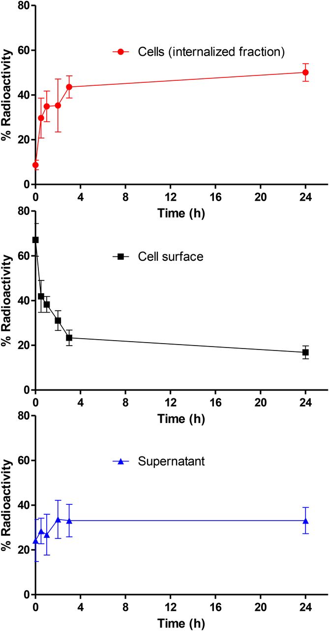
    Membrane binding and internalization of 89Zr-PRS-110 after binding MET on H441 cells. Cell surface (acid buffer), intracellular, and supernatant fraction radioactivity, expressed as percentage of total activity. Data were obtained in 3 independent experiments.
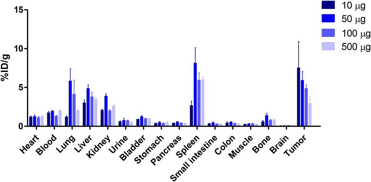
  • FIGURE 2.
    • Download figure
    • Open in new tab
    • Download powerpoint
    FIGURE 2.

    Biodistribution of 89Zr-PRS-110 at 96 h as determined by ex vivo analysis. Four dose groups of 10, 50, 100, and 500 μg of PRS-110 (with 10 μg of 89Zr-PRS-110 per group) were included in dose-escalation biodistribution in H441 human tumor–bearing mice (4–6 mice per group). Data are expressed as %ID/g.

  • FIGURE 3.
    • Download figure
    • Open in new tab
    • Download powerpoint
    FIGURE 3.

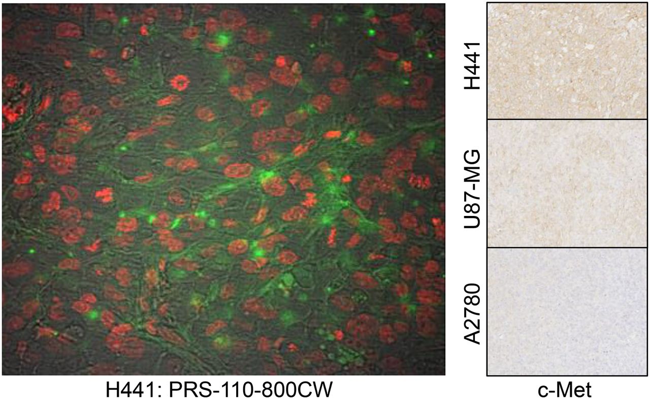
    Ex vivo tumor analysis of PRS-110 distribution 96 h after tracer injection in H441 tumor visualized by fluorescence microscopy. Green is from IRDye 800CW conjugated to PRS-110, and nuclei (in red) are visualized. In background, tumor structure is visible. MET status of H441, U87-MG, and A2780 was determined by immunohistochemistry, and representative picture is included.

  • FIGURE 4.
    • Download figure
    • Open in new tab
    • Download powerpoint
    FIGURE 4.

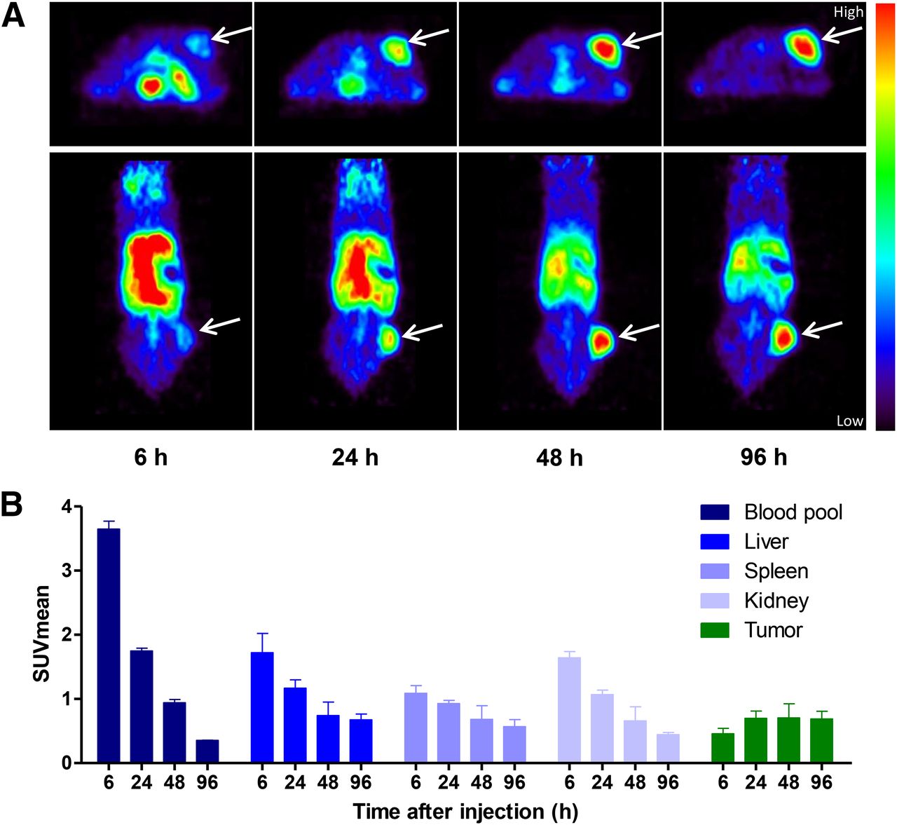
    89Zr-PRS-110 small-animal PET imaging of H441-bearing mice. (A) Representative transversal and coronal small-animal PET images are shown at 6, 24, 48, and 96 h after tracer injection. (B) Small-animal PET data quantification was performed for blood pool, liver, spleen, kidney, and tumor uptake in all mice (4–6 mice per group). Data are expressed as mean standardized uptake value (SUVmean).

  • FIGURE 5.
    • Download figure
    • Open in new tab
    • Download powerpoint
    FIGURE 5.

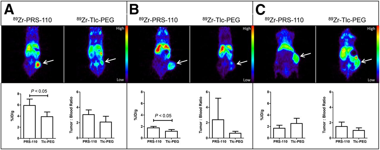
    89Zr-PRS-110 small-animal PET imaging of H441- (A), U87-MG- (B), and A2780-bearing mice (C). Representative transversal and coronal small-animal PET images are shown in the upper panel at 96 h after tracer injection of 89Zr-PRS-110 for MET-driven tumor uptake and 89Zr-Tlc-PEG for nonspecific tumor uptake. Ex vivo tumor uptake of 89Zr-PRS-110 and 89Zr-Tlc-PEG is shown in the lower panel. To compare specific tumor uptake between tumor models, tumor-to-blood ratios are provided (4–6 mice per group).

Additional Files

  • Figures
  • Supplemental Data

    Files in this Data Supplement:

    • Supplemental Data
PreviousNext
Back to top

In this issue

Journal of Nuclear Medicine: 55 (4)
Journal of Nuclear Medicine
Vol. 55, Issue 4
April 1, 2014
  • Table of Contents
  • Table of Contents (PDF)
  • About the Cover
  • Index by author
Print
Download PDF
Article Alerts
Sign In to Email Alerts with your Email Address
Email Article

Thank you for your interest in spreading the word on Journal of Nuclear Medicine.

NOTE: We only request your email address so that the person you are recommending the page to knows that you wanted them to see it, and that it is not junk mail. We do not capture any email address.

Enter multiple addresses on separate lines or separate them with commas.
In Vivo Visualization of MET Tumor Expression and Anticalin Biodistribution with the MET-Specific Anticalin 89Zr-PRS-110 PET Tracer
(Your Name) has sent you a message from Journal of Nuclear Medicine
(Your Name) thought you would like to see the Journal of Nuclear Medicine web site.
Citation Tools
In Vivo Visualization of MET Tumor Expression and Anticalin Biodistribution with the MET-Specific Anticalin 89Zr-PRS-110 PET Tracer
Anton G.T. Terwisscha van Scheltinga, Marjolijn N. Lub-de Hooge, Marlon J. Hinner, Remy B. Verheijen, Andrea Allersdorfer, Martin Hülsmeyer, Wouter B. Nagengast, Carolien P. Schröder, Jos G.W. Kosterink, Elisabeth G.E. de Vries, Laurent Audoly, Shane A. Olwill
Journal of Nuclear Medicine Apr 2014, 55 (4) 665-671; DOI: 10.2967/jnumed.113.124941

Citation Manager Formats

  • BibTeX
  • Bookends
  • EasyBib
  • EndNote (tagged)
  • EndNote 8 (xml)
  • Medlars
  • Mendeley
  • Papers
  • RefWorks Tagged
  • Ref Manager
  • RIS
  • Zotero
Share
In Vivo Visualization of MET Tumor Expression and Anticalin Biodistribution with the MET-Specific Anticalin 89Zr-PRS-110 PET Tracer
Anton G.T. Terwisscha van Scheltinga, Marjolijn N. Lub-de Hooge, Marlon J. Hinner, Remy B. Verheijen, Andrea Allersdorfer, Martin Hülsmeyer, Wouter B. Nagengast, Carolien P. Schröder, Jos G.W. Kosterink, Elisabeth G.E. de Vries, Laurent Audoly, Shane A. Olwill
Journal of Nuclear Medicine Apr 2014, 55 (4) 665-671; DOI: 10.2967/jnumed.113.124941
Twitter logo Facebook logo LinkedIn logo Mendeley logo
  • Tweet Widget
  • Facebook Like
  • Google Plus One
Bookmark this article

Jump to section

  • Article
    • Abstract
    • MATERIALS AND METHODS
    • RESULTS
    • DISCUSSION
    • CONCLUSION
    • DISCLOSURE
    • Acknowledgments
    • Footnotes
    • REFERENCES
  • Figures & Data
  • Supplemental
  • Info & Metrics
  • PDF

Related Articles

  • This Month in JNM
  • PubMed
  • Google Scholar

Cited By...

  • Emerging Opportunities for c-MET Visualization in the Clinic
  • Biodistribution and PET Imaging of Labeled Bispecific T Cell-Engaging Antibody Targeting EpCAM
  • c-Met PET Imaging Detects Early-Stage Locoregional Recurrence of Basal-Like Breast Cancer
  • Feasibility of Affibody-Based Bioorthogonal Chemistry-Mediated Radionuclide Pretargeting
  • ADAPT, a Novel Scaffold Protein-Based Probe for Radionuclide Imaging of Molecular Targets That Are Expressed in Disseminated Cancers
  • PET of c-Met in Cancer with 64Cu-Labeled Hepatocyte Growth Factor
  • Google Scholar

More in this TOC Section

  • Tumor-Specific Binding of Radiolabeled PEGylated GIRLRG Peptide: A Novel Agent for Targeting Cancers
  • PET/MRI of Hypoxic Atherosclerosis Using 64Cu-ATSM in a Rabbit Model
  • Tumor Uptake of Anti-CD20 Fabs Depends on Tumor Perfusion
Show more Basic Science Investigations

Similar Articles

Keywords

  • anticalins
  • 89Zr
  • PET
  • MET
SNMMI

© 2025 SNMMI

Powered by HighWire