


Article Figures & Data
Figures
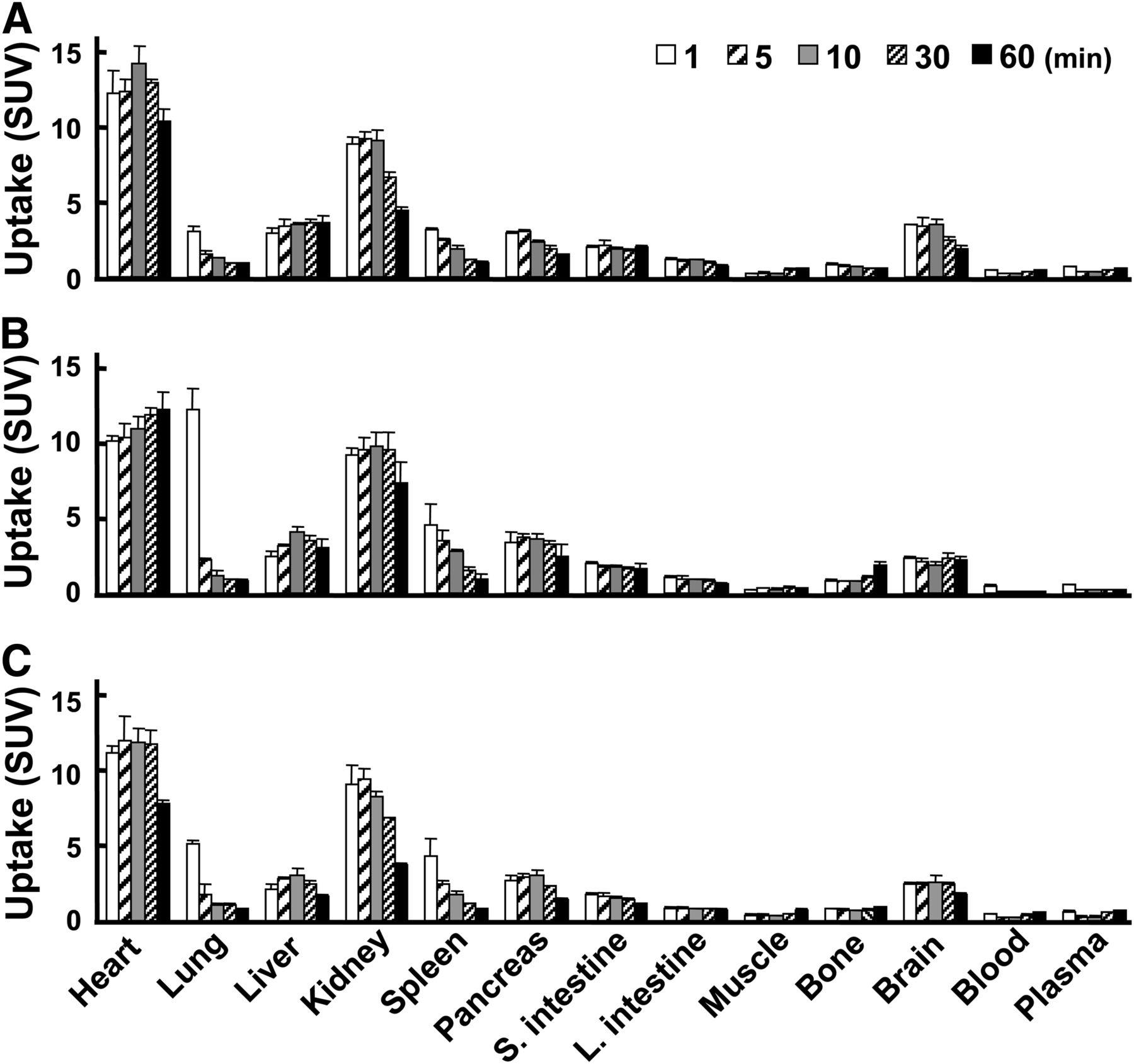
- FIGURE 1.
Distribution and kinetics of 18F-BCPP-EF (A), 18F-BCPP-BF (B), and 18F-BMS (C) in rats. Rats were intravenously injected with 5 MBq of 18F-BCPP-EF, 18F-BCPP-BF, or 18F-BMS via tail vein and sacrificed 1, 5, 10, 30, and 60 min after injection to calculate SUV. All values are expressed as mean ± SD for 3 animals.
- FIGURE 2.
Effects of preadministration of rotenone on uptake of 18F-BCPP-EF (A), 18F-BCPP-BF (B), and 18F-BMS (C) in rats imaged by small-animal PET and X-CT. After continuous infusion of vehicle or rotenone at dose of 0.1 mg/kg/h for 1 h, PET scanning was conducted for 60 min after injection of each PET probe. Summation PET data from 10 to 30 min for A, B, and C were reconstructed for SUV images, and then PET images were superimposed on individual X-CT images.
- FIGURE 3.
Effects of preadministration of rotenone on time–activity curves of 18F-BCPP-EF (A), 18F-BCPP-BF (B), and 18F-BMS (C) in rat brain. PET scans were conducted as described in Figure 2, and then summation data from 10 to 30 min were reconstructed for SUV images. ROIs for brain were set on reconstructed PET images to obtain time–activity curves of each PET probe in these regions. ○ = cortex-vehicle; ● = cortex-rotenone; △ = striatum-vehicle; ▲ = striatum-rotenone; □ = cerebellum-vehicle; ■ = cerebellum-rotenone.
- FIGURE 4.
Effects of preadministration of rotenone on time–activity curves of 18F-BCPP-EF (A), 18F-BCPP-BF (B), and 18F-BMS (C) in rat heart. PET scans were conducted as described in Figure 2, and then summation data from 10 to 30 min were reconstructed for SUV images. ROIs for heart (myocardium) were set on reconstructed PET images to obtain time–activity curves of each PET probe in these regions. ○ = vehicle; ● = rotenone.
- FIGURE 5.
Temporal changes in PET images of 18F-FDG and 18F-BCPP-EF in rat brains of PIT model. PET scans were conducted for 60 min with each PET probe before (normal), 1 (day 1), and 7 d (day 7) after PIT operation.
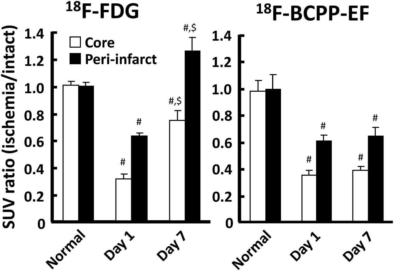
- FIGURE 6.
Temporal changes in uptake (SUV ratio) of 18F-FDG and 18F-BCPP-EF in rat brains of PIT model. PET scans were conducted for 60 min with each PET probe before (normal), 1 (day 1), and 7 d (day 7) after PIT operation. #P < 0.01 vs. corresponding normal condition. $P < 0.01 vs. corresponding day 1 condition.
- FIGURE 7.
Immunohistochemical analyses in rat brains of PIT model. Brains were dissected at day 7 for immunohistochemistry with Iba1 for inflammation and NeuN for neurons.


Tables
- TABLE 1
Uptake Ratios of Brain to Blood, Brain to Plasma, Heart to Blood, and Heart to Plasma of 18F-BCPP-EF, 18F-BCPP-BF, and 18F-BMS in Rat
Probe 1 min 5 min 10 min 30 min 60 min 18F-BCPP-EF Brain to blood 7.643 ± 0.495 15.058 ± 1.358 15.525 ± 2.178 7.866 ± 0.453 4.411 ± 0.435 Brain to plasma 5.280 ± 0.217 11.703 ± 1.361 11.879 ± 1.559 5.941 ± 0.502 3.328 ± 0.642 Heart to blood 26.785 ± 1.387 54.629 ± 3.596 63.098 ± 6.226 41.310 ± 4.125 24.705 ± 1.546 Heart to plasma 18.554 ± 1.699 40.049 ± 1.853 48.291 ± 4.328 31.229 ± 3.950 18.638 ± 1.245 18F-BCPP-BF Brain to blood 4.432 ± 0.371 13.362 ± 1.321 12.567 ± 0.708 14.817 ± 3.156 14.978 ± 3.006 Brain to plasma 3.926 ± 0.319 10.986 ± 1.042 10.039 ± 0.786 11.658 ± 2.918 11.112 ± 2.304 Heart to blood 19.617 ± 0.680 65.826 ± 5.418 72.429 ± 1.703 76.204 ± 3.909 83.791 ± 18.636 Heart to plasma 17.391 ± 1.020 54.127 ± 4.272 57.818 ± 2.271 59.739 ± 5.148 62.153 ± 14.094 18F-BMS Brain to blood 5.483 ± 1.289 11.318 ± 1.328 11.568 ± 2.916 6.141 ± 0.399 3.169 ± 0.124 Brain to plasma 4.291 ± 1.030 8.804 ± 1.116 9.039 ± 2.329 4.657 ± 0.128 2.511 ± 0.119 Heart to blood 25.195 ± 4.438 56.286 ± 5.319 53.204 ± 8.714 29.393 ± 2.996 14.379 ± 0.442 Heart to plasma 19.720 ± 3.658 43.831 ± 5.400 41.555 ± 6.982 22.268 ± 1.422 11.385 ± 0.172 Rats were intravenously injected with 5 MBq of 18F-BCPP-EF, 18F-BCPP-BF, or 18F-BMS via tail vein and sacrificed at 1, 5, 10, 30, and 60 min after injection to obtain tissue samples for uptake ratio analyses. All values are expressed as mean ± SD for 3 animals.
- TABLE 2
Metabolic Profiles of 18F-BCPP-EF, 18F-BCPP-BF, and 18F-BMS in Rat Brain and Plasma
Probe 1 min 5 min 10 min 30 min 60 min 18F-BCPP-EF Brain ND 98.5 ± 0.1 ND 90.5 ± 0.2 84.4 ± 0.1 Plasma 84.5 ± 3.2 46.1 ± 8.0 40.4 ± 4.8 17.3 ± 2.6 8.3 ± 0.9 18F-BCPP-BF Brain ND 99.3 ± 0.1 ND 97.1 ± 0.1 95.3 ± 0.1 Plasma 92.5 ± 0.3 69.5 ± 1.6 39.8 ± 2.0 18.4 ± 3.5 12.1 ± 3.6 18F-BMS Brain ND 97.9 ± 0.3 ND 87.2 ± 0.5 76.3 ± 1.2 Plasma 89.8 ± 1.5 39.9 ± 3.5 24.3 ± 9.3 7.5 ± 1.8 2.9 ± 0.7 Rats were intravenously injected with 5 MBq of 18F-BCPP-EF, 18F-BCPP-BF, or 18F-BMS via tail vein and sacrificed at 1, 5, 10, 30, and 60 min after injection to obtain brain and plasma for metabolic analyses. All values are expressed as mean ± SD for 3 animals.
ND = not determined.
Supplemental Data
Files in this Data Supplement:


