


Article Figures & Data
Figures
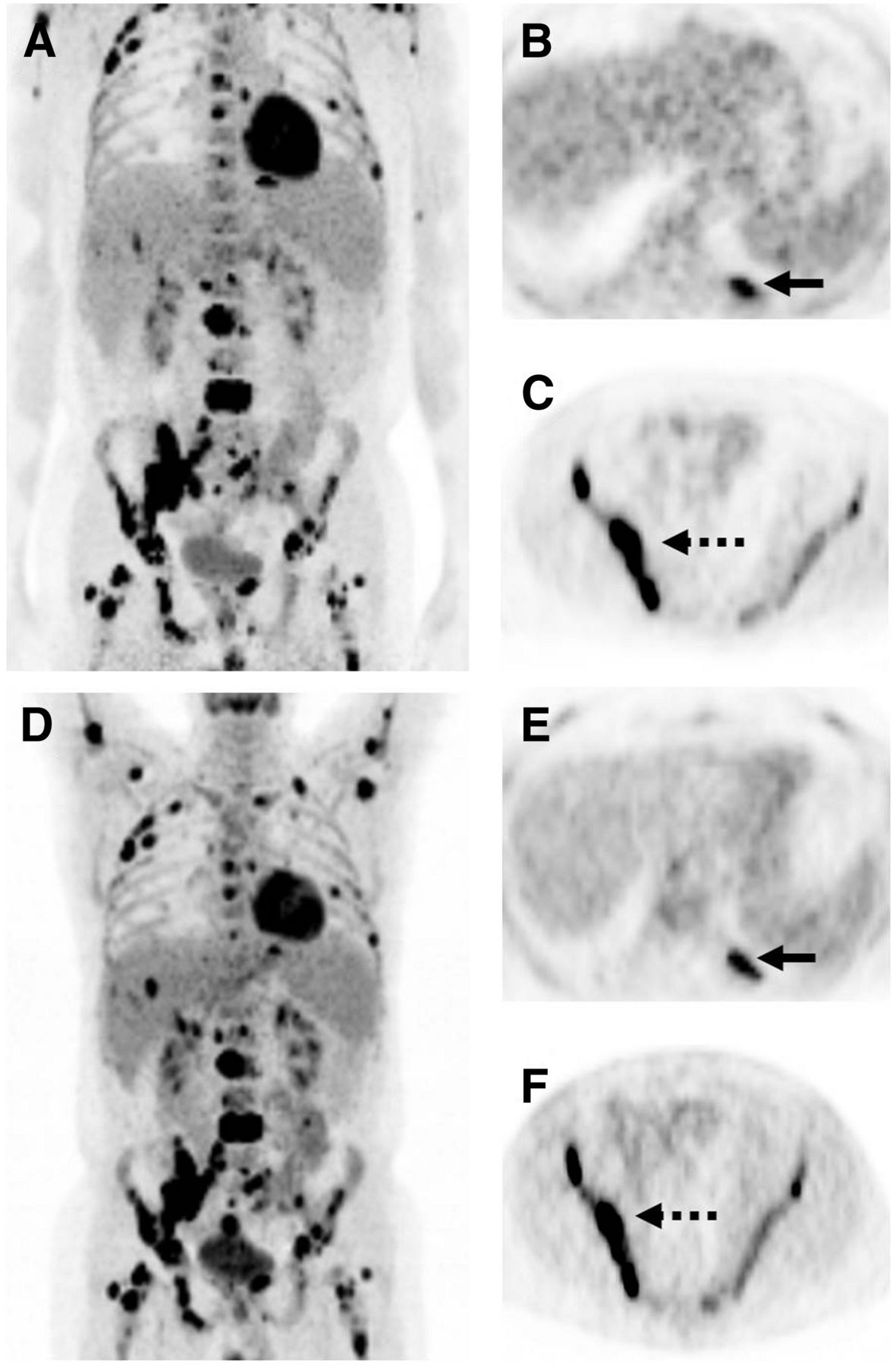
- FIGURE 1.
Images of bone metastases (arrows) in 69-y-old man presenting for staging of oropharyngeal cancer. (A–C) PET/MR examination with coronal PET (A), coronal T1-weighted Dixon in-phase MR sequence (B), and coronal T1-weighted TSE MR sequence (C). (D–F) PET/CT examination with coronal PET (D) and coronal bone (E) and soft-tissue (F) windows of CT dataset. Two metastases in spine show intense uptake in both PET datasets (A and D). Replacement of bone marrow is seen in both T1-weighted Dixon in-phase MR sequence (B) and T1-weighted TSE MR sequence (C), with better lesion delineation in TSE than in VIBE Dixon because of higher in-plane resolution. (E and F) Faint sclerosis is present as anatomic correlate of caudal metastases in both bone window (E) and soft-tissue window (F) on CT, whereas cranial metastasis is depicted only in soft-tissue window.
- FIGURE 2.
PET/MR (A–C) and PET/CT (D–F) images from patient with diffuse osseous manifestation of non-Hodgkin lymphoma. Visually, both datasets provide excellent image quality, as demonstrated by maximum-intensity projection (A and D). Patient is imaged with arms down in PET/MR (A) and arms up in PET/CT (B). This approach is often used because imaging time in PET/MR is lengthy and too uncomfortable for a patient lying with arms up. Quantitative analysis showed lower SUVmean for lesions on PET/MR (121 min after injection) than on PET/CT (85 min after injection). Moderate difference (29.4%) was found for lesions in left rib (PET/MR, 5.99; PET/CT, 7.75; arrows in B and E). Slight difference (10.4%) was observed for lesion in right ilium (PET/MR, 19.86; PET/CT, 22.16; arrows in C and F).
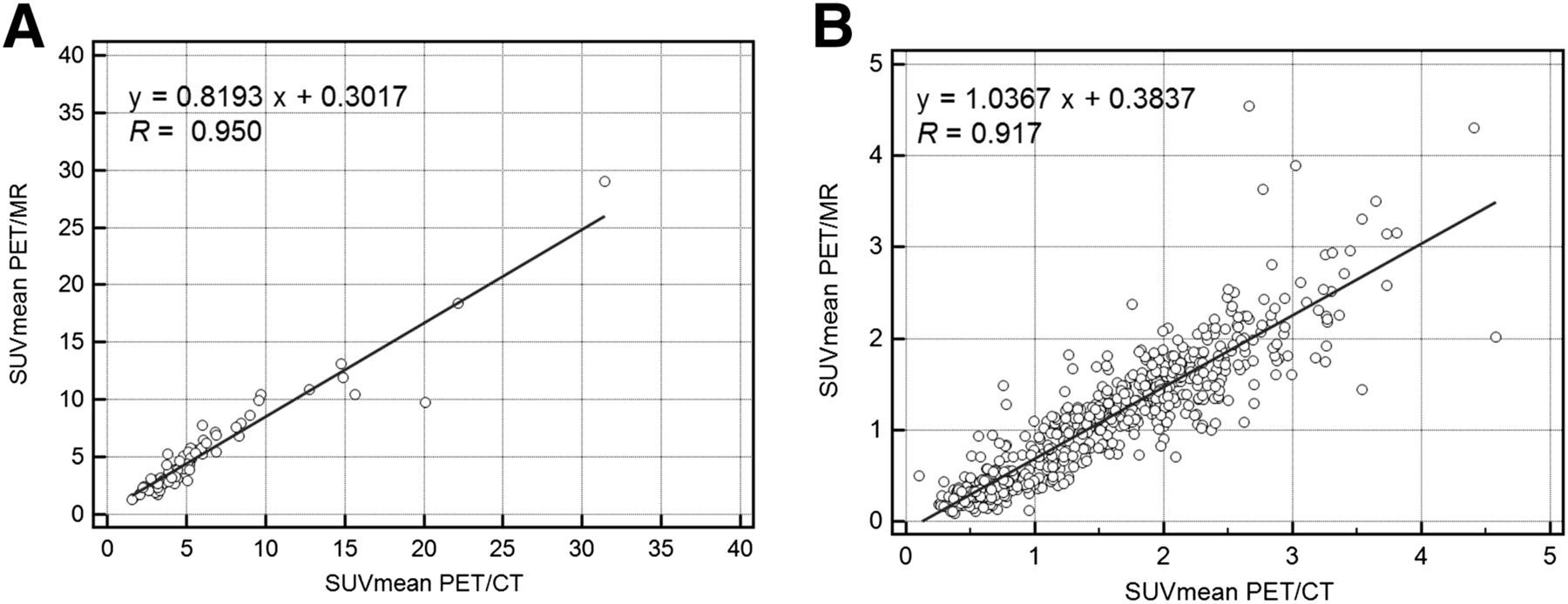
- FIGURE 3.
Correlation of tracer uptake between PET/CT and subsequent PET/MR as assessed by SUVmean. x-axis displays quantitative values obtained by PET/CT, and y-axis displays corresponding values by PET/MR. Both for bone lesions (A) and for regions of normal bone (B), high correlation as expressed by Spearman correlation coefficient is found (R = 0.950 and R = 0.917, P < 0.0001, respectively) between the 2 modalities.


Tables
Malignancy All patients (n = 119) Patients suitable for SUV measurement (n = 84) Head and neck squamous cell carcinoma 20 20 Breast cancer 19 9 Gastrointestinal tract cancer 17 12 Sarcoma 15 13 Malignant lymphoma/leukemia 11 6 Primary unknown cancer 10 4 Genitourinary cancer 9 8 Malignant melanoma 9 5 Thyroid cancer 7 7 Lung 3 — Other 4 3 Total 125* 87† Sequence T1-weighted VIBE Dixon T1-weighted TSE coronal TR/TE (ms) 3.60/1.23–2.46* 600/8.7 Slice thickness (mm) 3.12 5 Gap (%) 0 30 Matrix 192 × 121 384 × 230 Field of view (mm) 500 450 % phase field of view 100 67.2 Acquisition time (min:s) 0:19 1:11 Number of excitations 1 1 iPAT factor 2 2 ↵* Fat-saturation techniques with Dixon require 2 repetition times.
TR/TE = repetition time/echo time; iPAT = integrated parallel acquisition technique.
Location n Lytic/mixed/sclerotic CT Dixon T1-weighted TSE Cervical spine 4 1/3/0 2.00 ± 0.82 2.25 ± 0.50 3.00* Thoracic spine 16 0/15/1 2.50 ± 0.52 2.81 ± 0.40 2.94 ± 0.25 Lumbar spine 20 3/14/3 2.60 ± 0.51 2.70 ± 0.47 3.00* Pelvis 27 5/13/9 2.59 ± 0.51 2.76 ± 0.55 2.85 ± 0.36 Upper extremity or shoulder 12 1/10/1 2.75 ± 0.45 2.58 ± 0.52 2.92 ± 0.29 Legs 6 1/4/1 2.50 ± 0.84 2.66 ± 0.82 2.67 ± 0.82 Ribs or sternum 11 2/6/3 2.63 ± 0.51 1.72 ± 0.79 2.36 ± 0.67 Other 2 0/1/1 2.50 ± 0.71 1.50 ± 0.71 2.50 ± 0.71 Total 98 13/66/19 2.57 ± 0.54 2.54 ± 0.65 2.84 ± 0.42 ↵* Same results for all lesions, SD = 0.
Data are mean ± SD.
Technique SUVmean SUVmax PET/CT 5.70 ± 4.70 8.54 ± 6.96 PET/MR 4.97 ± 4.03 7.55 ± 6.02 P <0.0001 <0.0001 Data are mean ± SD.
Technique SUVmean SUVmax PET/CT 1.520 ± 0.658 2.300 ± 1.070 PET/MR 1.092 ± 0.705 1.820 ± 0.998 P <0.0001 <0.0001 Data are mean ± SD.
Supplemental Data
Files in this Data Supplement:


Jump to section
Related Articles
Cited By...
- 18F-FDG PET/CT in the Management of Osteosarcoma
- 18F-FDG PET/CT in the Management of Osteosarcoma
- Molecular Imaging of Bone Metastases and Their Response to Therapy
- Relationship Between Ktrans and K1 with Simultaneous Versus Separate MR/PET in Rabbits with VX2 Tumors
- 18F-FDG PET/CT and PET/MRI Perform Equally Well in Cancer: Evidence from Studies on More Than 2,300 Patients
- Imaging Bone Metastases in Breast Cancer: Staging and Response Assessment
- Quantitative Evaluation of Segmentation- and Atlas-Based Attenuation Correction for PET/MR on Pediatric Patients
- Evaluation of 18F-Fluoride PET/MR and PET/CT in Patients with Foot Pain of Unclear Cause
- PET/MR in Oncology: Non-18F-FDG Tracers for Routine Applications