


Abstract
Affibody molecules are small (7 kDa) nonimmunoglobulin scaffold proteins with favorable tumor-targeting properties. Studies concerning the influence of chelators on biodistribution of 99mTc-labeled Affibody molecules demonstrated that the variant with a C-terminal glycyl-glycyl-glycyl-cysteine peptide–based chelator (designated ZHER2:V2) has the best biodistribution profile in vivo and the lowest renal retention of radioactivity. The aim of this study was to evaluate 188Re-ZHER2:V2 as a potential candidate for radionuclide therapy of human epidermal growth factor receptor type 2 (HER2)–expressing tumors. Methods: ZHER2:V2 was labeled with 188Re using a gluconate-containing kit. Targeting of HER2-overexpressing SKOV-3 ovarian carcinoma xenografts in nude mice was studied for a dosimetry assessment. Results: Binding of 188Re-ZHER2:V2 to living SKOV-3 cells was demonstrated to be specific, with an affinity of 6.4 ± 0.4 pM. The biodistribution study showed a rapid blood clearance (1.4 ± 0.1 percentage injected activity per gram [%ID/g] at 1 h after injection). The tumor uptake was 14 ± 2, 12 ± 2, 5 ± 2, and 1.8 ± 0.5 %IA/g at 1, 4, 24, and 48 h after injection, respectively. The in vivo targeting of HER2-expressing xenografts was specific. Already at 4 h after injection, tumor uptake exceeded kidney uptake (2.1 ± 0.2 %IA/g). Scintillation-camera imaging showed that tumor xenografts were the only sites with prominent accumulation of radioactivity at 4 h after injection. Based on the biokinetics, a dosimetry evaluation for humans suggests that 188Re-ZHER2:V2 would provide an absorbed dose to tumor of 79 Gy without exceeding absorbed doses of 23 Gy to kidneys and 2 Gy to bone marrow. This indicates that future human radiotherapy studies may be feasible. Conclusion: 188Re-ZHER2:V2 can deliver high absorbed doses to tumors without exceeding kidney and bone marrow toxicity limits.
Overexpression of the human epidermal growth factor receptor type 2 (HER2) is associated with malignant transformation of cells and provides a growth advantage for tumors. Treatment of HER2-expressing disseminated tumors with the anti-HER2 monoclonal antibody trastuzumab improves survival in patients with metastatic breast (1) and gastric cancer (2). However, many tumors have a primary trastuzumab resistance or develop resistance during therapy despite preserved HER2 expression (2). Conjugation of cytotoxic payloads (e.g., drugs or radionuclides) to tumor-targeting antibodies may enhance their antitumor effects and hence improve the response duration and overall response rate (3,4). β-emitting radionuclides are considered a promising form of cytotoxic payload, because of the cross-fire effect. The use of full-length antibodies for radionuclide therapy, however, is associated with several issues. The major obstacles are insufficient penetration in the tumor tissue, limiting the absorbed dose to tumors and slow clearance from the body, causing second-organ (mainly bone marrow) toxicity (5). The use of smaller targeting agents, such as antibody fragments, would provide more efficient extravasation, better tumor penetration, and more rapid clearance of an unbound tracer (6,7).
A promising approach for development of high-affinity small tumor-targeting agents is the use of engineered scaffold proteins (8). Affibody (Affibody AB) molecules are the most studied class of scaffold proteins for in vivo radionuclide targeting. These high-affinity ligands are based on a 58-amino-acid (7 kDa) triple α-helical scaffold derived from domain B of staphylococcal protein A (9). An Affibody with picomolar affinity (22 pM) to the extracellular domain of HER2 has been selected earlier (10). Affibody molecules labeled with different radionuclides have demonstrated efficient tumor targeting and high-contrast imaging of HER2-expressing tumors (11). Two clinical studies have demonstrated that Affibody molecules are nontoxic and nonimmunogenic in humans (12,13). However, Affibody molecules undergo renal excretion followed by substantial renal reabsorption (14). The use of residualizing labels has therefore resulted in a high retention of radionuclides in kidneys, with a radioactivity concentration exceeding severalfold the radioactivity concentration in tumors. This would make the use of most radiometals (e.g., 177Lu or 90Y) unsuitable for radionuclide therapy (11). An important feature of the HER2-targeting Affibody molecules is their slow internalization by HER2-expressing cells (15), enabling the use of nonresidualizing labels without appreciable reduction of tumor retention. In contrast, rapid internalization of the Affibody in proximal tubuli results in rapid clearance of nonresidualizing labels from kidneys (11,16).
Earlier, we evaluated how the composition of peptide-based N3S chelators can influence the targeting and biodistribution properties of 99mTc-labeled Affibody molecules. Mercaptoacetyl-containing chelators on the N terminus (17–20) and cysteine-containing chelators on the C terminus (21–23) were evaluated. We showed that biodistribution of Affibody molecules can be altered by varying the amino acid composition of such chelators. Importantly, residualizing properties of the 99mTc label can be modulated in a wide range. For example, a 99mTc-labeled variant having a -GGGC peptide chelator at the C terminus (designated 99mTc-ZHER2:V2) demonstrated tumor uptake exceeding renal uptake appreciably because of weak residualizing properties (22).
Rhenium is a chemical analog to technetium. The β-emitting rhenium isotope 188Re (half-life, 17.0 h; maximum β energy, 2.1 MeV) has a potential for therapeutic applications. Production via a generator with the long-lived mother nuclide 188W (half-life, 70 d) makes this radionuclide readily available with high specific radioactivity. A low-abundance (15%) low-energy (155 keV) γ emission enables imaging of the biodistribution of 188Re-labeled therapeutic agents in patients for patient-specific dosimetry. We expected that substitution of 99mTc for 188Re in Affibody molecules might provide a conjugate with a high tumor uptake and low renal retention. However, the chemical properties of rhenium and technetium are similar but not identical, and biodistribution of 99mTc- and 188Re-labeled peptides can be different (24,25). Therefore, an evaluation of 188Re-labeled ZHER2:V2 was necessary.
The goal of the current study was to evaluate the tumor-targeting properties and biokinetics of 188Re-ZHER2:V2 in mice bearing HER2-expressing xenografts and, based on dosimetry evaluation, determine whether 188Re-ZHER2:V2 might be a candidate for treatment of HER2-expressing tumors without severe second-organ toxicity.
MATERIALS AND METHODS
188Re was obtained as perrhenate by elution of a 188W/188Re generator (29.6 GBq) with 0.9% sodium chloride (Polatom). Affibody molecule ZHER2:V2 was produced and purified as described previously (26). All chemicals were purchased from Sigma-Aldrich. The HER2-expressing ovarian cancer cell line SKOV-3, 1.6 × 10⁶ HER2 receptors per cell (ATCC), was used in cell studies. Cells were cultured in RPMI medium supplemented with 10% fetal calf serum, 2 mM l-glutamine, and PEST (penicillin, 100 IU/mL, and streptomycin, 100 μg/mL).
Labeling and Stability
ZHER2:V2 Affibody molecules were site-specifically labeled with 188Re using freeze-dried labeling kits containing 5 mg of sodium α-d-gluconate, 100 μg of disodium ethylenediaminetetraacetic acid, and 1 mg of tin(II) chloride dihydrate. After reconstitution, labeling was performed at pH 4.2 and 90°C for 1 h. Ascorbic acid and bovine serum albumin were introduced as antioxidants, as described in the supplemental materials (available at http://jnm.snmjournals.org).
To evaluate serum stability, freshly labeled 188Re-ZHER2:V2 (10 μL) was diluted in a serum sample (200 μL) to a concentration similar to the concentration in blood at the moment of injection and was incubated for 1 h at 37°C. The mixture was analyzed using radio–sodium dodecylsulfate polyacrylamide gel electrophoresis. A sample of perrhenate was used as a reference. The analysis was performed in duplicate.
In Vitro Evaluation
The specificity of binding and cellular processing of 188Re-ZHER2:V2 by SKOV-3 cells were evaluated as described previously (15). The affinity of 188Re-ZHER2:V2 binding to SKOV-3 was measured at 4°C using LigandTracer (Ridgeview Instruments AB) with 2 concentrations: 49 pM and 98 pM (details of the in vitro experiments are provided in the supplemental materials).
In Vivo Studies
The animal experiments were planned and performed in accordance with national legislation on laboratory animals’ protection. The animal study was approved by the local Ethics Committee for Animal Research in Uppsala.
SKOV-3 cells (107 cells per mouse) were implanted on the right hind leg of female NMRI (Naval Medical Research Institute) nu/nu mice. The average animal weight was 27 ± 2 g, and the average tumor weight was 0.8 ± 0.2 g at the start of the experiment. Six groups of mice (4 per group) were injected intravenously with 188Re-ZHER2:V2 in 100 μL of phosphate-buffered saline. The injected radioactivity was 53 kBq/mouse for 6 groups. The injected protein dose was adjusted to 1 μg/mouse by adding nonlabeled ZHER2:V2. The animals were euthanized at 20 min, 1 h, 4 h, 8 h, and 24 h after injection. One group was injected with 140 kBq/mouse and euthanized at 48 h after injection. For specificity control, 1 group of 3 mice was preinjected with 500 μg of ZHER2:342 45 min before injection of 188Re-ZHER2:V2 and euthanized at 4 h after injection. Organs and tissue samples (Table 1), gastrointestinal tract, and the remaining carcass were collected and weighed, and their radioactivity was measured in a NaI(Tl) well counter (PerkinElmer). Biodistribution data were corrected for decay and self-attenuation in samples, and organ uptake values were calculated as percentage injected activity per gram (%IA/g), except for the intestinal content, thyroid, and carcass, which were calculated as %IA per whole sample.
Biodistribution of 188Re-ZHER2:V2 in NMRI nu/nu Mice Bearing HER2-Expressing SKOV-3 Xenografts
In vivo imaging was performed to visually confirm the biodistribution data. Three SKOV-3–bearing mice were injected with 0.7 MBq (8 μg) of 188Re-ZHER2:V2. Two mice were injected 4 h before the imaging experiment and 1 mouse 1 h before. Immediately before imaging, the mice were sacrificed by cervical dislocation and the bladders were excised. The imaging was performed using an Infinia scintillation camera (GE Healthcare) equipped with a high-energy general-purpose collimator. Static images (30 min) were obtained with a zoom factor of 2 in a 256 × 256 matrix.
Dosimetry
Animal Dosimetry
To evaluate the feasibility of experimental therapy in mice, absorbed doses to tumors and normal tissues were calculated using the anatomically realistic murine Moby phantom developed by Larsson et al. (27,28) based on the biokinetics results. Absorbed doses per injected activity (Gy/MBq) for 188Re-ZHER2:V2 were calculated for 3 tumor locations: the right hind leg, left hind leg, and left flank.
Human Dosimetry
For dosimetry estimations in humans, uptake was extrapolated from animal data according to the percentage kg/g method (29).
where TB is total body. Organ time–activity curves were calculated using the organ weights of the 58-kg reference adult female (ICRP publication 23). Residence times were calculated as the area under the curve of biexponential fits to the animal organ time–activity curves. Remainder-of-body residence time was based on radioactivity in the carcass. Red marrow activity concentrations were conservatively assumed to equal whole-blood concentrations. Absorbed doses were estimated using OLINDA/EXM 1.0. For calculation of absorbed dose to the intestines, either the ICRP 30 gastrointestinal model was applied, using a fraction of 0.35 %IA (the measured value at 1 h after injection) entering the small intestine, or residence times calculated as above for small intestine and large intestine were used.
In tumor, standardized uptake value was assumed to be identical in humans and rodents, and tumor-absorbed doses were calculated assuming deposition of all β energy within the tumor, neglecting any self- or cross-dose from γ radiation. Dependence of absorbed dose on tumor volume was assessed using the OLINDA/EXM 1.0 sphere model.
RESULTS
Labeling and Stability
The labeling method provided high yield (95.8% ± 1%), with radiocolloid content below 1%. Purification using disposable NAP-5 columns (GE Healthcare) provided radiochemical purity over 99%. Up-scaling experiments demonstrated that specific activity of 17.5 MBq/μg (116 GBq/μmol) can be obtained. 188Re-ZHER2:V2 was stable in the formulation containing 6 mM ascorbic acid in phosphate-buffered saline with 2% bovine serum albumin (radiochemical purity of 99% ± 1% during 4 h). In the absence of bovine serum albumin, radiochemical purity was 94% ± 2% after 4 h.
Sodium dodecylsulfate polyacrylamide gel electrophoresis was performed after incubation in murine blood plasma at 37°C for 60 min. The main peak corresponded to the monomeric Affibody molecule. No peaks indicating aggregation of Affibody molecules or transchelation of the radionuclide to blood proteins were observed. The only other radioactivity peak had a path length equal to the path length of 188Re-perrhenate. This peak contained less than 2% of the total activity at the end of the stability test (Supplemental Fig. 1).
In Vitro Evaluation
The in vitro specificity experiment showed that binding of 188Re-ZHER2:V2 to SKOV-3 ovarian carcinoma cells can be reduced from 52% ± 1% to 0.84% ± 0.05% of added activity (P < 0.0001) by presaturation of HER2 with nonlabeled Affibody molecule. This indicates that the binding of 188Re-ZHER2:V2 was specific. The affinity of 188Re-ZHER2:V2 binding to SKOV3 cells at 4°C was 6.4 ± 0.4 pM (Supplemental Fig. 2).
The cellular retention profile (Fig. 1) showed a rapid drop in activity during the first hour followed by a slow decline with a biologic half-life of 19.6 h. The total cellular retention of the radioactivity was 34.6% ± 1.3% after 24 h of incubation at 37°C. The internalized radioactivity of 188Re-ZHER2:V2 was relatively low, less than 4% of total radioactivity after 24 h.
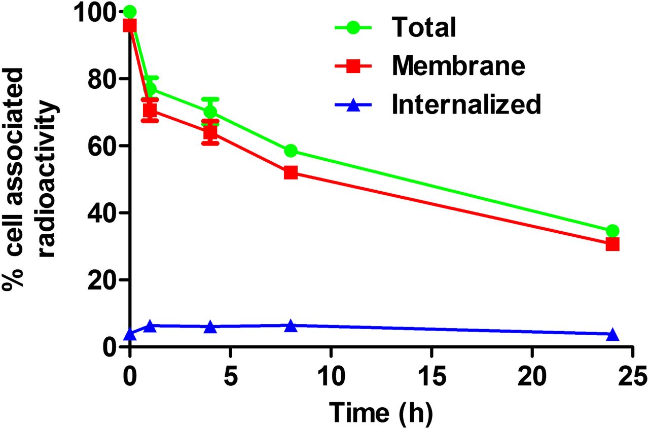
Cell-associated radioactivity as function of time after interrupted incubation of SKOV-3 cells with 188Re-ZHER2:V2. Data are presented as average value from 3 cell dishes ± SD. Error bars might not be seen because they are smaller than point symbols.
In Vivo Studies
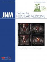
The results of the in vivo specificity test (Fig. 2) showed that presaturation of HER2 in xenografts with a parental ZHER2:342 decreased tumor uptake of 188Re-ZHER2:V2 4.5-fold (P < 0.005). This demonstrates a saturable character of the tumor accumulation and suggests its HER2 specificity. A small but statistically significant decrease in uptake in liver and an increase in kidneys was also detected in this experiment.

In vivo binding specificity of 188Re-ZHER2:V2 in mice bearing ovarian cancer SKOV-3 xenografts at 4 h after injection. Blocked group was subcutaneously preinjected with excess amount of nonlabeled ZHER2:342. Results are presented as %IA/g.
Biodistribution of 188Re-ZHER2:V2 in tumor-bearing mice (Table 1) was characterized by rapid clearance of radioactivity from all organs and tissues. Low radioactivity in the content of the gastrointestinal tract (<2% of injected activity; data not shown) suggested that the hepatobiliary pathway played a minor role in the excretion of 188Re-ZHER2:V2 or its radiometabolites. Low radioactivity uptake in organs accumulating free perrhenate, that is, salivary gland, thyroid, and stomach, suggests high stability of 188Re-ZHER2:V2 to reoxidation in vivo. The kidneys demonstrated high initial uptake, which decreased rapidly. At 1 h after injection, renal uptake was approximately equal to tumor uptake, and at 4 h after injection, tumor uptake was 5-fold higher.
Uptake was prominent in tumor as early as 20 min after injection and was higher than in any normal organ except the kidneys. Tumor uptake maximized at 1 h after injection (14 ± 2 %IA/g), followed by a decrease with a half-life of 15 h.
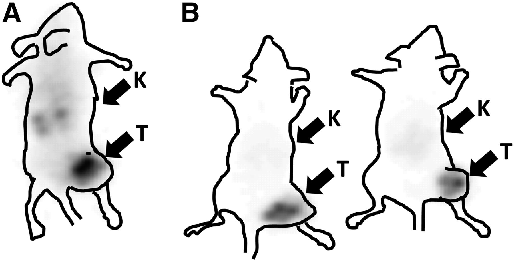
Scintillation-camera imaging, performed at 1 and 4 h after injection (Fig. 3), confirmed the results of the biodistribution experiments. The tumor xenografts were the only sites with prominent accumulation of radioactivity. The kidneys were visualized at 1 h after injection, but at 4 h after injection only the HER2-expressing tumor xenografts were clearly visible.

Imaging of HER2 expression in SKOV-3 ovarian cancer xenografts (high HER2 expression) in NMRI nu/nu mice using 188Re-ZHER2:V2. Planar scintillation-camera images were acquired at 1 h (A) and 4 h (B) after injection. Arrows point to tumors (T) and kidneys (K).
Dosimetry
The evaluation of absorbed doses in mice for 188Re-ZHER2:V2 is presented in Table 2. The self-doses to most organs and tissues were much smaller than absorbed doses to tumors. However, the size of a mouse body is comparable to the range of the β particles emitted by 188Re (∼10 mm). For this reason, the total absorbed doses became much higher because of cross-irradiation. For example, the total absorbed dose to bone marrow was approximately 22% of the total absorbed dose to tumor. Thus, it was impossible to deliver a therapeutically meaningful absorbed dose to tumors (>50 Gy) without delivering a lethal absorbed dose to the red marrow (∼8 Gy for mice). Performing experiments with188Re-ZHER2:V2 therapy in mice was thus considered unethical under these circumstances, and because of dimensional differences between mice and humans and the range of β particles, experiments in murine models will generate irrelevant results.
Calculated Absorbed Dose (Gy/MBq) for 188Re-ZHER2:V2 in Mice Using Murine Moby Phantom
The results of estimated absorbed dose calculations of 188Re-ZHER2:V2 in humans using OLINDA/EXM 1.0 are presented in Table 3. In brief, tumor standardized uptake value was approximately constant after 1 h, with a mean value of 3.3. Assuming local deposition of all β energy, and no self- or cross-dose from γ photons, this would result in an absorbed dose to tumor of 0.65 mGy/MBq. As shown in Figure 4, absorbed doses to tumors with a weight of less than 10 g would be slightly lower because of incomplete absorption of β particles, whereas slightly higher absorbed doses are expected in larger tumors due to self-dose from γ radiation. Absorbed dose to tumor exceeded absorbed dose to kidney and bone marrow by 3.4-fold and 79-fold, respectively.
Calculated Absorbed Dose (mGy/MBq) for 188Re-ZHER2:V2 in Humans Using OLINDA/EXM 1.0

Tumor-absorbed dose vs. mass for 188Re-ZHER2:V2.
DISCUSSION
Radioimmunotherapy of radiosensitive hematologic malignancies has proven to be successful, but treatment of more radioresistant solid tumors has so far been inefficient. The main reason is a failure to reach tumor-absorbed doses above 50 Gy, which are typically required to achieve a response to external-beam therapy (7).
Clinical experience with targeted radionuclide therapy using short peptides, particularly somatostatin analogs, demonstrated that the use of small targeting agents can solve the problem of both poor tumor penetration and exposure of bone marrow (30). Unfortunately, all small HER2-targeting radiolabeled agents, including antibody fragments, diabodies, designed ankyrin repeat proteins, and Affibody molecules, undergo considerable renal reabsorption (31). In the case of residualizing radiometal labels, the renal uptake would be higher than tumor uptake for all these targeting proteins.
Our approach is based on knowledge of the cellular processing of Affibody molecules. The main hypothesis was that the use of a nonresidualizing label would cause rapid clearance from kidneys, where Affibody molecules are rapidly internalized and degraded and their radiocatabolites excreted from cells. At the same time, processing of Affibody molecules bound to HER2-expressing cancer cells is slow, and cellular retention is good even in the case of nonresidualizing labels. Our previous structure–property relationship studies demonstrated that the use of a -GGGC peptide–based chelator yields nonresidualizing 99mTc labels (22) with low accumulation in kidneys. The present study has shown that ZHER2:V2 can be efficiently labeled with 188Re. The high-fidelity refolding of Affibody molecules under physiologic conditions (32) permitted their direct rhenium labeling at high temperature with high yields. In fact, 188Re-ZHER2:V2 preserved high-affinity binding (6.4 ± 0.4 pM) to HER2-expressing cells after labeling at 90°C and pH 4.2. Although an in vitro processing study (Fig. 1) showed low intracellular retention of radiocatabolites, the overall retention of radioactivity was reasonable, with a biologic half-life of 19.6 h. 188Re-ZHER2:V2 has shown specific uptake in HER2-expressing xenografts in vivo (Fig. 2). The biodistribution study confirmed that unbound radioactivity cleared quickly from blood, and release of retained radioactivity was much more rapid from kidneys than from tumors (Table 1). The area under the curve for tumor was 47-fold greater than that for blood, 70-fold greater than that for bone, and 2.8-fold greater than that for kidneys. This finding suggests that the goal of delivering an efficient absorbed dose to tumors while sparing critical organs may be achievable.
We performed dosimetry calculations for 188Re-ZHER2:V2 in mice using the anatomically realistic murine Moby phantom (27,28). The self-doses agreed with area-under-curve data, but the total absorbed dose to bone marrow was much higher than the self-dose. A high total absorbed dose to bone marrow is a phenomenon associated with mice as an animal model. The maximum range of β particles from 188Re is 10.4 mm (7), which is comparable to the dimensions of a mouse. For this reason, the cross-dose to bone marrow is much higher in mice than in humans. Delivery of therapeutically meaningful absorbed doses to tumors in mice would be associated with lethal absorbed doses to bone marrow.
Human dosimetry is much different because of the larger dimensions of the human body. Upscaling of murine biodistribution data to humans suggests that much lower absorbed doses will be delivered to radiosensitive organs in relation to tumor-absorbed doses. A maximum absorbed dose of 2 Gy for bone marrow is generally accepted when radionuclide therapy is being planned, as such an absorbed dose is associated with a low risk that leukemia will develop (33) and a low risk for acute bone marrow toxicity. An absorbed dose of 23 Gy is a commonly used limit for kidneys in peptide receptor radionuclide therapy (34). In the case of 188Re-ZHER2:V2, an absorbed dose of 23 Gy to the kidneys would correspond to a tumor-absorbed dose of 79 Gy, whereas an absorbed dose of 2 Gy to the bone marrow would correspond to a tumor-absorbed dose of 130 Gy. Thus, an absorbed dose of well above 50 Gy could be delivered without exceeding commonly accepted absorbed dose limits to critical organs. However, upscaling from mice to humans is associated with apparent uncertainties, and a clinical imaging study would be required to more reliably assess human dosimetry.
In addition to dosimetry estimations, there are other factors to consider when planning future therapy applications. 188Re is produced via a generator in no-carrier-added form, which provides high specific activity. The short half-life of 188Re enables irradiation of tumors with a high dose rate. Close matching of physical and biologic half-lives and generator-mediated production from the long-lived mother nuclide 188W (half-life, 70 d) permits fractionated therapy. Both the absorbed dose rate during tumor irradiation and treatment fractionation are considered important radiobiologic factors for increasing the efficacy of targeted radionuclide therapy (7).
One important lesson from this study concerns the selection of labeling strategy. It is common knowledge that the selection of radionuclide and chelator influences the stability of nuclide attachment to a targeting protein and intracellular retention of radioactivity after internalization. Our earlier studies demonstrated that modification of the physicochemical properties of Affibody molecules by incorporation of a radionuclide–chelator complex also modifies off-target interactions, changing blood clearance rate, the predominant excretion pathway, and biodistribution (11). These factors can be used to optimize targeting properties. We demonstrated earlier that the 186Re-maGSG-ZHER2:342 Affibody molecule, having an N-terminal mercaptoacetyl-glycyl-seryl-glycyl chelator, shows better retention of radioactivity in tumors than in kidneys (25). However, appreciable hepatobiliary excretion of 186Re-maGSG-ZHER2:342 (20% of injected radioactivity was measured in the intestinal content at 4 h after injection) caused a risk of high absorbed dose to intestines. Through reengineering of Affibody molecules by modifying the chelator and its placement to the C terminus, as well as by increasing the hydrophilicity of the N terminus by amino acid substitution, we suppressed hepatobiliary excretion of 188Re-ZHER2:V2 while keeping good tumor retention, low renal uptake, and rapid clearance of unbound radioactivity. Thus, optimal molecular design, including labeling strategy, may appreciably improve the properties of scaffold-protein–based conjugates for radionuclide therapy.
CONCLUSION
The Affibody molecule ZHER2:V2 can be labeled with 188Re with a high yield and preserved picomolar affinity to HER2. 188Re-ZHER2:V2 provides efficient targeting of HER2-expressing xenografts, rapid blood clearance, and low uptake in kidneys and bones. Dosimetry calculations in humans suggest that 188Re-ZHER2:V2 may provide an absorbed dose to tumors of more than 70 Gy while keeping the absorbed dose to kidneys below 23 Gy and the absorbed dose to bone marrow below 2 Gy. Thus, 188Re-ZHER2:V2 is a promising targeting agent for radionuclide therapy against HER2-expressing tumors.
DISCLOSURE
The costs of publication of this article were defrayed in part by the payment of page charges. Therefore, and solely to indicate this fact, this article is hereby marked “advertisement” in accordance with 18 USC section 1734. This research was supported in part by grants from the Swedish Cancer Society (Cancerfonden), Swedish Research Council (Vetenskapsrådet), and Governmental Funding of Clinical Research within the National Health Service (ALF). No other potential conflict of interest relevant to this article was reported.
Footnotes
Published online Oct. 2, 2014.
- © 2014 by the Society of Nuclear Medicine and Molecular Imaging, Inc.
REFERENCES
- Received for publication March 13, 2014.
- Accepted for publication August 6, 2014.


Jump to section
Related Articles
Cited By...
- Same-Day Imaging Using Small Proteins: Clinical Experience and Translational Prospects in Oncology
- Radionuclide Tumor Targeting Using ADAPT Scaffold Proteins: Aspects of Label Positioning and Residualizing Properties of the Label
- Feasibility of Affibody-Based Bioorthogonal Chemistry-Mediated Radionuclide Pretargeting