


Article Figures & Data
Figures
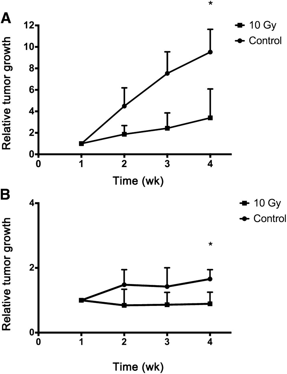
- FIGURE 1.
Relative tumor volume measured between −7 d (week 1) and 14 d (week 4) after irradiation for SCCNij202 (A) and SCCNij167 (B) in controls and irradiated (10 Gy) groups. Values represent mean ± SD. *P < 0.05.
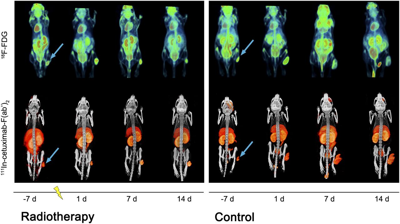
- FIGURE 2.
Longitudinal visualization with 18F-FDG PET imaging and 111In-cetuximab-F(ab′)2 SPECT imaging in irradiated and control SCCNij202 mice at day −7, 1, 7 and 14. Images were acquired at 24 and 1 h after injection for SPECT and PET imaging, respectively. Tumors were irradiated at time zero (represented as lightning bolt). Arrow = subcutaneous tumor location in right hind leg.
- FIGURE 3.
18F-FDG PET SUVmax (A) and 111In-cetuximab-F(ab′)2 SPECT Tmax/L ratio (B) in SCCNij202 and SCCNij167 xenografts. Values represent mean ± SD. *P < 0.05. **P < 0.001.
- FIGURE 4.
Example of autoradiography image of SCCNij202 (A) and corresponding EGFR immunofluorescence staining (red) and vessels (blue) (magnification, ×10) (B). (C) Intratumor distribution of 111In-cetuximab-F(ab′)2 as determined by autoradiography correlated well with immunohistochemical distribution of EGFR (r = 0.85; range, 0.69–0.95). Round symbols = unirradiated tumors; square symbols = irradiated tumors.
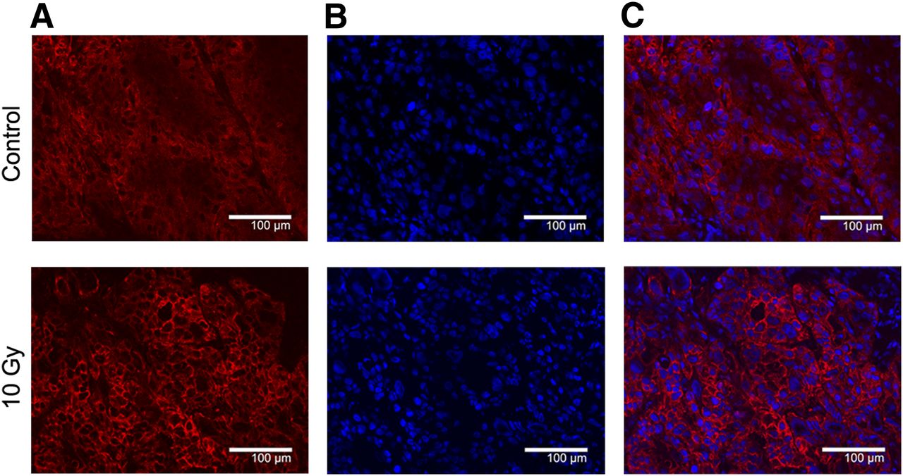
- FIGURE 5.
Typical example of EGFR immunofluorescent staining (A), nuclei (B), and combination image (C) of control and 10-Gy irradiated SCCNij202 tumor. Control xenografts display predominantly cytoplasmatic staining whereas irradiated xenografts show increased membranous staining intensity. Magnification, ×200.
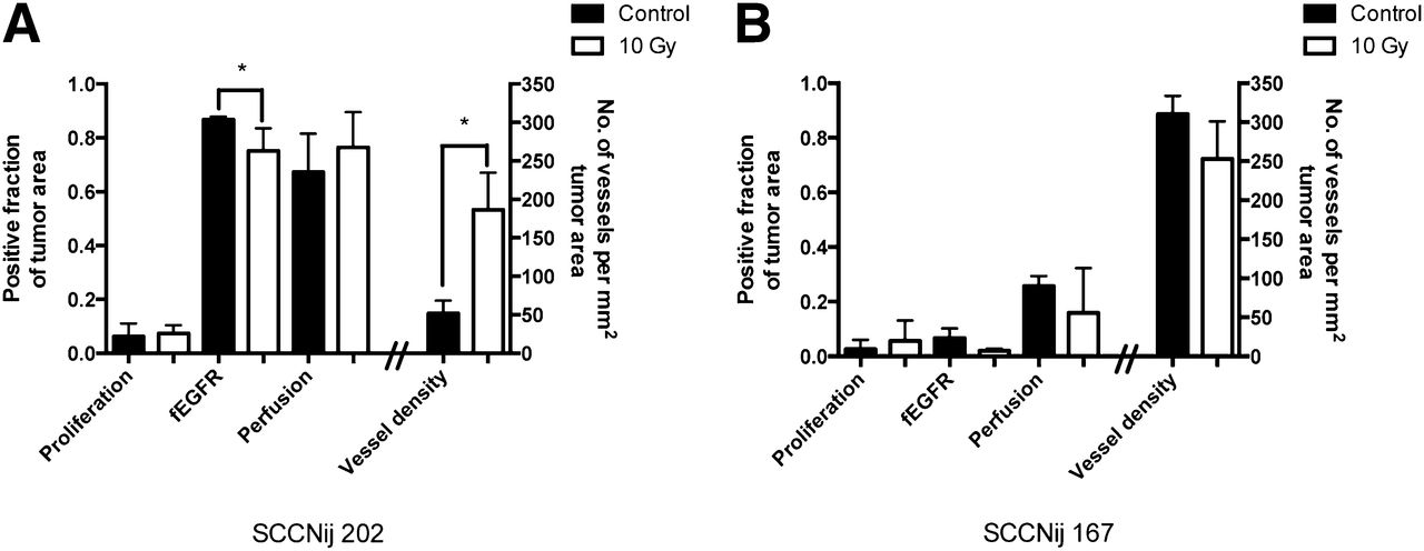
- FIGURE 6.
Immunofluorescent markers evaluated for SCCNij202 (A) and SCCNij167 (B) for control and irradiated groups. Markers were proliferation fraction (BrdUrd), fEGFR, fraction of perfused vessels (Hoechst), and vessel density per mm2 (9F1). Values are presented as mean ± SD. *P < 0.05.


Additional Files
Supplemental Data
Files in this Data Supplement:


