


Article Figures & Data
Figures
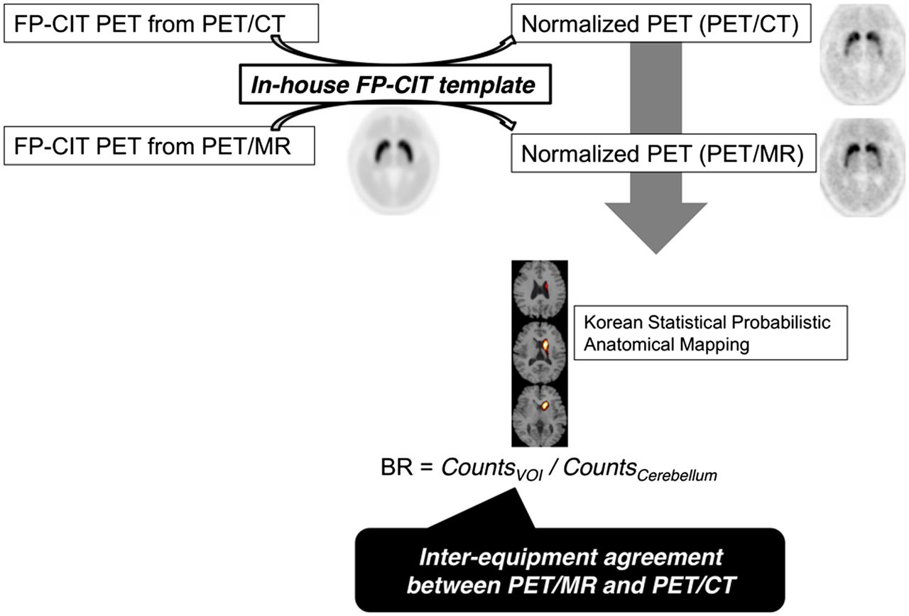
- FIGURE 1.
Schematic workflow for image processing and quantification. PET scans acquired from PET/MR and PET/CT were spatially normalized to Korean Statistical Probabilistic Anatomic Mapping template. BR was calculated using statistical probabilistic maps of putamen and caudate nucleus. Interequipment agreement between PET/MR and PET/CT was calculated. VOI = volume of interest.
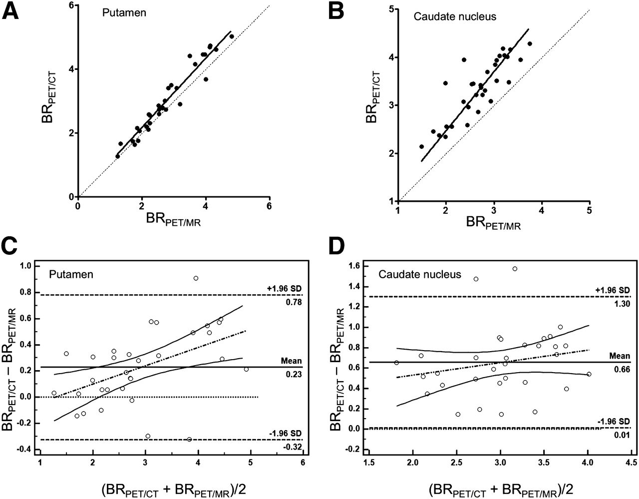
- FIGURE 2.
(A) DAT BR of PET/MR and PET/CT. BR of putamen calculated from PET/MR and PET/CT showed excellent interequipment agreement. ICC of BR was 0.967 (95% CI, 0.841–0.989). (B) BR of caudate nucleus calculated from PET/MR was underestimated, compared with PET/CT. ICC of BR was 0.682 (95% CI, −0.185–0.908). Bland–Altman plots show interequipment agreement in putamen (C) and caudate nucleus (D). Bland–Altman plot of caudate nucleus shows comparable variability between BR calculated from PET/MR and PET/CT, compared with putamen, which showed considerable mean difference of BR (0.23 ± 0.28 for putamen and 0.66 ± 0.33 for caudate nucleus).
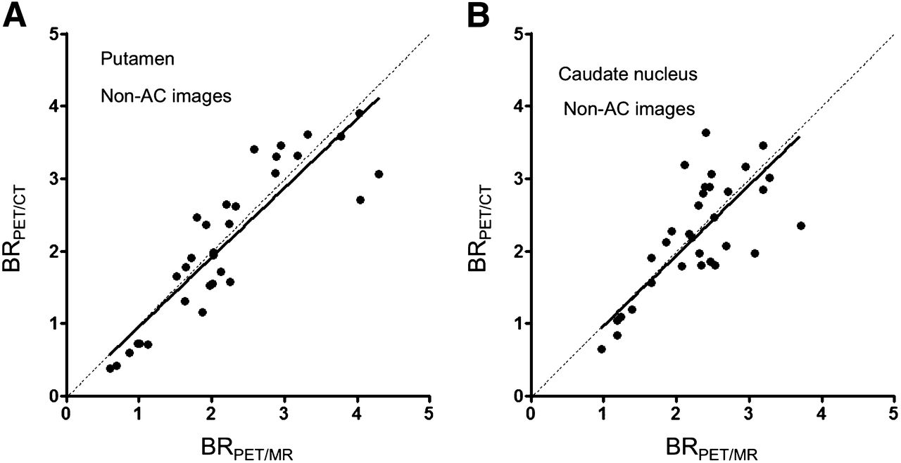
- FIGURE 3.
BR of PET/MR and PET/CT calculated from non-AC images. BR of putamen (A) and caudate nucleus (B) shows excellent interequipment agreement when non-AC images are applied (ICC, 0.937; 95% CI, 0.873–0.969, and ICC, 0.832; 95% CI, 0.655–0.918 for putamen and caudate nucleus, respectively).
- FIGURE 4.
Voxelwise comparison between PET/MR and PET/CT. Image shows underestimated voxels in PET/MR using UTE sequence–based AC, compared with PET/CT (uncorrected P < 0.001 as a threshold with an extent threshold of 100 contagious voxels).
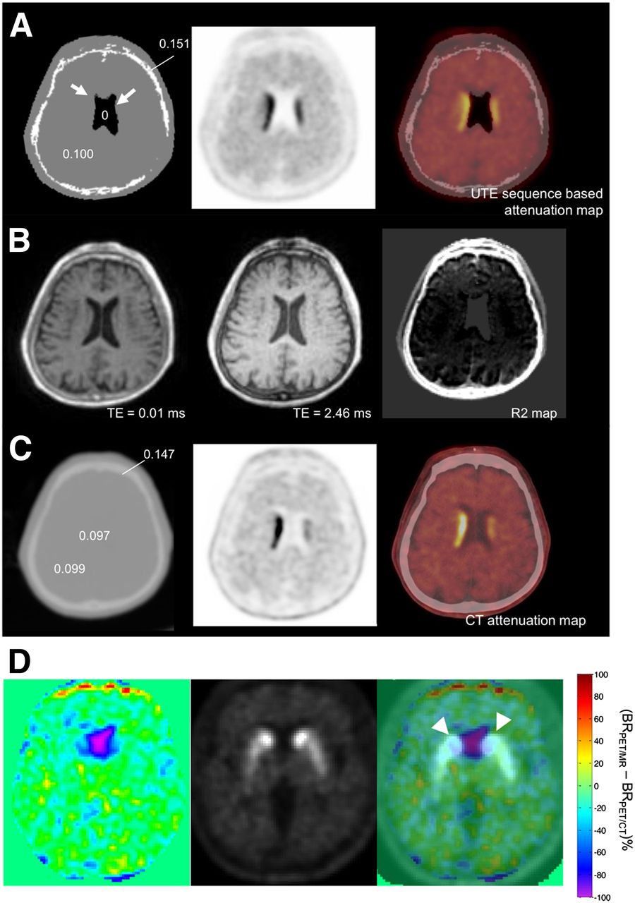
- FIGURE 5.
Representative images of UTE sequence–based AC and CT-based AC. (A) UTE sequence–based attenuation maps show misclassified voxels in lateral ventricle as air (arrow). (B) UTE images and R2 maps for tissue classification. UTE images were acquired at echo times TE1 (0.07 ms)/TE2 (2.46 ms), and R2 map is derived from 2 images after air mask. (C) CT-based attenuation map shows water attenuation in lateral ventricle. Difference in attenuation maps of PET/MR and PET/CT affects quantification of DAT BR. Numbers on attenuation map of PET/MR (A) and PET/CT (C) reveal attenuation coefficients of each region (cm−1). Representative 1-cm-sized circular regions of interest were drawn on lateral ventricle, cortical bone, and brain tissue on CT-based attenuation map. (D) Difference image in percentage BR between PET images from PET/MR and PET/CT showed underestimation in lateral ventricles. Fusion images of difference map and 18F-FP-CIT PET image showed that underestimation of BR was found in lateral ventricle, which overlapped caudate nucleus (arrowheads).


Tables
Patient no. Sex Age (y) Initial clinical assessment Visual assessment of DAT density 1 M 56 Parkinson disease Reduced in both striata 2 M 59 Parkinson disease Reduced in both striata 3 F 71 Parkinson disease Reduced in both striata 4 M 68 Parkinson disease Reduced in both striata 5 F 50 Parkinson disease Reduced in both striata 6 F 67 Nonparkinsonian tremor Preserved in both striata 7 M 39 Parkinson disease Reduced in both striata 8 F 68 Parkinson disease Reduced in both striata 9 M 69 Parkinson disease Preserved in both striata 10 F 64 Nonparkinsonian tremor Preserved in both striata 11 M 54 Parkinson disease Reduced in both striata 12 M 58 Parkinson disease Reduced in both striata 13 M 54 Parkinson disease Reduced in both striata 14 F 61 Parkinson disease Reduced in both striata 15 F 65 Parkinson disease Reduced in both striata 16 F 77 Parkinson disease Preserved in both striata
Supplemental Data
Files in this Data Supplement:


