
Main menu

  • Home
  • Content
    • Current
    • Ahead of print
    • Past Issues
    • JNM Supplement
    • SNMMI Annual Meeting Abstracts
    • Continuing Education
    • JNM Podcasts
  • Subscriptions
    • Subscribers
    • Institutional and Non-member
    • Rates
    • Journal Claims
    • Corporate & Special Sales
  • Authors
    • Submit to JNM
    • Information for Authors
    • Assignment of Copyright
    • AQARA requirements
  • Info
    • Reviewers
    • Permissions
    • Advertisers
  • About
    • About Us
    • Editorial Board
    • Contact Information
  • More
    • Alerts
    • Feedback
    • Help
    • SNMMI Journals
  • SNMMI
    • JNM
    • JNMT
    • SNMMI Journals
    • SNMMI

User menu

  • Subscribe
  • My alerts
  • Log in
  • My Cart

Search

  • Advanced search
Journal of Nuclear Medicine
  • SNMMI
    • JNM
    • JNMT
    • SNMMI Journals
    • SNMMI
  • Subscribe
  • My alerts
  • Log in
  • My Cart
Journal of Nuclear Medicine

Advanced Search

  • Home
  • Content
    • Current
    • Ahead of print
    • Past Issues
    • JNM Supplement
    • SNMMI Annual Meeting Abstracts
    • Continuing Education
    • JNM Podcasts
  • Subscriptions
    • Subscribers
    • Institutional and Non-member
    • Rates
    • Journal Claims
    • Corporate & Special Sales
  • Authors
    • Submit to JNM
    • Information for Authors
    • Assignment of Copyright
    • AQARA requirements
  • Info
    • Reviewers
    • Permissions
    • Advertisers
  • About
    • About Us
    • Editorial Board
    • Contact Information
  • More
    • Alerts
    • Feedback
    • Help
    • SNMMI Journals
  • View or Listen to JNM Podcast
  • Visit JNM on Facebook
  • Join JNM on LinkedIn
  • Follow JNM on Twitter
  • Subscribe to our RSS feeds
Research ArticleBasic Science Investigations

Implementation and Validation of an Adaptive Template Registration Method for 18F-Flutemetamol Imaging Data

Roger Lundqvist, Johan Lilja, Benjamin A. Thomas, Jyrki Lötjönen, Victor L. Villemagne, Christopher C. Rowe and Lennart Thurfjell
Journal of Nuclear Medicine August 2013, 54 (8) 1472-1478; DOI: https://doi.org/10.2967/jnumed.112.115006
Roger Lundqvist
1GE Healthcare, Uppsala, Sweden
  • Find this author on Google Scholar
  • Find this author on PubMed
  • Search for this author on this site
Johan Lilja
1GE Healthcare, Uppsala, Sweden
  • Find this author on Google Scholar
  • Find this author on PubMed
  • Search for this author on this site
Benjamin A. Thomas
2Institute of Nuclear Medicine, University College London, London, United Kingdom
  • Find this author on Google Scholar
  • Find this author on PubMed
  • Search for this author on this site
Jyrki Lötjönen
3VTT Technical Research Centre of Finland, Tampere, Finland; and
  • Find this author on Google Scholar
  • Find this author on PubMed
  • Search for this author on this site
Victor L. Villemagne
4Department of Nuclear Medicine and Centre for PET, Austin Health, Heidelberg, Australia
  • Find this author on Google Scholar
  • Find this author on PubMed
  • Search for this author on this site
Christopher C. Rowe
4Department of Nuclear Medicine and Centre for PET, Austin Health, Heidelberg, Australia
  • Find this author on Google Scholar
  • Find this author on PubMed
  • Search for this author on this site
Lennart Thurfjell
1GE Healthcare, Uppsala, Sweden
  • Find this author on Google Scholar
  • Find this author on PubMed
  • Search for this author on this site
  • Article
  • Figures & Data
  • Info & Metrics
  • PDF
Loading

Article Figures & Data

Figures

  • FIGURE 1.
    • Download figure
    • Open in new tab
    • Download powerpoint
    FIGURE 1.

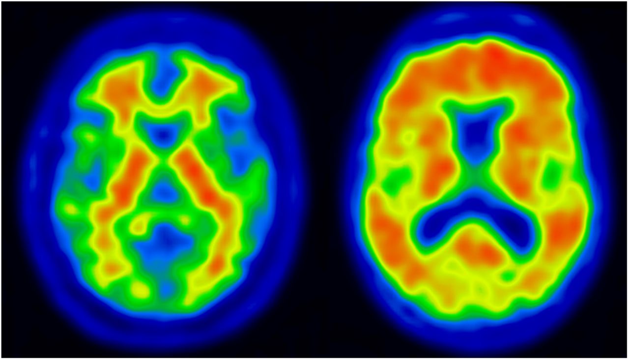
    Typical patterns of 18F-flutemetamol uptake in negative scan (left) and positive scan (right). White matter uptake is similar in both scans, but there is considerably more uptake in gray matter in the positive scan.

  • FIGURE 2.
    • Download figure
    • Open in new tab
    • Download powerpoint
    FIGURE 2.

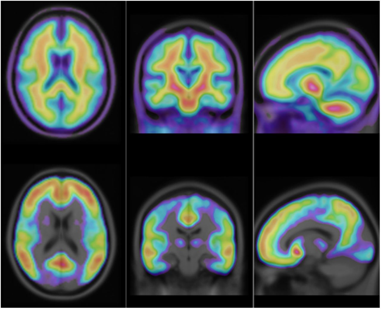
    Intercept image (top) and slope image (bottom) from linear regression of input images on SUVRs for a neocortical composite region. Images are overlaid on MR T1 template image.
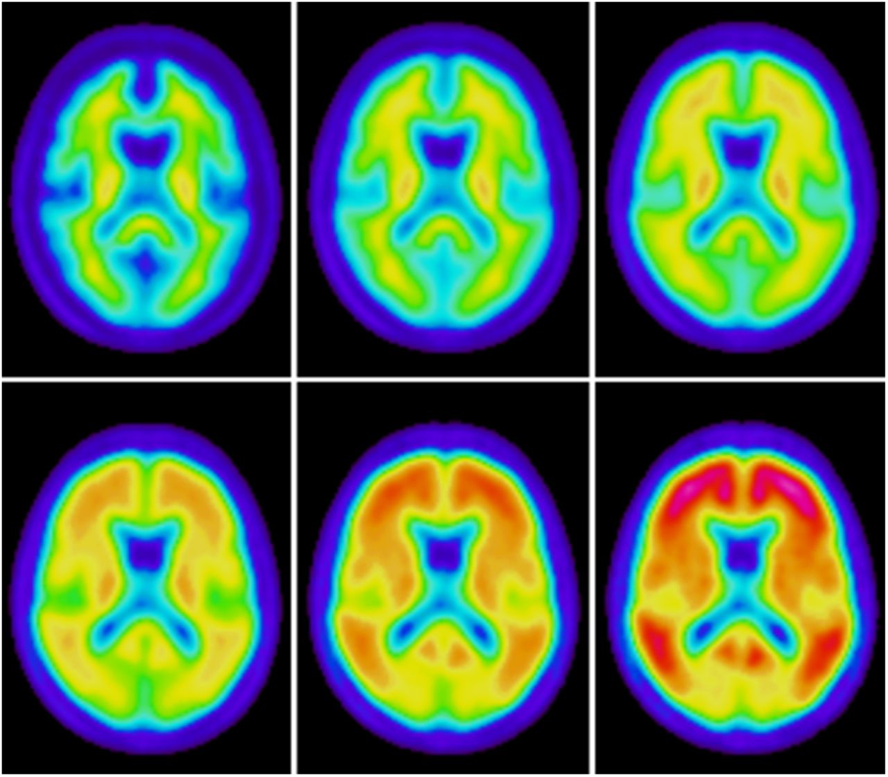
  • FIGURE 3.
    • Download figure
    • Open in new tab
    • Download powerpoint
    FIGURE 3.

    Synthetic images showing typical 18F-flutemetamol patterns going from most negative (upper left) to most positive case (lower right). Value of x is increased in steps by 0.2 going from left to right, top to bottom.
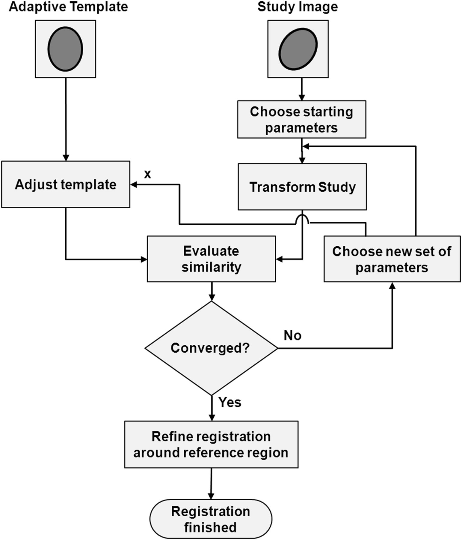
  • FIGURE 4.
    • Download figure
    • Open in new tab
    • Download powerpoint
    FIGURE 4.

    Flowchart illustrating spatial normalization using the adaptive template approach. Optimization method will change both transformation parameters and adaptive template parameter until maximal similarity between study image and template is achieved. Refined registration of reference region area is performed as a second step.

  • FIGURE 5.
    • Download figure
    • Open in new tab
    • Download powerpoint
    FIGURE 5.

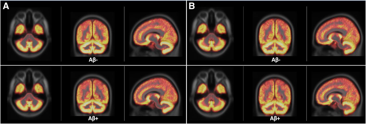
    Average gray matter probabilistic maps for Aβ− and Aβ+ groups superimposed on MNI template obtained using adaptive template registration (A) and SPM (B). Visually, there was no obvious difference between Aβ− and Aβ+, and adaptive template registration and SPM showed similar performance in cerebral cortex whereas adaptive template registration gave sharper cerebellar cortex.

  • FIGURE 6.
    • Download figure
    • Open in new tab
    • Download powerpoint
    FIGURE 6.

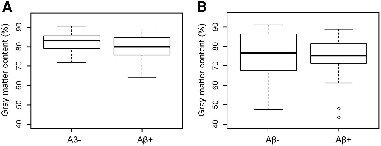
    Box plots showing amount of gray matter contained in reference region for Aβ− and Aβ+ groups. Adaptive template registration (A) shows higher amounts of gray matter in cerebellar reference region and less variability than does SPM (B).

  • FIGURE 7.
    • Download figure
    • Open in new tab
    • Download powerpoint
    FIGURE 7.

    Box plots showing amount of brain tissue contained in pons reference region for Aβ− and Aβ+ groups. Adaptive template registration (A) shows higher amounts of brain tissue in pons reference region and less variability than does SPM (B).
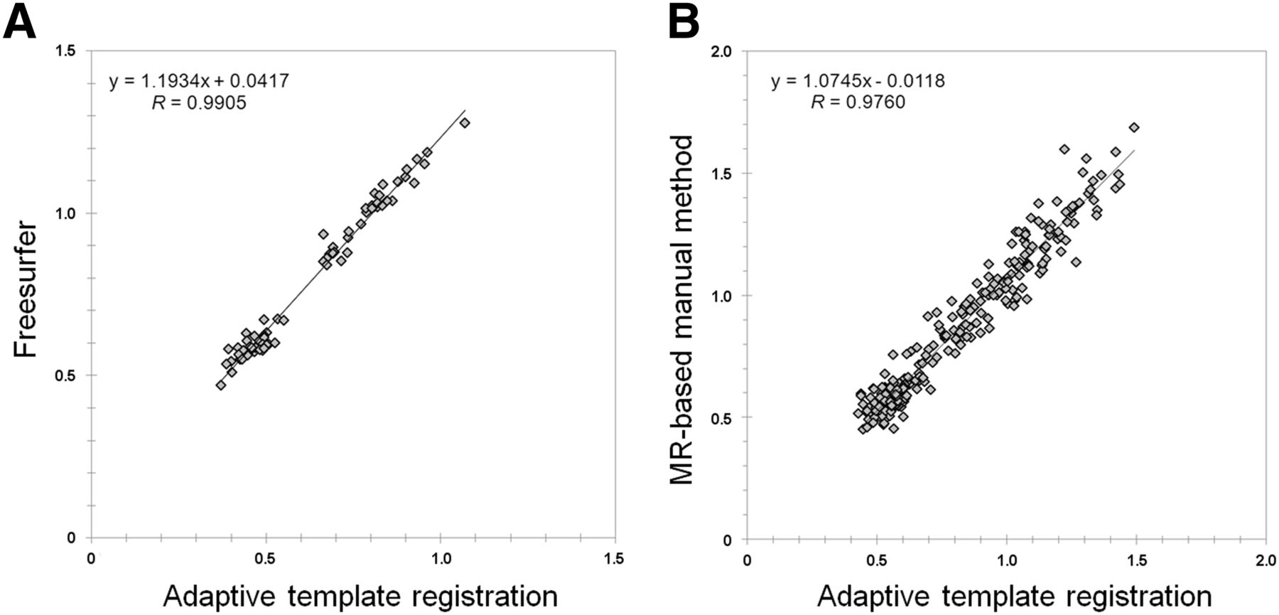
  • FIGURE 8.
    • Download figure
    • Open in new tab
    • Download powerpoint
    FIGURE 8.

    Correlation between 18F-flutemetamol phase II SUVRs for neocortical composite region computed with adaptive template registration and FreeSurfer (A) and corresponding correlation between AIBL PIB SUVRs computed with adaptive template registration and independent manual method (B). Pons or brain stem was used as reference region for both comparisons.

PreviousNext
Back to top

In this issue

Journal of Nuclear Medicine: 54 (8)
Journal of Nuclear Medicine
Vol. 54, Issue 8
August 1, 2013
  • Table of Contents
  • Table of Contents (PDF)
  • About the Cover
  • Index by author
Print
Download PDF
Article Alerts
Sign In to Email Alerts with your Email Address
Email Article

Thank you for your interest in spreading the word on Journal of Nuclear Medicine.

NOTE: We only request your email address so that the person you are recommending the page to knows that you wanted them to see it, and that it is not junk mail. We do not capture any email address.

Enter multiple addresses on separate lines or separate them with commas.
Implementation and Validation of an Adaptive Template Registration Method for 18F-Flutemetamol Imaging Data
(Your Name) has sent you a message from Journal of Nuclear Medicine
(Your Name) thought you would like to see the Journal of Nuclear Medicine web site.
Citation Tools
Implementation and Validation of an Adaptive Template Registration Method for 18F-Flutemetamol Imaging Data
Roger Lundqvist, Johan Lilja, Benjamin A. Thomas, Jyrki Lötjönen, Victor L. Villemagne, Christopher C. Rowe, Lennart Thurfjell
Journal of Nuclear Medicine Aug 2013, 54 (8) 1472-1478; DOI: 10.2967/jnumed.112.115006

Citation Manager Formats

  • BibTeX
  • Bookends
  • EasyBib
  • EndNote (tagged)
  • EndNote 8 (xml)
  • Medlars
  • Mendeley
  • Papers
  • RefWorks Tagged
  • Ref Manager
  • RIS
  • Zotero
Share
Implementation and Validation of an Adaptive Template Registration Method for 18F-Flutemetamol Imaging Data
Roger Lundqvist, Johan Lilja, Benjamin A. Thomas, Jyrki Lötjönen, Victor L. Villemagne, Christopher C. Rowe, Lennart Thurfjell
Journal of Nuclear Medicine Aug 2013, 54 (8) 1472-1478; DOI: 10.2967/jnumed.112.115006
Twitter logo Facebook logo LinkedIn logo Mendeley logo
  • Tweet Widget
  • Facebook Like
  • Google Plus One
Bookmark this article

Jump to section

  • Article
    • Abstract
    • MATERIALS AND METHODS
    • RESULTS
    • DISCUSSION
    • CONCLUSION
    • DISCLOSURE
    • Footnotes
    • REFERENCES
  • Figures & Data
  • Info & Metrics
  • PDF

Related Articles

  • This Month in JNM
  • PubMed
  • Google Scholar

Cited By...

  • Quantification Supports Amyloid PET Visual Assessment of Challenging Cases: Results from the AMYPAD Diagnostic and Patient Management Study
  • Quantification supports amyloid-PET visual assessment of challenging cases: results from the AMYPAD-DPMS study
  • Fixel-Based Analysis Reveals Tau-Related White Matter Changes in Early Stages of Alzheimer's Disease
  • Fixel-based analysis reveals macrostructural white matter changes associated with tau pathology in early stages of Alzheimers disease
  • The RSNA QIBA Profile for Amyloid PET as an Imaging Biomarker for Cerebral Amyloid Quantification
  • Comparing the Clinical Utility and Diagnostic Performance of CSF P-Tau181, P-Tau217, and P-Tau231 Assays
  • Improved Accuracy of Amyloid PET Quantification with Adaptive Template-Based Anatomic Standardization
  • Cerebral hypoperfusion is a late pathological event in the course of Alzheimers disease
  • Improving prediction of amyloid deposition in Mild Cognitive Impairment with a timed motor task
  • Association of Enlarged Perivascular Spaces and Measures of Small Vessel and Alzheimer Disease
  • Derivation and utility of an A{beta}-PET pathology accumulation index to estimate A{beta} load
  • Biomarker Localization, Analysis, Visualization, Extraction, and Registration (BLAzER) Workflow for Research and Clinical Brain PET Applications
  • Spatial Normalization of 18F-Flutemetamol PET Images Using an Adaptive Principal-Component Template
  • Generation of Structural MR Images from Amyloid PET: Application to MR-Less Quantification
  • Amyloid blood biomarker detects Alzheimer's disease
  • Increased midlife triglycerides predict brain {beta}-amyloid and tau pathology 20 years later
  • Visualization and Quantification of 3-Dimensional Stereotactic Surface Projections for 18F-Flutemetamol PET Using Variable Depth
  • Myo-inositol changes precede amyloid pathology and relate to APOE genotype in Alzheimer disease
  • Detailed comparison of amyloid PET and CSF biomarkers for identifying early Alzheimer disease
  • Automated Quantification of 18F-Flutemetamol PET Activity for Categorizing Scans as Negative or Positive for Brain Amyloid: Concordance with Visual Image Reads
  • Google Scholar

More in this TOC Section

  • Tumor-Specific Binding of Radiolabeled PEGylated GIRLRG Peptide: A Novel Agent for Targeting Cancers
  • PET/MRI of Hypoxic Atherosclerosis Using 64Cu-ATSM in a Rabbit Model
  • Tumor Uptake of Anti-CD20 Fabs Depends on Tumor Perfusion
Show more Basic Science Investigations

Similar Articles

Keywords

  • image registration
  • 18F-flutemetamol
  • Amyloid Imaging
SNMMI

© 2025 SNMMI

Powered by HighWire