


Article Figures & Data
Figures
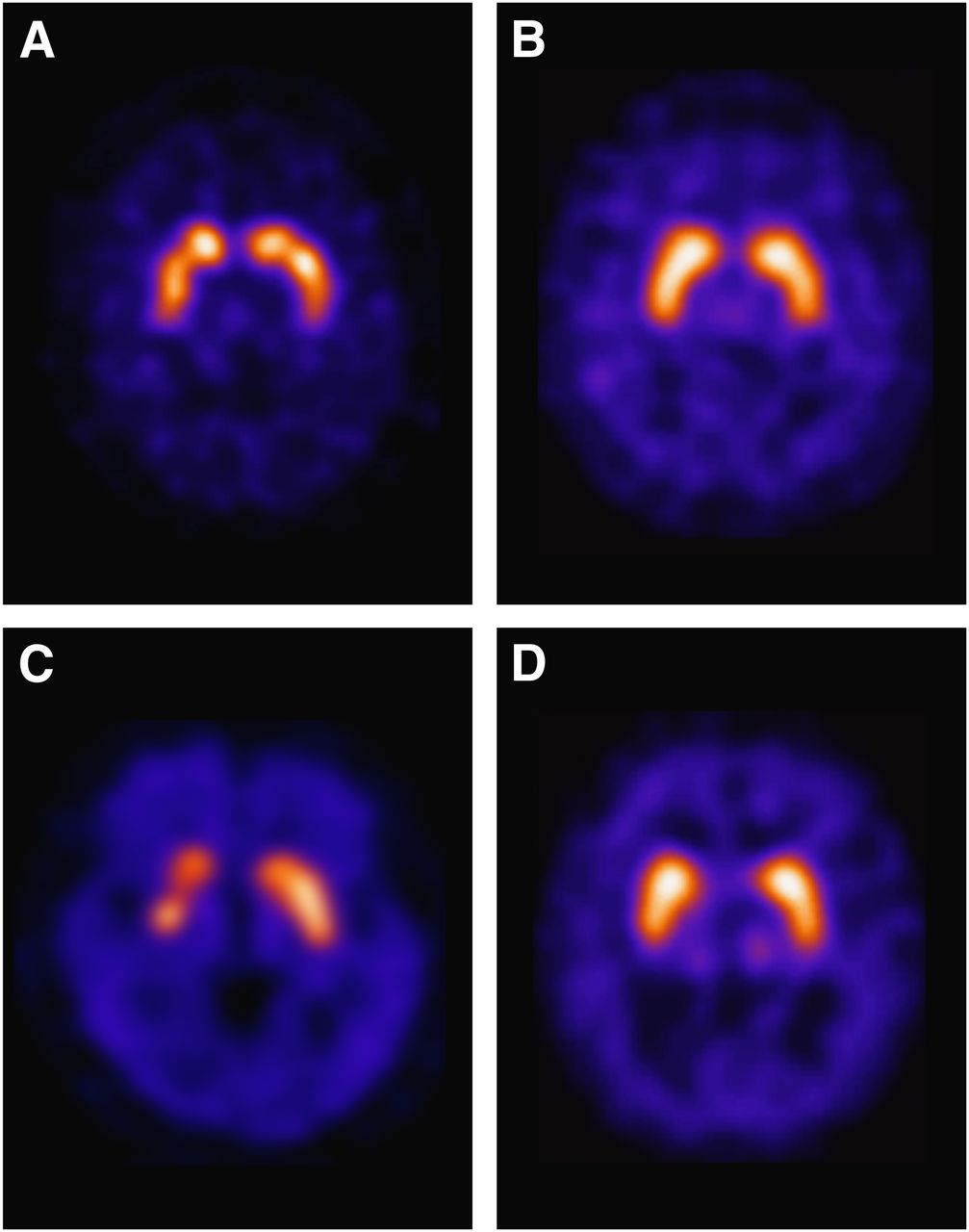
- FIGURE 1.
Representative examples of patients with neurodegenerative parkinsonian syndromes: PD (A), multiple-system atrophy (B), progressive supranuclear palsy (C), and dementia with DLB (D). Each case shows marked pathologic reduction of striatal 123I-FP-CIT binding representing loss of presynaptic neuron integrity in these disorders. In all cases, predominant involvement of putamen is seen, and extent of reduced binding advances from posterior to anterior putamen and finally caudate. Somewhat accentuated striatal asymmetry is evident. Reliable distinction between different entities (PD vs. various shown forms of aPS) based on DAT imaging is not possible (39,40).
- FIGURE 2.
Representative examples of patients with tremor syndromes and symptomatic parkinsonism: ET (A), drug-induced parkinsonism (B), vascular parkinsonism (C), and normal-pressure hydrocephalus (D). Each case shows normal preserved striatal 123I-FP-CIT binding. Vascular PS case reveals small striatal defect in right anterior putamen corresponding to infarction at this location and slightly reduced binding in caudate compared with putamen, but pattern is clearly different from cases with neurodegeneration. Case with normal-pressure hydrocephalus shows, apart from normal DAT binding, marked enlargement of ventricles that increases the distance between striata. All these entities are easily distinguishable from the various forms of neurodegenerative PS shown in Fig. 1.


Jump to section
Related Articles
Cited By...
- Pearls & Oy-sters: Idiopathic Normal Pressure Hydrocephalus With Synucleinopathy: Diagnosis and Treatment
- Determining the Degree of Dopaminergic Denervation Based on the Loss of Nigral Hyperintensity on SMWI in Parkinsonism
- A dominant-negative variant in the dopamine transporter PDZ-binding motif is linked to parkinsonism and neuropsychiatric disease
- Optimization of Parameters for Quantitative Analysis of 123I-Ioflupane SPECT Images for Monitoring Progression of Parkinson Disease
- Impairment of cross-modality of vision and olfaction in Parkinson disease
- Diagnostic Performance of the Visual Reading of 123I-Ioflupane SPECT Images With or Without Quantification in Patients With Movement Disorders or Dementia
- Is ioflupane I123 injection diagnostically effective in patients with movement disorders and dementia? Pooled analysis of four clinical trials