
Main menu

  • Home
  • Content
    • Current
    • Ahead of print
    • Past Issues
    • JNM Supplement
    • SNMMI Annual Meeting Abstracts
    • Continuing Education
    • JNM Podcasts
  • Subscriptions
    • Subscribers
    • Institutional and Non-member
    • Rates
    • Journal Claims
    • Corporate & Special Sales
  • Authors
    • Submit to JNM
    • Information for Authors
    • Assignment of Copyright
    • AQARA requirements
  • Info
    • Reviewers
    • Permissions
    • Advertisers
  • About
    • About Us
    • Editorial Board
    • Contact Information
  • More
    • Alerts
    • Feedback
    • Help
    • SNMMI Journals
  • SNMMI
    • JNM
    • JNMT
    • SNMMI Journals
    • SNMMI

User menu

  • Subscribe
  • My alerts
  • Log in
  • My Cart

Search

  • Advanced search
Journal of Nuclear Medicine
  • SNMMI
    • JNM
    • JNMT
    • SNMMI Journals
    • SNMMI
  • Subscribe
  • My alerts
  • Log in
  • My Cart
Journal of Nuclear Medicine

Advanced Search

  • Home
  • Content
    • Current
    • Ahead of print
    • Past Issues
    • JNM Supplement
    • SNMMI Annual Meeting Abstracts
    • Continuing Education
    • JNM Podcasts
  • Subscriptions
    • Subscribers
    • Institutional and Non-member
    • Rates
    • Journal Claims
    • Corporate & Special Sales
  • Authors
    • Submit to JNM
    • Information for Authors
    • Assignment of Copyright
    • AQARA requirements
  • Info
    • Reviewers
    • Permissions
    • Advertisers
  • About
    • About Us
    • Editorial Board
    • Contact Information
  • More
    • Alerts
    • Feedback
    • Help
    • SNMMI Journals
  • View or Listen to JNM Podcast
  • Visit JNM on Facebook
  • Join JNM on LinkedIn
  • Follow JNM on Twitter
  • Subscribe to our RSS feeds
Research ArticleSpecial Contribution

MIRD Pamphlet No. 24: Guidelines for Quantitative 131I SPECT in Dosimetry Applications

Yuni K. Dewaraja, Michael Ljungberg, Alan J. Green, Pat B. Zanzonico, Eric C. Frey, SNMMI MIRD Committee, Wesley E. Bolch, A. Bertrand Brill, Mark Dunphy, Darrell R. Fisher, Roger W. Howell, Ruby F. Meredith, George Sgouros and Barry W. Wessels
Journal of Nuclear Medicine December 2013, 54 (12) 2182-2188; DOI: https://doi.org/10.2967/jnumed.113.122390
Yuni K. Dewaraja
1Department of Radiology, University of Michigan Medical School, Ann Arbor, Michigan
  • Find this author on Google Scholar
  • Find this author on PubMed
  • Search for this author on this site
Michael Ljungberg
2Department of Medical Radiation Physics, Lund University, Lund, Sweden
  • Find this author on Google Scholar
  • Find this author on PubMed
  • Search for this author on this site
Alan J. Green
3University College London Cancer Institute, London, United Kingdom
  • Find this author on Google Scholar
  • Find this author on PubMed
  • Search for this author on this site
Pat B. Zanzonico
4Memorial Sloan-Kettering Cancer Center, New York, New York; and
  • Find this author on Google Scholar
  • Find this author on PubMed
  • Search for this author on this site
Eric C. Frey
5Department of Radiology and Radiological Science, Johns Hopkins University, Baltimore, Maryland
  • Find this author on Google Scholar
  • Find this author on PubMed
  • Search for this author on this site
Wesley E. Bolch
  • Find this author on Google Scholar
  • Find this author on PubMed
  • Search for this author on this site
A. Bertrand Brill
  • Find this author on Google Scholar
  • Find this author on PubMed
  • Search for this author on this site
Mark Dunphy
  • Find this author on Google Scholar
  • Find this author on PubMed
  • Search for this author on this site
Darrell R. Fisher
  • Find this author on Google Scholar
  • Find this author on PubMed
  • Search for this author on this site
Roger W. Howell
  • Find this author on Google Scholar
  • Find this author on PubMed
  • Search for this author on this site
Ruby F. Meredith
  • Find this author on Google Scholar
  • Find this author on PubMed
  • Search for this author on this site
George Sgouros
  • Find this author on Google Scholar
  • Find this author on PubMed
  • Search for this author on this site
Barry W. Wessels
  • Find this author on Google Scholar
  • Find this author on PubMed
  • Search for this author on this site
  • Article
  • Figures & Data
  • Info & Metrics
  • PDF
Loading

Article Figures & Data

Figures

  • Tables
  • FIGURE 1.
    • Download figure
    • Open in new tab
    • Download powerpoint
    FIGURE 1.

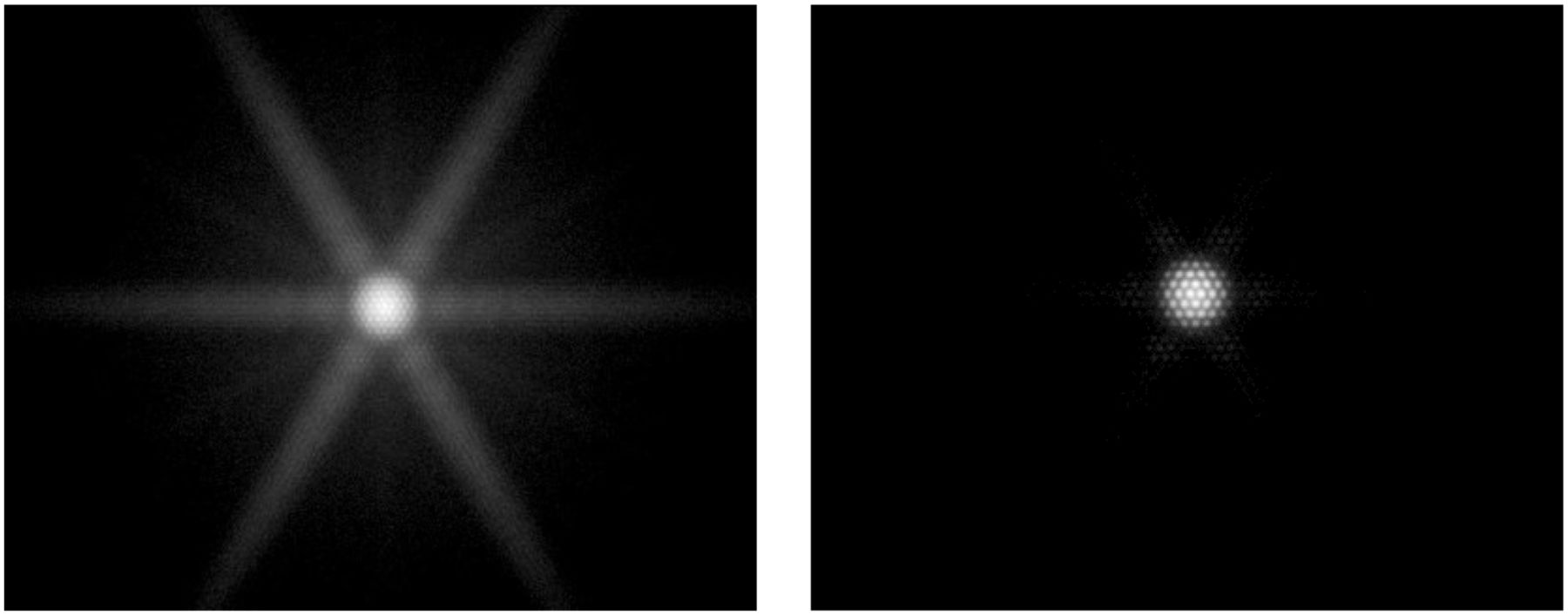
    Images corresponding to 131I pointlike source measured in air at 20 cm with medium-energy (left) and high-energy (right) collimators. Images are shown on a logarithmic gray scale (individually normalized). System planar sensitivities for a 364-keV window were 319 cps/MBq for the medium-energy collimator and 82 cps/MBq for the high-energy collimator, but the fraction of unwanted penetration and scatter events was much higher with the medium-energy collimator than with the high-energy collimator, 85% versus 49%, based on Monte Carlo simulation.

  • FIGURE 2.
    • Download figure
    • Open in new tab
    • Download powerpoint
    FIGURE 2.

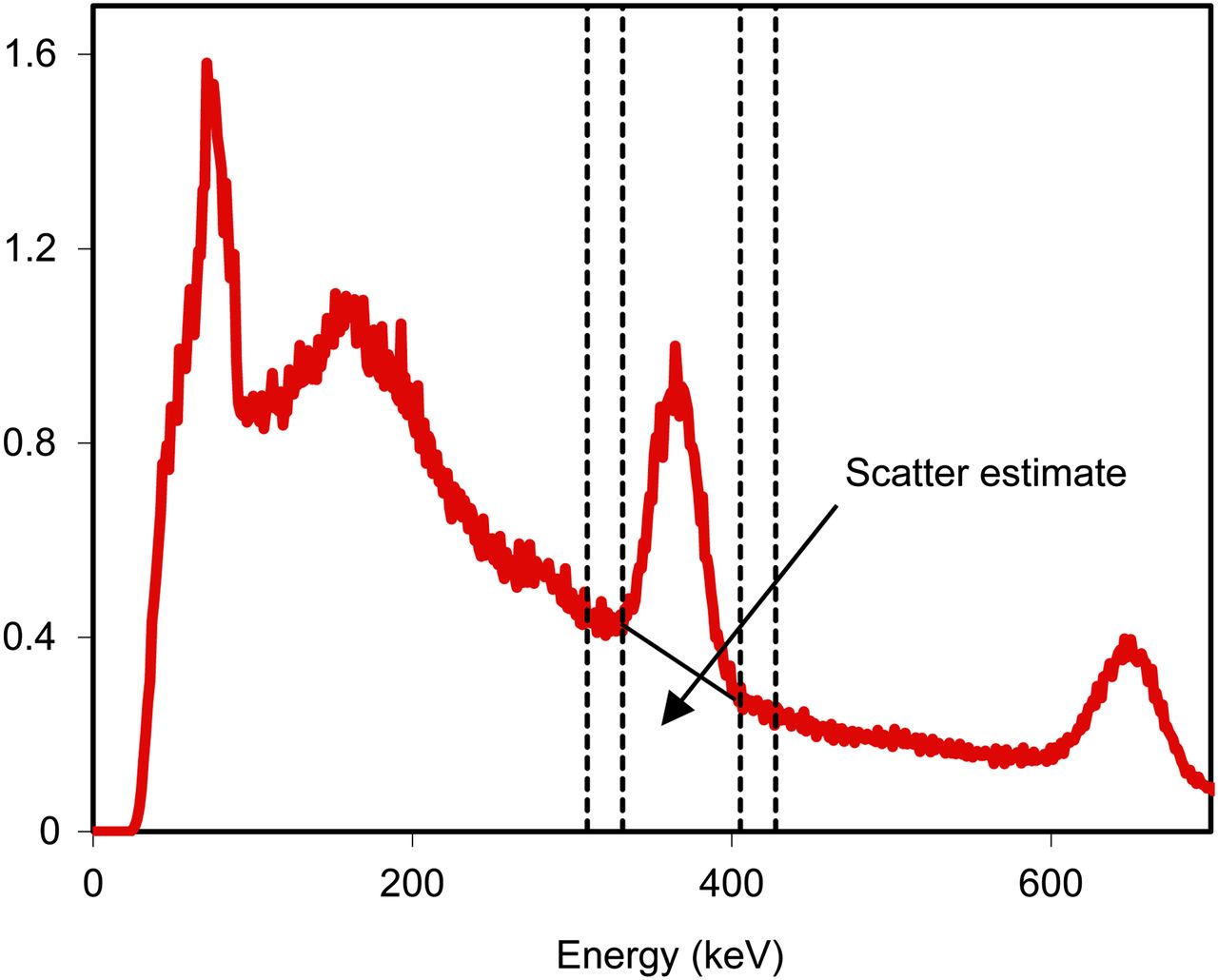
    Energy spectrum corresponding to a radioimmunotherapy patient imaged 2 d after administration of 2.8 GBq of 131I. Windows for TEW (310–332 keV and 405–427 keV) are shown by dashed lines, and the trapezoidal scatter estimate is indicated in the 332–405 keV photopeak window.

  • FIGURE 3.
    • Download figure
    • Open in new tab
    • Download powerpoint
    FIGURE 3.

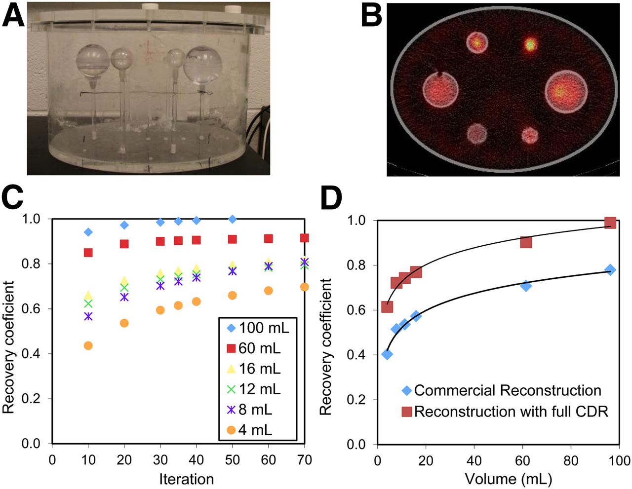
    Measurement of RCs discussed in patient example 1. (A) Phantom set-up. (B) SPECT/CT image. (C) RC as function of OSEM iteration number. (D) RC as function of volume at 35 iterations. RCs that were determined with commercial OSEM reconstruction are also shown.
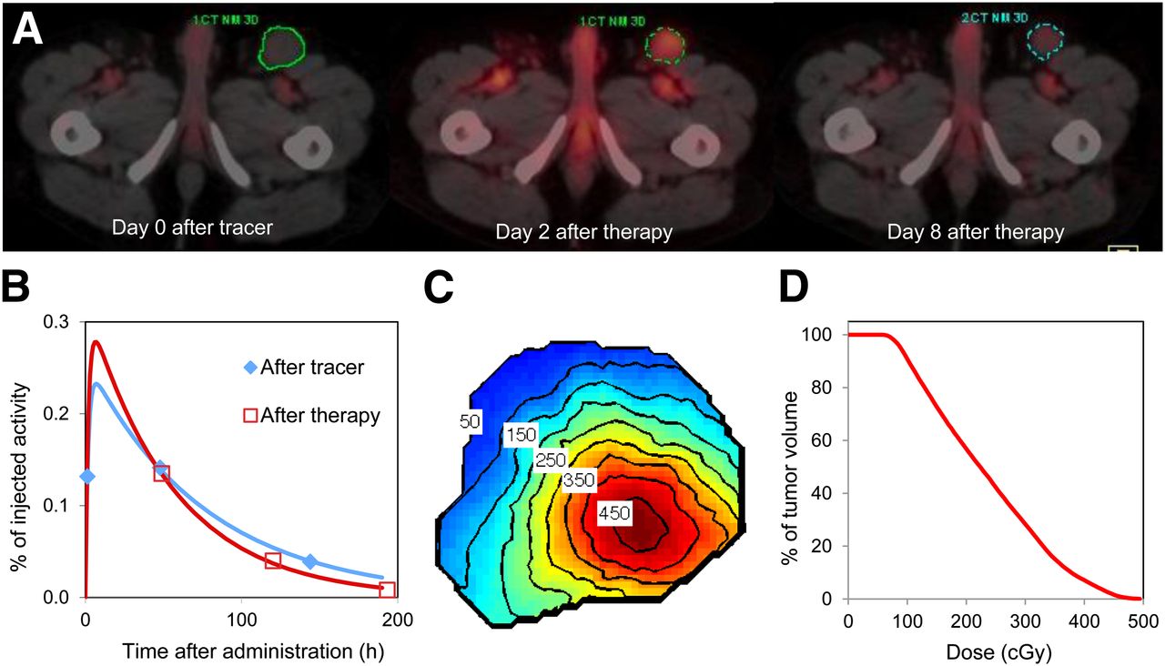
  • FIGURE 4.
    • Download figure
    • Open in new tab
    • Download powerpoint
    FIGURE 4.

    SPECT/CT imaging based tumor dosimetry in a non-Hodgkin lymphoma patient undergoing 131I radioimmunotherapy. (A) Inguinal tumor outline shown on superimposed SPECT/CT images. Tumor volumes at the 3 time points were 77, 63, and 39 mL. (B) Tumor time–activity curves. (C) Tumor-absorbed dose map, showing isodose contours in units of cGy. (D) Tumor dose–volume histogram.

  • FIGURE 5.
    • Download figure
    • Open in new tab
    • Download powerpoint
    FIGURE 5.

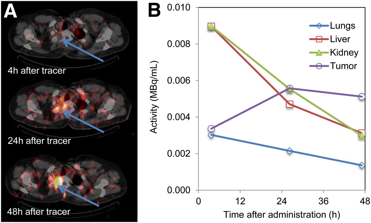
    SPECT/CT imaging–based biodistribution measurement in sarcoma patient undergoing radioimmunotherapy with 131I-L19SIP. (A) SPECT/CT images of upper thorax, with tumor indicated by arrows. (B) Time–activity concentration curves for tumor and normal organs.

Tables

  • Figures
    • View popup
    TABLE 1

    Phantom and In Vivo Validation Studies of Quantitative 131I SPECT

    StudyApplication/potential applicationStudySystemReconstructionQuantification accuracy*
    Song 2011 (14)RIT dosimetryPhantom simulationsSimulation studyOSEM with ESSE SC, AC, CDRC<5% error for larger organs; 10%–15% for smaller organs
    Dewaraja 2010 (4)RIT dosimetryPhantom measurements/simulationsSPECT/CTOSEM with TEW SC, CT-based AC, CDRC<17% error for 8- to 95-mL spheres; 31% for 4-mL sphere
    Pereira 2010 (28)DosimetryPhantom measurementsSPECT/CTOSEM with TEW SC, CT-based ACMeasured-to-true ratios of >90% for 11.5-mL sphere, 13%–63% for 1.4- and 2.2-mL spheres
    Shcherbinin 2008 (29)MIBG dosimetryPhantom measurementsSPECT/CTOSEM with analytic scatter model, CT-based AC, CDRC3%–4% error for 32-mL volumes
    Koral 2005 (24)RIT dosimetryPhantom measurementsSPECT and CT separatelyOSEM with TEW SC, CT-based AC, CDRC<7% average error for 100-mL sphere
    Gonzalez Trotter 2001 (13)Brain tumor RITPhantom measurementsSPECT with specialized collimatorOSEM with TEW SC, AC, CDRC<20% error for 6- to 11-mL spheres
    Alaamer 1993 (18)MIBG dosimetryPhantom measurementsSPECT and CT separatelyReconstruction with SC and ACSE = 0.24 MBq for 6–600 mL
    Israel 1990 (19)MIBG and thyroid carcinoma dosimetryPhantom measurements and in vivo patient study of 131I concentration in urinary bladderSPECTFBPPhantom: good correlation with truth (r = 0.98, SEE = 20.94 counts/voxel); patients: good correlation with concentration in urine (r = 0.98, SEE = 25.049 kBq/mL)
    Riggs 1988 (30)RITPhantom measurements and in vivo patient study of 131I concentration in heartSPECTFBP with DEW SC, Chang ACPhantom: <10% error; patients: good correlation with concentration in serial blood
    • ↵* Percentage difference between SPECT estimated activity and truth.

    • RIT = radioimmunotherapy; ESSE = effective scatter source estimation; SC = scatter correction; AC = attenuation correction; CDRC = CDR compensation; MIBG = metaiodobenzylguanidine; FBP = filtered backprojection; DEW = dual energy windows, SE = standard error; SEE = standard error of the estimate.

PreviousNext
Back to top

In this issue

Journal of Nuclear Medicine: 54 (12)
Journal of Nuclear Medicine
Vol. 54, Issue 12
December 1, 2013
  • Table of Contents
  • Table of Contents (PDF)
  • About the Cover
  • Index by author
Print
Download PDF
Article Alerts
Sign In to Email Alerts with your Email Address
Email Article

Thank you for your interest in spreading the word on Journal of Nuclear Medicine.

NOTE: We only request your email address so that the person you are recommending the page to knows that you wanted them to see it, and that it is not junk mail. We do not capture any email address.

Enter multiple addresses on separate lines or separate them with commas.
MIRD Pamphlet No. 24: Guidelines for Quantitative 131I SPECT in Dosimetry Applications
(Your Name) has sent you a message from Journal of Nuclear Medicine
(Your Name) thought you would like to see the Journal of Nuclear Medicine web site.
Citation Tools
MIRD Pamphlet No. 24: Guidelines for Quantitative 131I SPECT in Dosimetry Applications
Yuni K. Dewaraja, Michael Ljungberg, Alan J. Green, Pat B. Zanzonico, Eric C. Frey, SNMMI MIRD Committee, Wesley E. Bolch, A. Bertrand Brill, Mark Dunphy, Darrell R. Fisher, Roger W. Howell, Ruby F. Meredith, George Sgouros, Barry W. Wessels
Journal of Nuclear Medicine Dec 2013, 54 (12) 2182-2188; DOI: 10.2967/jnumed.113.122390

Citation Manager Formats

  • BibTeX
  • Bookends
  • EasyBib
  • EndNote (tagged)
  • EndNote 8 (xml)
  • Medlars
  • Mendeley
  • Papers
  • RefWorks Tagged
  • Ref Manager
  • RIS
  • Zotero
Share
MIRD Pamphlet No. 24: Guidelines for Quantitative 131I SPECT in Dosimetry Applications
Yuni K. Dewaraja, Michael Ljungberg, Alan J. Green, Pat B. Zanzonico, Eric C. Frey, SNMMI MIRD Committee, Wesley E. Bolch, A. Bertrand Brill, Mark Dunphy, Darrell R. Fisher, Roger W. Howell, Ruby F. Meredith, George Sgouros, Barry W. Wessels
Journal of Nuclear Medicine Dec 2013, 54 (12) 2182-2188; DOI: 10.2967/jnumed.113.122390
Twitter logo Facebook logo LinkedIn logo Mendeley logo
  • Tweet Widget
  • Facebook Like
  • Google Plus One
Bookmark this article

Jump to section

  • Article
    • Abstract
    • GUIDELINES
    • PATIENT EXAMPLE 1: QUANTITATIVE SPECT/CT FOR 3-DIMENSIONAL TUMOR DOSIMETRY IN 131I-TOSITUMOMAB RADIOIMMUNOTHERAPY
    • PATIENT EXAMPLE 2: QUANTITATIVE SPECT/CT FOR BIODISTRIBUTION MEASUREMENT IN 131I-L19SIP RADIOIMMUNOTHERAPY
    • DISCLOSURE
    • Footnotes
    • REFERENCES
  • Figures & Data
  • Info & Metrics
  • PDF

Related Articles

  • This Month in JNM
  • PubMed
  • Google Scholar

Cited By...

  • Computational Nuclear Oncology Toward Precision Radiopharmaceutical Therapies: Current Tools, Techniques, and Uncharted Territories
  • The MIRD Schema for Radiopharmaceutical Dosimetry: A Review
  • Analysis of Residence Time, Effective Half-Life, and Internal Dosimetry Before Radioiodine Therapy
  • The Relevance of Dosimetry in Precision Medicine
  • Optimizing Image Quantification for 177Lu SPECT/CT Based on a 3D Printed 2-Compartment Kidney Phantom
  • Alternative Means of Estimating 131I Maximum Permissible Activity to Treat Thyroid Cancer
  • Design and Fabrication of Kidney Phantoms for Internal Radiation Dosimetry Using 3D Printing Technology
  • Quantitative Comparison of 124I PET/CT and 131I SPECT/CT Detectability
  • Google Scholar

More in this TOC Section

  • Theranostics in Perspective: White Paper
  • Consensus Nomenclature for Radionuclide Therapy: Initial Recommendations from Nuclear Medicine Global Initiative
  • Computational Nuclear Oncology Toward Precision Radiopharmaceutical Therapies: Ethical, Regulatory, and Socioeconomic Dimensions of Theranostic Digital Twins
Show more SPECIAL CONTRIBUTION

Similar Articles

Keywords

  • MIRD
  • 131I
  • quantitative SPECT
  • SPECT/CT, dosimetry
SNMMI

© 2025 SNMMI

Powered by HighWire