


Article Figures & Data
Figures
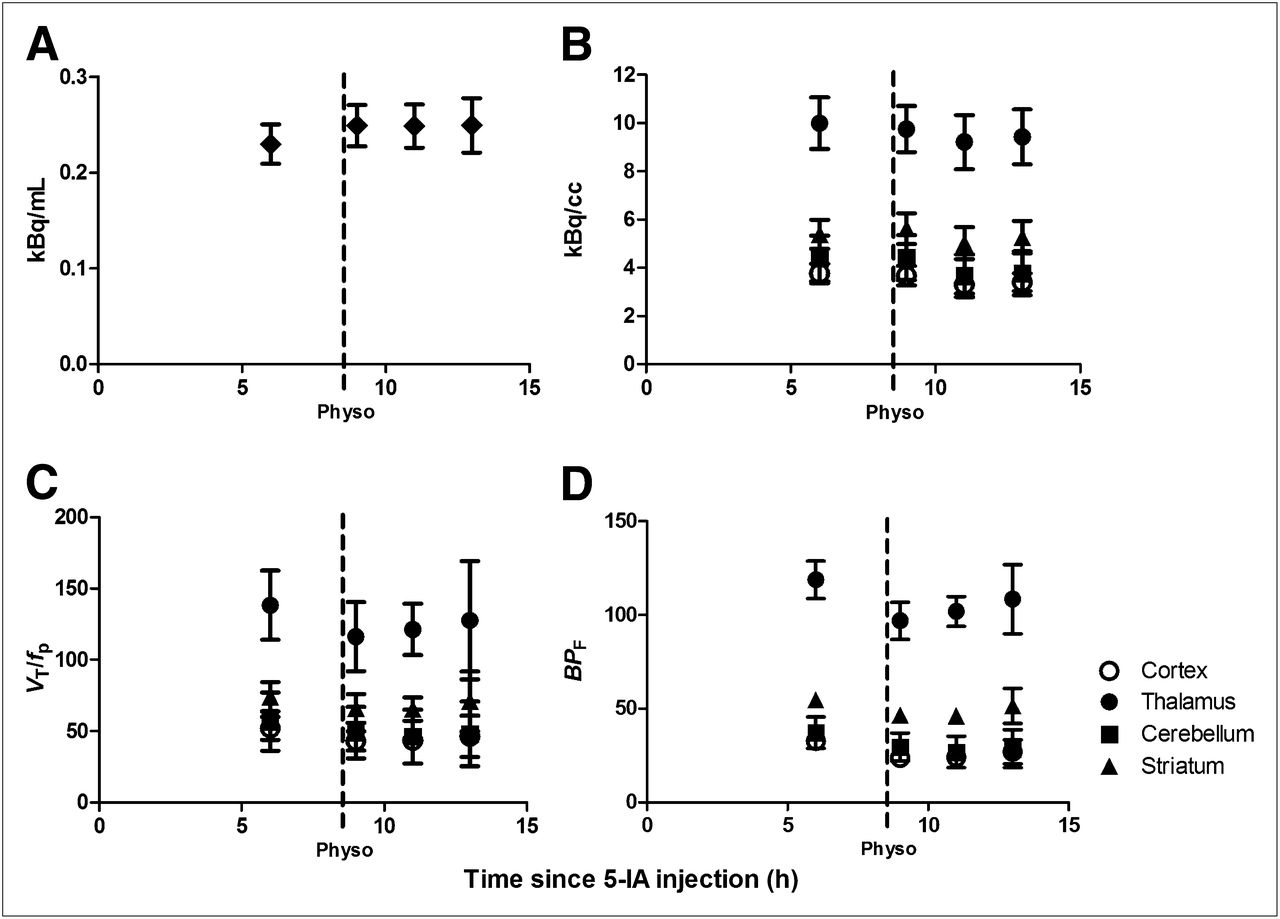
- FIGURE 1.
First point in each graph represents baseline data obtained starting 6 h after beginning of tracer infusion, when state of equilibrium was achieved, and provided baseline specific binding. After completion of baseline scans, physostigmine was administered intravenously (1.0–1.5 mg over 1 h). At onset of physostigmine infusion, scanning was resumed for up to 9 h. Bars represent SEM. (A) Plasma 123I-5-IA concentration (kBq/mL) (total parent) measured during 123I-5-IA constant infusion in healthy volunteers. After physostigmine administration, there was significant 9% increase in mean plasma 123I-5-IA concentration as compared with before physostigmine administration. (B) Tissue 123I-5-IA concentration (kBq/cm3) in thalamus, striatum, cortex, and cerebellum measured during 123I-5-IA constant infusion. We observed 7%–16% region-specific decrease in 123I-5-IA tissue concentration after physostigmine challenge. (C) 123I-5-IA VT/fp in thalamus, striatum, cortex, and cerebellum measured during 123I-5-IA constant infusion. VT/fp values measured after physostigmine infusion were significantly reduced (14%–18% region-specific), compared with baseline values. (D) 123I-5-IA BPf in thalamus, striatum, cortex, and cerebellum measured during 123I-5-IA constant infusion. BPf values measured after physostigmine infusion were significantly reduced (19%–36% region-specific), compared with baseline values.
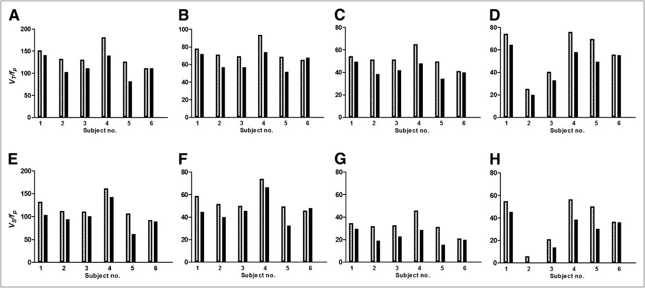
- FIGURE 2.
(A–D) β2*-nAChR availability (VT/fP) before (hatched bars) and after (black bars) physostigmine injection for each subject. For thalamus (A), percentage displacement of 5-IA for subjects 1–6 was −7%, −22%, −14%, −22%, −35%, and −0%. For striatum (B), percentage displacement was −8%, −20%, −18%, −20%, −25%, and −4%. For cortex (C), percentage displacement was −9%, −25%, −18%, −26%, −30%, and −2%. For cerebellum (D), percentage displacement was −12%, −20%, −18%, −23%, −28%, and 0%. (E–H) BPf before (hatched bars) and after (black bars) physostigmine injection for each subject. For thalamus (E), percentage displacement of 5-IA for subjects 1–6 was −8%, −25%, −17%, −25%, −41%, and 0%. For striatum (F), percentage displacement was −10%, −27%, −24%, −26%, −34%, and +5%. For cortex (G), percentage displacement was −13%, −40%, −29%, −36%, −50%, and −6%. For cerebellum (H), percentage displacement was −17%, −90%, −35%, −32%, −40%, and −1%.


Tables
- TABLE 1
Outcome Values for Each Subject at Baseline and 2–4 Hours After Physostigmine Injection
fp at baseline fp before physostigmine fp after physostigmine* fp at end of study VT/fp at baseline VT/fp after physostigmine* BPf at baseline BPf after physostigmine* Subject no. Thal CB Cort Str Thal CB Cort Str Thal CB Cort Str Thal CB Cort Str 1 32.6% 37.0% 36.4% 39.2% 151.0 73.9 54.0 77.8 140.9 64.7 49.4 71.9 131.6 54.5 34.6 58.4 121.5 45.3 30.0 52.5 2 37.0% 36.1% 36.5% 37.5% 131.4 25.2 51.3 70.9 102.9 20.0 38.4 56.9 112.0 5.8 31.9 51.5 83.5 0.6 19.0 37.5 3 35.9% 34.7% 32.9% 28.6% 129.9 40.3 51.3 69.0 111.5 33.0 42.0 56.9 110.5 20.9 31.9 49.6 92.1 13.6 22.6 37.5 4 25.8% 25.0% 25.9% 34.2% 180.5 75.8 64.9 93.1 140.2 58.0 48.1 74.1 161.1 56.4 45.5 73.7 120.8 38.6 28.7 54.7 5 33.7% 37.4% 44.3% 47.3% 125.4 69.4 49.4 68.5 81.5 49.6 34.4 51.7 106.0 50.0 30.0 49.1 62.1 30.2 15.0 32.3 6 36.6% 35.4% 39.3% 111.1 55.6 40.9 65.1 111.0 55.4 40.0 67.8 91.7 36.2 21.5 45.7 91.6 36.0 20.6 48.4 Mean 33.6% 34.3% 35.9% 37.4% 138.2 56.7 52.0 74.1 114.7 46.8 42.0 63.2 118.8 37.3 32.6 54.7 95.3 27.4 22.6 43.8 SD 4.17% 4.65% 6.19% 6.87% 24.4 20.4 7.8 10.22 22.8 17.0 5.8 9.2 24.4 20.4 7.8 10.2 22.8 17.0 5.8 9.2 ↵* Value at 2–4 h after physostigmine administration, at time of greatest displacement of radioligand by acetylcholine.
CB = cerebellum; Cort = mean cortex; Str = striatum; Thal = thalamus.
Final blood sample could not be drawn for subject 6.


