


Article Figures & Data
Figures
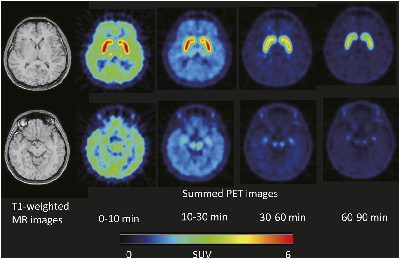
- FIGURE 1.
Representative dynamic PET images of healthy subject injected with 18F-FE-PE2I. PET images were created at level of striatum (top) and midbrain (bottom). SUV = standardized uptake value.
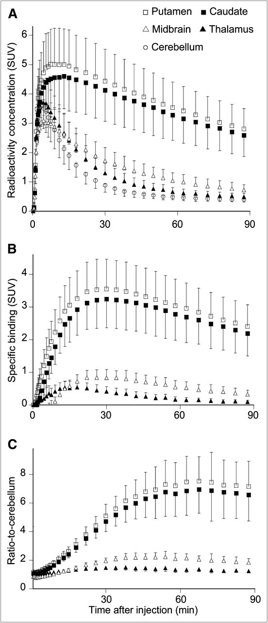
- FIGURE 2.
Average time course of radioactivity in brain regions after injection of 18F-FE-PE2I. Time course for regional radioactivity (A), specific binding (B), and ratio to cerebellum (C). Data represent mean ± SD of all 10 subjects. SUV = standardized uptake value.
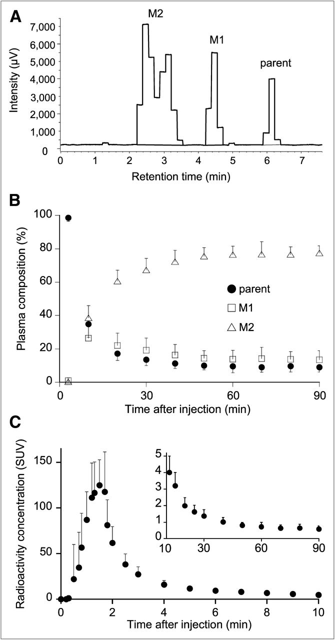
- FIGURE 3.
Concentration of 18F-FE-PE2I and its composition in arterial plasma after injection of 18F-FE-PE2I. (A) Representative radiochromatogram at 30 min after injection of 18F-FE-PE2I. (B) Plasma composition of parent, M1, and M2. (C) Concentration of 18F-FE-PE2I in plasma. Values from 0 to 10 and 10 to 90 min are shown in each graph with different ranges of y-axis. Data represent mean ± SD of all 10 subjects. SUV = standardized uptake value.
- FIGURE 4.
Representative fitted model curves of 1-TCM and 2-TCM. Time–activity curves in putamen, midbrain, and cerebellum were fitted to 1-TCM and 2-TCM using parent in plasma as input function. The 2-TCM (solid line) provided better fittings than 1-TCM (dotted line) for all 3 regions. SUV = standardized uptake value.
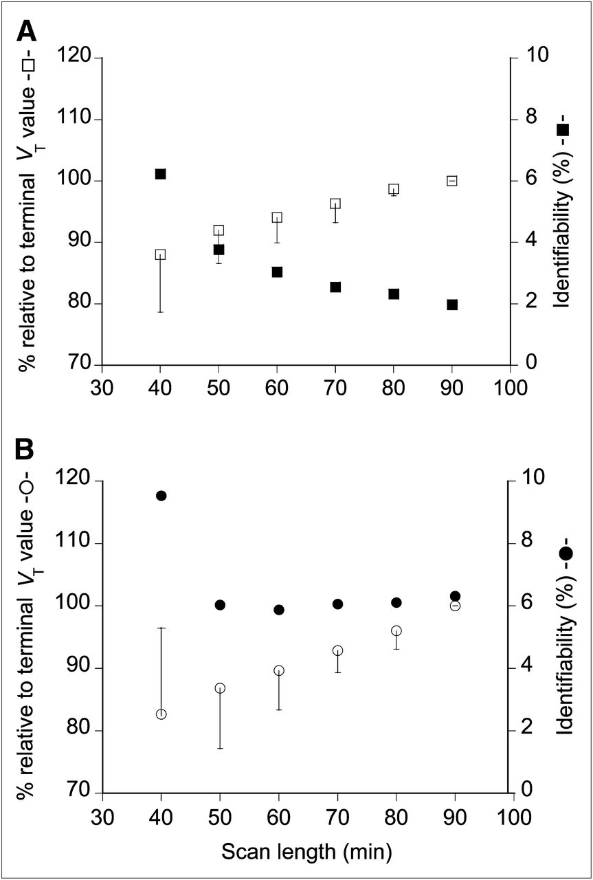
- FIGURE 5.
Value of VT and identifiability as function of scan length. VT and its corresponding SE (%) (SE (%)) were estimated in putamen (A) and cerebellum (B) with 2-TCM using parent as input function with truncating scan length from 90 to 40 min. VT values are expressed as percentage of terminal value and plotted along left y-axis for putamen (□) and cerebellum (○). Corresponding SE (%), which is inversely related to identifiability, is plotted along right y-axis for putamen (▪) and cerebellum (•). Error bar represents SD (n = 10).
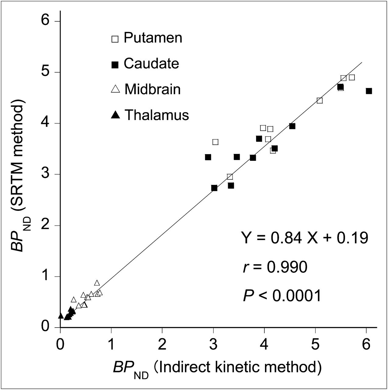
- FIGURE 6.
Correlation of BPND values estimated by indirect kinetic method using parent as input function and by SRTM method. BPND values showed significant correlation between 2 methods. Each data point represents BPND values in respective regions of each subject.
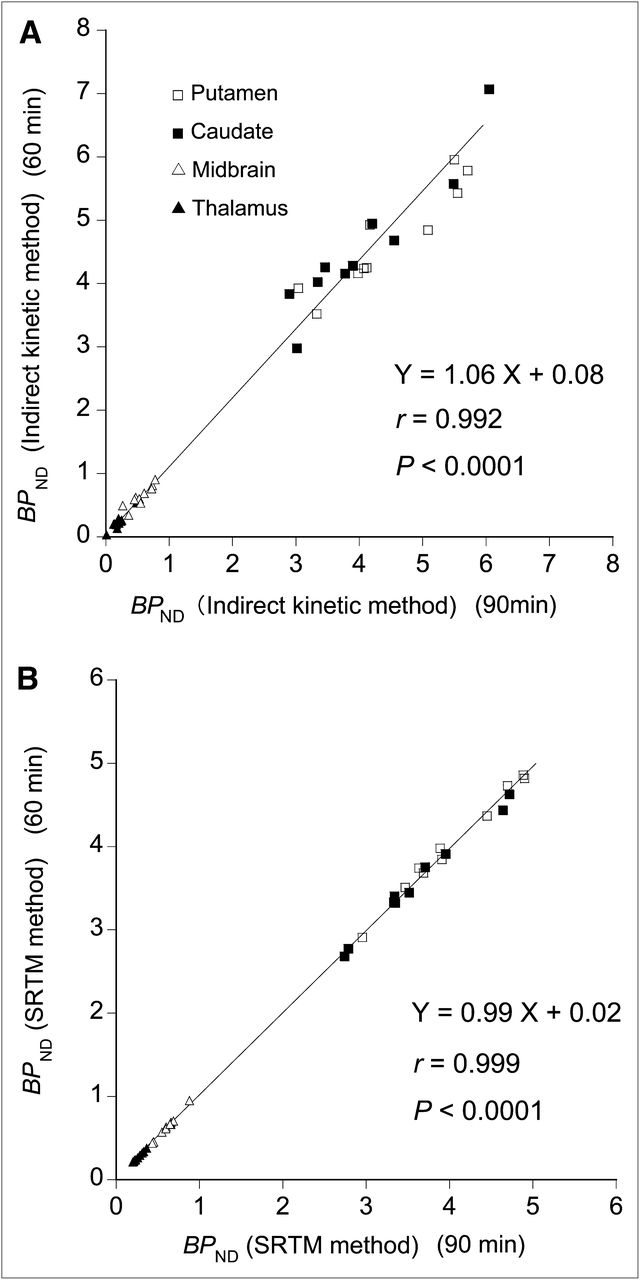
- FIGURE 7.
Correlation of BPND values estimated with 60- and 90-min data. (A) BPND values estimated by indirect kinetic method. (B) BPND values estimated by SRTM method. Significant correlations were observed in both methods between estimates with 60- and 90-min data.


Tables
Region K1 (mL⋅cm-3⋅min−1) k2 (min−1) k3 (min−1) k4 (min−1) K1/k2 (mL⋅cm−3) k3/k4 VT (mL⋅cm−3) AIC Putamen 0.292 ± 0.053 (1.7) 0.073 ± 0.022 (15) 0.133 ± 0.030 (21) 0.043 ± 0.007 (8.6) 4.25 ± 1.07 (13) 3.19 ± 0.97 (17) 17.3 ± 4.6 (2.0) −43 ± 21 Caudate 0.248 ± 0.047 (1.7) 0.051 ± 0.022 (19) 0.110 ± 0.063 (38) 0.051 ± 0.014 (17) 5.71 ± 2.69 (18) 2.09 ± 0.93 (27) 16.2 ± 5.5 (2.6) −38 ± 16 Midbrain 0.203 ± 0.044 (2.6) 0.095 ± 0.026 (13) 0.053 ± 0.028 (34) 0.042 ± 0.009 (22) 2.18 ± 0.37 (11) 1.29 ± 0.55 (18) 4.9 ± 1.1 (3.8) 20 ± 20 Thalamus 0.269 ± 0.042 (1.7) 0.123 ± 0.024 (6.0) 0.029 ± 0.018 (26) 0.041 ± 0.023 (23) 2.26 ± 0.55 (4.7) 0.71 ± 0.20 (12) 3.8 ± 0.8 (3.8) 10 ± 16 Cerebellum 0.265 ± 0.031 (1.2) 0.141 ± 0.025 (3.5) 0.013 ± 0.005 (23) 0.023 ± 0.013 (30) 1.94 ± 0.44 (2.6) 0.67 ± 0.32 (15) 3.2 ± 0.7 (6.3) −4 ± 29 Values are mean ± SD (n = 10), with percentage SE (which is inversely related to identifiability of parameters) in parentheses.
BPND Region Indirect kinetic SRTM Putamen 4.46 ± 0.95 4.05 ± 0.66 Caudate 4.06 ± 1.04 3.61 ± 0.67 Midbrain 0.55 ± 0.17 0.62 ± 0.13 Thalamus 0.20 ± 0.12 0.29 ± 0.08 Values are mean ± SD (n = 10).
Supplemental Data
Files in this Data Supplement:


Jump to section
Related Articles
Cited By...
- Locus coeruleus-related insula activation supports implicit learning
- In Vivo Visualization and Quantification of Brain Heat Shock Protein 90 with [11C]HSP990 in Healthy Aging and Neurodegeneration
- Cortical beta oscillations map to shared brain networks modulated by dopamine
- Concurrent assessment of neurometabolism and brain hemodynamics to comprehensively characterize the functional brain response to psychotropic drugs: an S-ketamine study
- Controlling the human connectome with spatially diffuse input signals
- Integrating brainstem and cortical functional architectures
- Mapping neurotransmitter systems to the structural and functional organization of the human neocortex
- Whole-Body Biodistribution and Dosimetry of the Dopamine Transporter Radioligand 18F-FE-PE2I in Human Subjects
- Optimal Acquisition Time Window and Simplified Quantification of Dopamine Transporter Availability Using 18F-FE-PE2I in Healthy Controls and Parkinson Disease Patients
- Dynamic Changes in Striatal mGluR1 But Not mGluR5 during Pathological Progression of Parkinson's Disease in Human Alpha-Synuclein A53T Transgenic Rats: A Multi-PET Imaging Study
- Quantitative Analysis of 18F-(E)-N-(3-Iodoprop-2-Enyl)-2{beta}-Carbofluoroethoxy-3{beta}-(4'-Methyl-Phenyl) Nortropane Binding to the Dopamine Transporter in Parkinson Disease