


Article Figures & Data
Figures
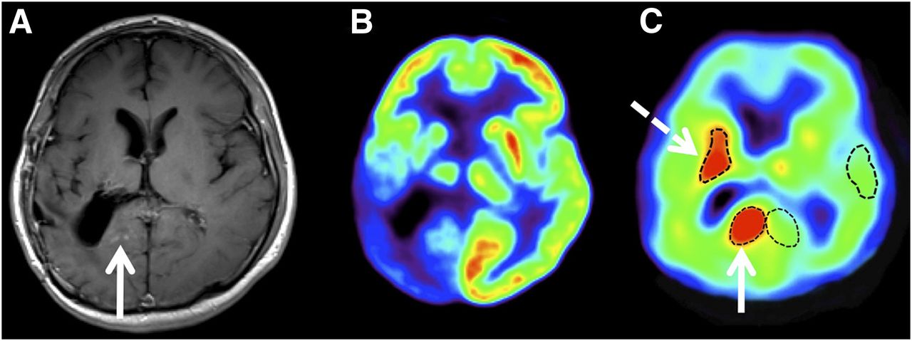
- FIGURE 1.
Coregistered T1-weighted postgadolinium MR (A), 18F-FDG PET (B), and 11C-AMT PET (C) images of 31-y-old man (patient 1) with surgically resected WHO grade II oligodendroglioma and suspected tumor recurrence. 18F-FDG PET showed marked glucose hypometabolism in neighboring cortex, whereas 11C-AMT PET demonstrated increased tryptophan uptake in area with mild contrast enhancement (solid arrows) and in more anterior region without contrast enhancement (dashed arrow). K and K lesion-to-cortex ratios of these 2 areas were similar (posterior area: 0.0070 mL/g/min and 1.60, respectively; anterior area: 0.0066 mL/g/min and 1.51, respectively). Repeated surgery demonstrated grade III oligodendroglioma in both areas.
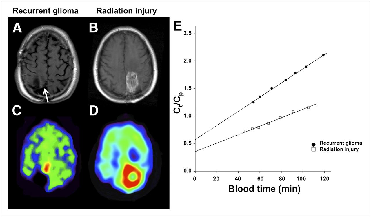
- FIGURE 2.
Representative T1-weighted postgadolinium MR and coregistered 11C-AMT PET images of patient with histologically verified glioma recurrence (A and C; patient 6) and patient with pure radiation injury, also verified by histology (B and D; patient 20). MR images showed contrast enhancement in both patients: small contrast-enhancing nodule medial to resection cavity in patient 6 (white arrow; A) and extensive contrast enhancement surrounding resection cavity in patient 20 (B). 11C-AMT PET summed images from 30 to 60 min after tracer injection demonstrated markedly increased uptake of 11C-AMT in both patients. However, kinetic analysis of dynamic PET images (E) revealed higher K (increased slope) and VD (higher y intercept) in recurrent glioma than in area of radiation injury. x-axis represents transformed time (blood time) in minutes. Ct = tracer concentration in tissue; Cp = tracer concentration in plasma.
Tables
No. Age (y) Sex Tumor type WHO grade Postsurgical treatment Histology Recurrence Time since surgery (y) SUV SUV ratio K (mL/g/min) K ratio VD VD ratio 1 31 M Oligo 2(3) Rad + chemo Yes Yes 1.5 2.18 1.35 0.007 1.60* 0.47 1.30 2a 31 M Astro 2(3) Rad + chemo Yes Yes 2 1.84 1.91 0.009 1.71 0.55 2.75 3 34 F AO 3(3) Rad Yes Yes 4 3.75 2.98 0.022 2.75 1.06 3.53 4 36 M GBM 4(4) Rad + chemo Yes Yes 3 2.37 1.60 0.008 2.40 0.42 1.50 5 45 M AO 3(3) Rad + chemo Yes Yes 2.8 3.82 3.23 0.019 3.30 1.75 5.00 6 47 M Oligo 2(3) Rad Yes Yes 4.2 1.45 2.55 0.013 2.60 0.57 2.85 7 47 M AO 3(4) Rad Yes Yes 2.2 1.87 1.67 0.009 1.70 0.56 2.70 8 56 M GBM 4(4) Rad + chemo Yes Yes 2.5 2.20 2.64 0.013 2.10 1.16 5.20 9 58 M OA 2 Rad No Yes 1 2.12 1.32 0.005 1.40 0.40 1.60 10 59 F Oligo 2(3) Rad Yes Yes 6 5.11 2.94 0.011 2.40† 0.85 4.20 11 61 M GBM 4 Rad + chemo No Yes 1 2.59 1.77 0.013 2.00 0.97 6.30 12 68 M GBM 4 Rad No Yes 1.3 2.84 1.78 0.014 2.00 0.67 3.00 13 30 M AOA 3 Rad + chemo No No 5 1.78 0.91 0.005 0.94 0.38 1.03 2b 32 M AA 3 Rad + chemo Yes No 3 1.65 1.43 0.007 1.18 0.55 2.00 14 41 F GBM 4 Rad + chemo Yes No 5 1.82 1.54 0.010 1.49 0.52 2.50 15 41 F GBM 4 Rad + chemo Yes No 3.1 2.59 1.41 0.007 1.20 0.81 2.45 16 43 M OA 2 Rad + chemo No No 2.2 1.16 1.02 0.004 0.89 0.23 1.10 17 48 M AA 3 Rad + chemo No No 5 1.33 0.99 0.004 1.15 0.30 0.97 18 53 F Oligo 2 Rad No No 6.3 0.95 0.81 0.003 1.05 0.18 0.66 19 58 F GBM 4 Rad No No 2.2 1.94 1.32 0.006 1.20 0.45 1.76 20 60 M GBM 4 Rad + chemo Yes No 1.8 1.84 1.45 0.008 1.30 0.36 2.15 21 63 M GBM 4 Rad No No 4 2.06 1.27 0.007 1.37 0.33 1.43 ↵* A second lesion (also histologically verified recurrent tumor) had K ratio of 1.51.
↵† A second lesion (also histologically verified recurrent tumor) had K ratio of 1.80.
Oligo = oligodendroglioma; astro = astrocytoma; AO = anaplastic oligodendroglioma; GBM = glioblastoma; OA = oligoastrocytoma; AOA = anaplastic oligoastrocytoma; AA = anaplastic astrocytoma; rad = radiation therapy; chemo = chemotherapy.
Ratios indicate lesion-to-contralateral cortex ratios. Histology refers to histologic verification of recurrent tumor vs. radiation injury. WHO grades in parenthesis indicate tumor grade determined by histologic assessment of recurrent glioma. Patient 2 had two 11C-AMT PET scans (2a and 2b), each followed by surgical resection and histopathologic examination (which showed tumor first but radiation injury the second time).
- TABLE 2
Comparison of 11C-AMT PET–Derived Parameters Between Recurrent Tumor and Radiation Injury Groups
All patients Patients with histology after PET (n = 13) 11C-AMT PET parameter Tumor Radiation injury P Tumor Radiation injury P Lesion SUV 2.68 ± 1.05 1.71 ± 0.48 0.014 2.73 ± 1.21 1.97 ± 0.42 0.26 Lesion-to-cortex SUV ratio 2.14 ± 0.68 1.22 ± 0.26 0.001 2.32 ± 0.69 1.45 ± 0.06 0.034 Lesion VD 0.79 ± 0.40 0.41 ± 0.18 0.012 0.82 ± 0.43 0.56 ± 0.19 0.28 Lesion-to-cortex VD ratio 3.33 ± 1.58 1.58 ± 0.66 0.004 3.22 ± 1.39 2.28 ± 0.24 0.21 Lesion K (mL/g/min) 0.012 ± 0.004 0.006 ± 0.002 0.002 0.012 ± 0.005 0.008 ± 0.001 0.10 Lesion-to-cortex K ratio 2.16 ± 0.55 1.18 ± 0.18 0.00003 2.28 ± 0.56 1.29 ± 0.14 0.006 Data are mean ± SD.


Jump to section
Related Articles
Cited By...
- Diagnostic Value of PET Tracers in Differentiating Glioma Tumor Recurrence from Treatment-Related Changes: A Systematic Review and Meta-Analysis
- Diagnostic Accuracy of PET Tracers for the Differentiation of Tumor Progression from Treatment-Related Changes in High-Grade Glioma: A Systematic Review and Metaanalysis
- Assessment of Tryptophan Uptake and Kinetics Using 1-(2-18F-Fluoroethyl)-L-Tryptophan and {alpha}-11C-Methyl-L-Tryptophan PET Imaging in Mice Implanted with Patient-Derived Brain Tumor Xenografts
- Clinical Significance of Tryptophan Metabolism in the Nontumoral Hemisphere in Patients with Malignant Glioma