


Abstract
Abnormal tryptophan metabolism via the kynurenine pathway is involved in the pathophysiology of a variety of human diseases including cancers. α-11C-methyl-l-tryptophan (11C-AMT) PET imaging demonstrated increased tryptophan uptake and trapping in epileptic foci and brain tumors, but the short half-life of 11C limits its widespread clinical application. Recent in vitro studies suggested that the novel radiotracer 1-(2-18F-fluoroethyl)-l-tryptophan (18F-FETrp) may be useful to assess tryptophan metabolism via the kynurenine pathway. In this study, we tested in vivo organ and tumor uptake and kinetics of 18F-FETrp in patient-derived xenograft mouse models and compared them with 11C-AMT uptake. Methods: Xenograft mouse models of glioblastoma and metastatic brain tumors (from lung and breast cancer) were developed by subcutaneous implantation of patient tumor fragments. Dynamic PET scans with 18F-FETrp and 11C-AMT were obtained for mice bearing human brain tumors 1–7 d apart. The biodistribution and tumoral SUVs for both tracers were compared. Results: 18F-FETrp showed prominent uptake in the pancreas and no bone uptake, whereas 11C-AMT showed higher uptake in the kidneys. Both tracers showed uptake in the xenograft tumors, with a plateau of approximately 30 min after injection; however, 18F-FETrp showed higher tumoral SUV than 11C-AMT in all 3 tumor types tested. The radiation dosimetry for 18F-FETrp determined from the mouse data compared favorably with the clinical 18F-FDG PET tracer. Conclusion: 18F-FETrp tumoral uptake, biodistribution, and radiation dosimetry data provide strong preclinical evidence that this new radiotracer warrants further studies that may lead to a broadly applicable molecular imaging tool to examine abnormal tryptophan metabolism in human tumors.
Tryptophan is an essential amino acid necessary for protein biosynthesis. Of the tryptophan not incorporated into proteins, more than 95% is metabolized via the kynurenine pathway (KP), with a smaller portion used for serotonin synthesis (Fig. 1) (1). The initial and rate-limiting step of the KP is the conversion of tryptophan to the central metabolite L-kynurenine and is mediated by 3 enzymes: indoleamine 2,3-dioxygenase 1 (IDO1), indoleamine 2,3-dioxygenase 2 (IDO2), and tryptophan 2,3-dioxygenase 2 (TDO2) (2). Kynurenine can be transformed to downstream KP metabolites including quinolinic acid, an excitotoxic glutamate receptor agonist (2,3). KP metabolites are implicated in the pathophysiology underlying neurodegenerative diseases such as Parkinson, Alzheimer, and Huntington disease (3,4). In cancer, local tryptophan depletion, kynurenine, and other KP metabolites foster an immunosuppressive tumor microenvironment (5,6). Recent studies have identified IDO1 in both hematologic cancers and solid tumors (e.g., breast cancer, lung cancer, glioblastoma) (7) with increased IDO1 expression linked to poorer outcomes (8–10). In addition to IDO1, TDO2 may also play a prominent role in glioma pathophysiology (11,12). Overall, alterations in KP enzymes and metabolites have been implicated in a wide spectrum of human diseases beyond tumors, including inflammation and psychiatric conditions (13,14).

KP of tryptophan metabolism. (A) Abridged KP overview. Enzymes in blue represent rate-limiting step. (B and C) Structures of 18F-FETrp and 11C-AMT, respectively.
To study tryptophan metabolism in vivo, the PET tracer α-11C-methyl-l-tryptophan (11C-AMT) can be used, because the radiolabeled methyl group prevents protein incorporation (15). 11C-AMT PET was originally designed to estimate brain serotonin synthesis in the context of neuropsychiatric disorders (15,16) but also demonstrated the ability to identify epileptic foci, epileptogenic tumors, and malformations (17,18). 11C-AMT PET studies were further extended to nonepileptogenic primary and metastatic brain tumors (19–22). These studies demonstrated variably high 11C-AMT uptake in gliomas (18,23,24) and meningiomas (21) and showed strong prognostic value for survival in patients with recurrent glioblastoma (20). Furthermore, 11C-AMT uptake characteristics distinguished recurrent gliomas from radiation injury (25) and glioblastoma from metastatic brain tumors (26), allowing tryptophan PET imaging as a possible clinical diagnostic tool. The development of a tryptophan analog with a longer half-life isotope such as 18F would permit widespread clinical application of tryptophan PET imaging.
Several novel 18F-labeled tryptophan derivatives have been developed and tested as potential PET tracers for tumor imaging (27–32). Although most compounds showed tumor uptake, they were not designed to target IDO1, IDO12, or TDO2 activity. Most studies focused on radiosynthesis, biodistribution, or tracer transport, which could be blocked by L-type amino acid transporter 1 inhibition. Recently, 1-(2-fluoroethyl)-l-tryptophan (FETrp) was tested as a potential substrate for human IDO1 and TDO2 in in vitro enzymatic assays (33,34). FETrp showed variable consumption depending on the human IDO1 concentration and incubation time and appeared to be a better substrate of human IDO1 than α-d,l-methyl-tryptophan.
In this study, we tested 18F-labeled FETrp (18F-FETrp) as a potential PET tracer for tumor imaging by examining 18F-FETrp uptake in patient-derived xenograft (PDX) mouse models of glioblastoma and brain metastases. We directly compared the tumoral uptake and tracer kinetics of 18F-FETrp with those of 11C-AMT using PDX models that showed 11C-AMT accumulation and variable protein expression of IDO1, IDO2, and TDO2 in our recent study (35).
MATERIALS AND METHODS
Patient Tumor Specimens
The Wayne State University Institutional Review Board approved this study, and all subjects signed a written informed consent form. Active tumor tissue (determined by the neurosurgeon) was acquired immediately after resection and histopathologic analysis. Fragments were formalin-fixed or implanted subcutaneously into immunocompromised mice. Tumors included 2 glioblastomas, 3 breast cancer brain metastases, and 1 non–small cell lung cancer brain metastasis.
Generation of Mouse Xenograft Models
The Wayne State University Institutional Animal Care and Use Committee approved all animal experiments. Female severe combined immunodeficient BALB/c background mice (Charles River), 4- to 6-wk old, were maintained on a 12-h light–dark cycle with ad libitum food and water. Tumor fragments (∼30 mg) were implanted subcutaneously, bilaterally, via trocar. Tumor growth and animal health were monitored twice weekly. Tumors were measured with Vernier calipers, and masses were estimated with the formula length × width2/2. When the tumor burden for each mouse reached approximately 5% of its body weight, mice were euthanized and harvested tumors were serially passaged into naïve mice. Tumors were passaged at least 3 times to ensure reproducible tumor growth. Harvested tumor tissues were formalin-fixed, paraffin-embedded, and sectioned at 5 μM for histology.
Radiopharmaceuticals
PET tracers were produced at the Children’s Hospital of Michigan Cyclotron Facility. 11C-AMT was produced as previously described (36). 18F-FETrp was produced following published methods (34,37) in which 18F-fluoride was produced using proton bombardment of an 18O-water target. The produced 18F-fluoride in 18O-water was delivered and trapped on a Sep-Pak light QMA cartridge (Waters Corp.), which was eluted with a K2CO3/Kryptofix (Sigma Aldrich Corp.) solution (1 mg of K2CO3 and 6 mg of Kryptofix in 0.9 mL of acetonitrile and 0.03 mL of water). The 18F solution was evaporated under a nitrogen stream at 110°C and repeated with 1.0 mL of acetonitrile. Then, a solution of 18F-FETrp precursor (1.0 mg) in acetonitrile (0.25 mL) was added to the reaction vial containing dried 18F ion and heated at 115°C for 5 min. After radiofluorination, hydrochloric acid (0.25 mL, 2N) was added to the vial and the mixture was heated at 115°C for 5 min. The resulting solution was diluted and passed through an alumina-N cartridge (Waters Corp.), then loaded for chiral high-performance liquid chromatography separation. The collected fraction was pH adjusted and filter sterilized. The final 18F-FETrp product was a clear, colorless solution free of particulates in 5% ethanol in buffer (final pH ∼5.5). Total synthesis time was approximately 50 min from the end of bombardment. The decay-corrected radiochemical yield of 18F-FETrp approximated 25% at the end of synthesis. The radiochemical purity of 18F-FETrp was more than 98% by the analytic chiral high-performance liquid chromatography analysis.
Small-Animal PET/CT
For each tumor model, the mouse with the greatest tumor burden from the group (n = 2–3) was imaged. Six mice were scanned with 18F-FETrp. Five of these were also scanned with 11C-AMT 1–7 d later (Table 1), as previously described (35). Mice were fasted 2–4 h before PET scanning. Scans were obtained between 11 am and 2 pm, when tryptophan plasma levels are at the nadir in mice maintained on a 12-h light–dark cycle (38). Radiotracers were administered via tail vein injection. The total injected activity in mice for the 18F-FETrp tracer was between 5.5 and 9.2 MBq (150–250 μCi), a dose commonly used in small-animal PET studies of mice using 18F-labeled PET tracers (39,40). Because of the shorter half-life of the 11C isotope (20 min), a higher activity was administered in the AMT study (20–22 MBq equivalent to 550–600 μCi). This dose was previously determined by our group as sufficient to obtain high-quality images in the PDX tumor models (35). Mice were anesthetized (1.5% isoflurane) and placed in a microPET R4 scanner (Concorde Microsystems Inc.) with an in-plane resolution of 1.76 mm in full width at half maximum in the center of field of view and a linear resolution of less than 2.0 mm in all 3 dimensions. Approximately 5 min after tracer injection, a 60-min list-mode data acquisition in 3-dimensional mode was initiated. The list-mode data were rebinned into discrete time frames (6 × 10 min) and reconstructed using measured attenuation correction and the ordered-subsets expectation maximization iterative algorithm, yielding an isotropic resolution of approximately 2 mm in full width at half maximum. After PET scanning, a spatially corresponding CT scan was obtained using an Inveon SPECT/CT small-animal imager (Siemens Medical Solutions USA, Inc.). Food was provided to mice immediately after anesthetic recovery.
Xenograft-Bearing Mouse Characteristics
PET images were analyzed using AMIDE software (A Medical Image Data Examiner version 1.0.4) (41). PET and CT image volumes were coregistered by manually matching body contours in both datasets. Three-dimensional regions of interest were defined on the basis of a combination of anatomic and functional data at the tumor locations (both sides). The tracer concentration at each time point was converted to SUVs. Because time–activity curves plateaued after 30 min, data between 30 and 60 min were taken as representative of maximum tryptophan uptake for both tracers. Therefore, to characterize tumoral tryptophan accumulation, the average SUVs from 30 to 60 min were compared.
Dosimetry Calculation for 18F-FETrp Radioactivity
Regions of interest for various organs were defined on serial images, and non–decay-corrected time–activity curves were obtained for the bladder, brain, gallbladder, heart, kidneys, liver, lungs, muscle, pancreas, red marrow and spleen, as identified from the CT. Time–activity curves were extended to 10 h (∼5 half-lives) by extrapolating the initial dynamic scan and considering only physical decay. The residence time in each organ was calculated by integration of the area under the time–activity curve, normalized to the administered activity and multiplying the result with the organ weight. The absorbed dose to the organs was calculated assuming homogeneous distribution of radioactivity throughout the organ. The residence time of the remainder activity was accounted for by subtracting the sum of residence times determined for the organs from the inverse of the decay constant for 18F (2.64 h−1). Because blood is not a source organ of medical internal radionuclide dose, blood activity was assigned to the remainder of the body. Subsequently, these residence times were used together with the OLINDA software (42) to estimate the dose to multiple organs. The OLINDA software considers all doses from a source to a specific target organ contributed by the various decay schemes of 18F (β+, electron capture) and yields the effective dose, which is representative of the overall radiation dose to a subject from PET imaging (42,43).
Statistical Analysis
Statistical analyses were performed with SPSS (version 22; IBM Corp.). A paired t test was applied to determine the significance of differences between tumor SUVs derived using both tracers. A P value of 0.05 or less was considered statistically significant.
RESULTS
Development and Characterization of Subcutaneous Flank Patient-Derived Xenografts
Six subcutaneous PDX models (14-038, 14-112S, 15-015, 15-017, 15-037, and 15-070) were established from patient tumor fragments (Table 1). Hematoxylin and eosin (Supplemental Fig. 1; supplemental materials are available at http://jnm.snmjournals.org) and immunohistochemical staining (Supplemental Fig. 2) were performed on all tumors, demonstrating minimal IDO1, low IDO2, moderate TDO2, and abundant L-type amino acid transporter 1 expression.
Biodistribution of Radioactivity
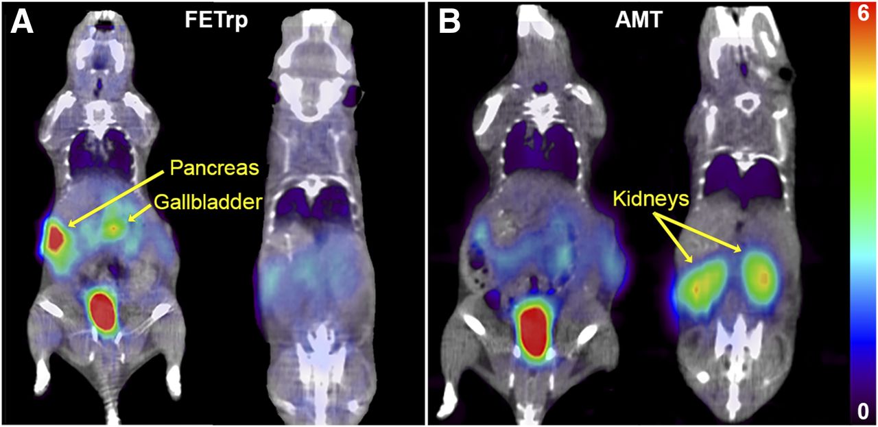
When at least 1 tumor on each mouse was 250 mg or more (Table 1), mice were imaged. Representative images of 11C-AMT and 18F-FETrp organ distribution are shown in Figure 2. For both tracers, there was prominent uptake in the bladder as they were being eliminated. 18F-FETrp showed high uptake in the pancreas followed by the gallbladder, whereas 11C-AMT showed prominent uptake in the kidneys. Assessment of the organ-specific time–activity curves for both tracers displayed some differences in tracer kinetics (Fig. 3). Whereas 11C-AMT was rapidly extracted by the kidneys with subsequent clearance into the bladder, 18F-FETrp was retained in both the pancreas and the gallbladder, and then quickly removed from the body with relatively low retention in the kidneys. Liver uptake was relatively low for both tracers, showing initially higher uptake with subsequent washout. Brain uptake was low and comparable for both tracers, showing a slow, steady increase during the scan.

Biodistribution of 18F-FETrp and 11C-AMT between 30 and 60 min. Two coronal slices of 14-038 glioblastoma PDX mouse showing pancreas, gallbladder, and bladder in one plane with kidneys in other. Color scale bar represents SUV range for both tracers. (A) For 18F-FETrp, high uptake is observed in both bladder and pancreas, with lesser uptake in gallbladder. Minimal uptake is seen in kidneys. (B) For 11C-AMT, tracer uptake is high in bladder and kidneys, with low uptake detected in pancreas and gallbladder.
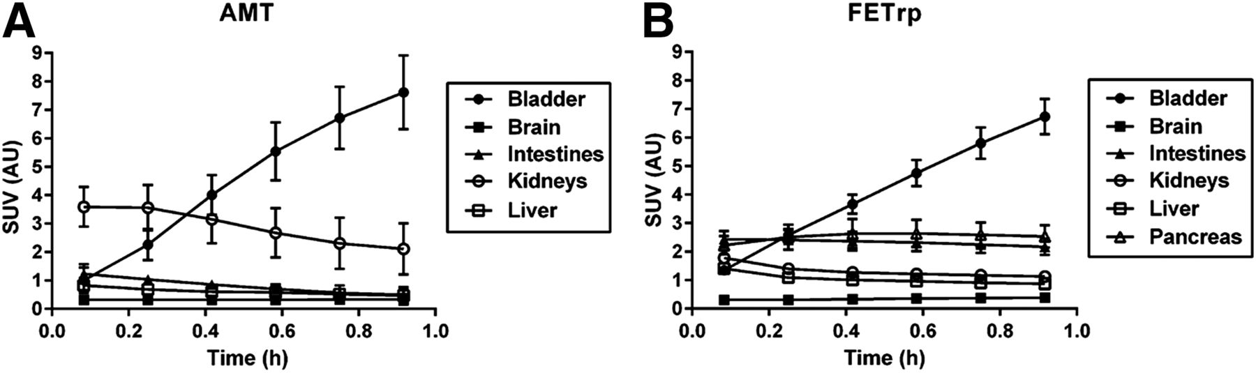
Time-dependent SUV curves characterizing 18F-FETrp and 11C-AMT tracers. Averaged SUVs for indicated organs (n = 5). Error bars represent SD. (A) 11C-AMT tracer uptake is initially high in kidneys and undergoes washout to bladder, where activity increases with time. (B) 18F-FETrp tracer shows increasing uptake in bladder, followed by relatively constant retention in pancreas and intestines. For both tracers, brain displays slow but steady accumulation.
Higher Tumoral 18F-FETrp Tracer Uptake as Compared with 11C-AMT
Both 11C-AMT and 18F-FETrp tracers showed PDX tumor uptake above background, with representative scans of the 14-038 glioblastoma mouse shown in Figure 4A. Corresponding SUVs demonstrated significantly higher tumor retention of 18F-FETrp than 11C-AMT (P < 0.01; Fig. 3B). Supplemental Table 1 compares SUVs obtained during the 30- to 60-min time period for 18F-FETrp and 11C-AMT. Overall there was higher tumoral uptake of 18F-FETrp in all 3 tumor types, with the highest SUVs in glioblastomas (Fig. 5).

Comparison of tumoral tracer uptake between 18F-FETrp and 11C-AMT in the 14-038 glioblastoma PDX mouse. Mouse was injected with 18F-FETrp, followed by 11C-AMT 7 d later. (A) 18F-FETrp tracer (left) and 11C-AMT tracer (right). 18F-FETrp shows increased tumoral tracer accumulation compared with 11C-AMT. Color scale bar represents SUV range for both tracers. (B) Corresponding time–activity curves confirm higher tracer retention for 18F-FETrp.
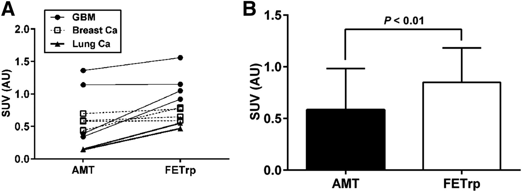
Comparison of 11C-AMT and 18F-FETrp PET SUVs for PDX tumors. (A) In glioblastoma, breast metastatic tumors, and non–small cell lung cancer metastatic tumors, 18F-FETrp SUVs were greater than 11C-AMT SUVs. (B) 18F-FETrp SUVs were significantly greater than 11C-AMT SUVs (paired t test).
Radiation Dosimetry for 18F-FETrp
The residence times were calculated from kinetic 18F-FETrp PET data (Supplemental Table 2), and organ-absorbed radiation dose estimates in humans were calculated (Supplemental Table 3). The results indicate that the critical organ is the pancreas (∼160 μSv/MBq), followed by the bladder (∼150 μSv/MBq). Finally, we determined that administration of 5 MBq/kg (0.14 mCi/kg) of 18F-FETrp for PET studies would result in a total PET dose of approximately 8 mSv in human subjects (Table 2).
Radiation Absorbed Dose from 18F-FETrp PET Studies in Adult Control Subjects
DISCUSSION
This study had 3 major findings. First, we demonstrated the biodistribution of 18F-FETrp in a mouse model, which was somewhat different from what is typically seen on 11C-AMT PET; second, we showed accumulation of 18F-FETrp in PDXs of different human tumors, with mean 18F-FETrp SUVs exceeding 11C-AMT uptake by 30% or more in the same tumors, depending on the tumor type. Finally, our radiation dosimetry data indicate pancreas and bladder to be the critical organs for 18F-FETrp, with the estimated total PET dose of 8 mSv in humans.
The most apparent differences in organ distribution between the 2 tracers include the higher pancreas and lower kidney uptake of 18F-FETrp than 11C-AMT. The reason for these differences is currently unclear and will require further studies. 18F-FETrp shows no appreciable bone accumulation, suggesting negligible defluorination (44). The observed organ radioactivities may arise mostly from unmetabolized radiotracers, although this will require further studies. Nevertheless, the high 18F-FETrp uptake in the bladder and pancreas suggests that this tracer would not allow visualization of tumors in these organs because of high background activity. Despite the differences in distribution, both tracers demonstrated similarly low and continuous brain accumulation, suggesting that 18F-FETrp will be suitable for the imaging of intracranial brain tumors as previously established with 11C-AMT (18–26).
The substantially higher uptake of 18F-FETrp than 11C-AMT in all 3 tumor types is promising, suggesting that 18F-FETrp could be useful for imaging tryptophan uptake and kinetics in these human tumors. Our previous PET studies demonstrated differential 11C-AMT transport and trapping in human gliomas, lung cancer, and breast cancer, tumors that expressed both the L-type amino acid transporter 1 and IDO1 (23,26,45–47). However, the widespread use of 11C-AMT for cancer imaging is not feasible because of its short half-life and tedious radiosynthesis. The current findings suggest that 18F-FETrp may not only overcome these limitations but also provide a better tumor-to-background ratio. Importantly, 18F-FETrp could also be useful to estimate tryptophan metabolism via IDO1, IDO2, or TDO2. This is supported by in vitro data demonstrating markedly higher 18F-FETrp accumulation in IDO1-expressing P815B-mIDO1 cl6 cells than in P815B-IDO1–negative cl1 cells (33,34). Notably, high 18F-FETrp accumulation in P815B-mIDO1 cl6 cells was blocked by 1-methyl-l-tryptophan, an IDO1 inhibitor, suggesting this radiotracer is a promising candidate for imaging in vivo IDO1 enzyme activity. Noninvasive estimation of tumoral IDO1 activity would be critical for patient stratification and treatment monitoring in clinical trials using IDO1 inhibitors developed to overcome tumoral immune resistance via the upregulated KP (48,49). A clinicaltrials.gov search (search terms IDO, cancer; May 11, 2016) yielded more than 20 open, active, or completed studies using IDO1-targeted drugs in cancer. Most of these included epacodostat or indoximod in conjunction with standard chemotherapies for the treatment of solid tumors such as glioblastoma, non–small cell lung cancer, and breast cancer, further underscoring the potential clinical utility of 18F-FETrp PET imaging. The tumor models in the present study showed stronger expression of IDO2 and TDO2 than IDO1, suggesting that 18F-FETrp PET may be useful to monitor activity of these enzymes. Whether or not in vivo uptake values can estimate KP enzyme activity will need to be determined.
Finally, the effective dose of the 18F-FETrp tracer was found to be approximately 20% lower than the effective dose of 18F-FDG PET tracer (8 vs. 10 mSv, respectively) (50), which is the standard clinical tracer for cancer PET imaging. The average 18F-FETrp dose to the critical organ, the pancreas, is only about 75% of 18F-FDG to its critical organ, the urinary bladder (160 μSv/MBq vs. 220 μSv/MBq, respectively). Collectively, our data indicate that the overall radiation dose for a PET study using 18F-FETrp compares favorably with the clinical 18F-FDG tracer. Importantly, a recent study reported a simplified, fully automated synthesis of 18F-FETrp (34), making this a potentially attractive radiotracer for widespread clinical use.
CONCLUSION
Although limited to PDX mouse models, our study provides strong preliminary evidence for the potential clinical value of 18F-FETrp PET. Future studies should evaluate this radiotracer for imaging in vivo tumoral IDO1 activity. Studies using 18F-FETrp PET in patients to image both brain and extracranial tumors are clearly warranted and may lead to a new, broadly available noninvasive diagnostic and prognostic clinical tool.
DISCLOSURE
The study was supported by grant R01CA123451 (Csaba Juhász and Sandeep Mittal) from the National Cancer Institute; by the Fund for Medical Research and Education, Wayne State University School of Medicine (Sandeep Mittal); and a Strategic Research Initiative Grant from Karmanos Cancer Institute (Sandeep Mittal and Csaba Juhász). Anthony R. Guastella is supported by the Initiative to Maximize Student Development Fellowship (Wayne State University). The Animal Model and Therapeutics Evaluation Core; the Microscopy, Imaging and Cytometry Resources Core; and Biobanking and Correlative Sciences Core are supported, in part, by NIH Center grant P30CA022453 to the Karmanos Cancer Institute at Wayne State University. No other potential conflict of interest relevant to this article was reported.
Acknowledgments
We thank Xin Lu, MS, and Kirk Douglas, BS, for performing the small-animal PET/CT studies.
Footnotes
↵* Contributed equally to this work.
Published online Oct. 20, 2016.
- © 2017 by the Society of Nuclear Medicine and Molecular Imaging.
REFERENCES
- Received for publication June 29, 2016.
- Accepted for publication September 14, 2016.


