
Main menu

  • Home
  • Content
    • Current
    • Ahead of print
    • Past Issues
    • JNM Supplement
    • SNMMI Annual Meeting Abstracts
    • Continuing Education
    • JNM Podcasts
  • Subscriptions
    • Subscribers
    • Institutional and Non-member
    • Rates
    • Journal Claims
    • Corporate & Special Sales
  • Authors
    • Submit to JNM
    • Information for Authors
    • Assignment of Copyright
    • AQARA requirements
  • Info
    • Reviewers
    • Permissions
    • Advertisers
  • About
    • About Us
    • Editorial Board
    • Contact Information
  • More
    • Alerts
    • Feedback
    • Help
    • SNMMI Journals
  • SNMMI
    • JNM
    • JNMT
    • SNMMI Journals
    • SNMMI

User menu

  • Subscribe
  • My alerts
  • Log in
  • My Cart

Search

  • Advanced search
Journal of Nuclear Medicine
  • SNMMI
    • JNM
    • JNMT
    • SNMMI Journals
    • SNMMI
  • Subscribe
  • My alerts
  • Log in
  • My Cart
Journal of Nuclear Medicine

Advanced Search

  • Home
  • Content
    • Current
    • Ahead of print
    • Past Issues
    • JNM Supplement
    • SNMMI Annual Meeting Abstracts
    • Continuing Education
    • JNM Podcasts
  • Subscriptions
    • Subscribers
    • Institutional and Non-member
    • Rates
    • Journal Claims
    • Corporate & Special Sales
  • Authors
    • Submit to JNM
    • Information for Authors
    • Assignment of Copyright
    • AQARA requirements
  • Info
    • Reviewers
    • Permissions
    • Advertisers
  • About
    • About Us
    • Editorial Board
    • Contact Information
  • More
    • Alerts
    • Feedback
    • Help
    • SNMMI Journals
  • View or Listen to JNM Podcast
  • Visit JNM on Facebook
  • Join JNM on LinkedIn
  • Follow JNM on Twitter
  • Subscribe to our RSS feeds
Research ArticleBasic Science Investigations

PET of HER2-Positive Pulmonary Metastases with 18F-ZHER2:342 Affibody in a Murine Model of Breast Cancer: Comparison with 18F-FDG

Gabriela Kramer-Marek, Marcelino Bernardo, Dale O. Kiesewetter, Ulas Bagci, Monika Kuban, Aras Omer, Rafal Zielinski, Jurgen Seidel, Peter Choyke and Jacek Capala
Journal of Nuclear Medicine June 2012, 53 (6) 939-946; DOI: https://doi.org/10.2967/jnumed.111.100354
Gabriela Kramer-Marek
  • Find this author on Google Scholar
  • Find this author on PubMed
  • Search for this author on this site
Marcelino Bernardo
  • Find this author on Google Scholar
  • Find this author on PubMed
  • Search for this author on this site
Dale O. Kiesewetter
  • Find this author on Google Scholar
  • Find this author on PubMed
  • Search for this author on this site
Ulas Bagci
  • Find this author on Google Scholar
  • Find this author on PubMed
  • Search for this author on this site
Monika Kuban
  • Find this author on Google Scholar
  • Find this author on PubMed
  • Search for this author on this site
Aras Omer
  • Find this author on Google Scholar
  • Find this author on PubMed
  • Search for this author on this site
Rafal Zielinski
  • Find this author on Google Scholar
  • Find this author on PubMed
  • Search for this author on this site
Jurgen Seidel
  • Find this author on Google Scholar
  • Find this author on PubMed
  • Search for this author on this site
Peter Choyke
  • Find this author on Google Scholar
  • Find this author on PubMed
  • Search for this author on this site
Jacek Capala
  • Find this author on Google Scholar
  • Find this author on PubMed
  • Search for this author on this site
  • Article
  • Figures & Data
  • Supplemental
  • Info & Metrics
  • PDF
Loading

Article Figures & Data

Figures

  • Tables
  • Additional Files
  • FIGURE 1.
    • Download figure
    • Open in new tab
    • Download powerpoint
    FIGURE 1.

    BLI monitoring of metastasis growth. (A) Strong linear correlation (R2 = 0.99) was found between absolute MDA-MB-231HER2-Luc cell number and BLI signal. (B) Total photon flux from BLI signals acquired 9 wk after intravenous injection of MDA-MB-231HER2-Luc cells. Highest ROI signal from either ventral or dorsal position is presented for each individual mouse. Animals indicated by oval shapes were chosen for further imaging studies. (C) Metastasis progression between 9 and 10 wk after cell injection. (D) Representative in vivo images of lungs of mouse number two 10 wk after cell injection.

  • FIGURE 2.
    • Download figure
    • Open in new tab
    • Download powerpoint
    FIGURE 2.

    Contiguous axial PET/MRI sections of mouse 2 collected 9 and 10 wk after cells injection showing rapid metastasis progression. HER2-positive lung metastases were visualized with high tumor-to-background ratio 1 h after 18F-ZHER2:342-Affibody injection. Tracer uptake 10 wk after cell inoculation, indicated by white arrows, was as follows: 6.2 %ID/g (lesion 1), 6.9 %ID/g (lesion 2), and 3.4 %ID/g (lesion 3). TBR = tumor-to-background ratio.

  • FIGURE 3.
    • Download figure
    • Open in new tab
    • Download powerpoint
    FIGURE 3.

    Representative autoradiography (left) with corresponding H&E staining (right) of lung tissue sections of mouse 2 injected with 18F-ZHER2:342-Affibody. Arrows indicate tumor nodules. HER2 expression is heterogeneous. N = normal lung tissue.
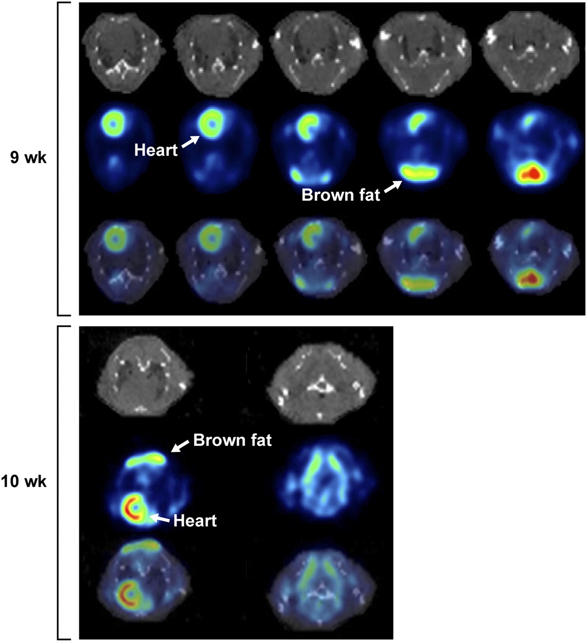
  • FIGURE 4.
    • Download figure
    • Open in new tab
    • Download powerpoint
    FIGURE 4.

    Contiguous transaxial PET/CT section of HER2-positive lung metastasis of mouse 2 at 1 h after 18F-FDG injection. Images were collected 9 and 10 wk after cells injection. Increased background activity made evaluation of small pulmonary nodules difficult. Heart and interscapular brown fat had prominent uptake that probably interfered with detection and delineation of most lesions.

  • FIGURE 5.
    • Download figure
    • Open in new tab
    • Download powerpoint
    FIGURE 5.

    Automated segmentation of tumor lesions from MRI slices acquired 10 wk after tumor cells inoculation. Six slices are arranged such that first column shows original images, second column shows delineated tumor regions, and last column shows fused delineated regions onto MR images. Corresponding intensity and geometry-based features are listed in Table 2.

  • FIGURE 6.
    • Download figure
    • Open in new tab
    • Download powerpoint
    FIGURE 6.

    H&E (top) and anti-HER2 (bottom) staining of lung tissue slices.

Tables

  • Figures
  • Additional Files
    • View popup
    TABLE 1

    Percentage of HER2 Immunohistochemistry Scoring from Overall Metastasis Areas That Were Annotated

    HER2 score
    Animal no.(3+) % cells(2+) % cells(1+) % cells(0+) % cells
    416.414.432.836.4
    54.511.438.845.3
    104.411.940.842.9
    1115.615.941.826.6
    257.49.129.254.3
    • For scoring, Aperio algorithm was used. Scoring system is same as for clinical HercepTest.

    • View popup
    TABLE 2

    Quantitative Measurements of Segmented Lesions Acquired by MRI

    Maximum intensity (8-bit scale: 0–255) (wk)Mean intensity (wk)Area (mm2) (wk)
    Slice no.910910910
    23809527.5832.2011.5015.39
    2519215276.4356.0915.1319.16
    6786039.5331.661.262.46
    • Maximum and mean intensity levels, size of lesions, and geometry of segmented regions are shown.

Additional Files

  • Figures
  • Tables
  • Supplemental Data

    Files in this Data Supplement:

    • Supplemental Data
PreviousNext
Back to top

In this issue

Journal of Nuclear Medicine: 53 (6)
Journal of Nuclear Medicine
Vol. 53, Issue 6
June 1, 2012
  • Table of Contents
  • Table of Contents (PDF)
  • About the Cover
  • Index by author
Print
Download PDF
Article Alerts
Sign In to Email Alerts with your Email Address
Email Article

Thank you for your interest in spreading the word on Journal of Nuclear Medicine.

NOTE: We only request your email address so that the person you are recommending the page to knows that you wanted them to see it, and that it is not junk mail. We do not capture any email address.

Enter multiple addresses on separate lines or separate them with commas.
PET of HER2-Positive Pulmonary Metastases with 18F-ZHER2:342 Affibody in a Murine Model of Breast Cancer: Comparison with 18F-FDG
(Your Name) has sent you a message from Journal of Nuclear Medicine
(Your Name) thought you would like to see the Journal of Nuclear Medicine web site.
Citation Tools
PET of HER2-Positive Pulmonary Metastases with 18F-ZHER2:342 Affibody in a Murine Model of Breast Cancer: Comparison with 18F-FDG
Gabriela Kramer-Marek, Marcelino Bernardo, Dale O. Kiesewetter, Ulas Bagci, Monika Kuban, Aras Omer, Rafal Zielinski, Jurgen Seidel, Peter Choyke, Jacek Capala
Journal of Nuclear Medicine Jun 2012, 53 (6) 939-946; DOI: 10.2967/jnumed.111.100354

Citation Manager Formats

  • BibTeX
  • Bookends
  • EasyBib
  • EndNote (tagged)
  • EndNote 8 (xml)
  • Medlars
  • Mendeley
  • Papers
  • RefWorks Tagged
  • Ref Manager
  • RIS
  • Zotero
Share
PET of HER2-Positive Pulmonary Metastases with 18F-ZHER2:342 Affibody in a Murine Model of Breast Cancer: Comparison with 18F-FDG
Gabriela Kramer-Marek, Marcelino Bernardo, Dale O. Kiesewetter, Ulas Bagci, Monika Kuban, Aras Omer, Rafal Zielinski, Jurgen Seidel, Peter Choyke, Jacek Capala
Journal of Nuclear Medicine Jun 2012, 53 (6) 939-946; DOI: 10.2967/jnumed.111.100354
Twitter logo Facebook logo LinkedIn logo Mendeley logo
  • Tweet Widget
  • Facebook Like
  • Google Plus One
Bookmark this article

Jump to section

  • Article
    • Abstract
    • MATERIALS AND METHODS
    • RESULTS
    • DISCUSSION
    • CONCLUSION
    • DISCLOSURE STATEMENT
    • Acknowledgments
    • Footnotes
    • REFERENCES
  • Figures & Data
  • Supplemental
  • Info & Metrics
  • PDF

Related Articles

  • This Month in JNM
  • Errata
  • PubMed
  • Google Scholar

Cited By...

  • Phase II Trial Assessing the Repeatability and Tumor Uptake of [68Ga]Ga-HER2 Single-Domain Antibody PET/CT in Patients with Breast Carcinoma
  • HER3-Mediated Resistance to Hsp90 Inhibition Detected in Breast Cancer Xenografts by Affibody-Based PET Imaging
  • Preclinical and clinical applications of specific molecular imaging for HER2-positive breast cancer
  • Targeting the Human Epidermal Growth Factor Receptors with Immuno-PET: Imaging Biomarkers from Bench to Bedside
  • Positron Emission Tomography Imaging with 18F-Labeled ZHER2:2891 Affibody for Detection of HER2 Expression and Pharmacodynamic Response to HER2-Modulating Therapies
  • Three Methods for 18F Labeling of the HER2-Binding Affibody Molecule ZHER2:2891 Including Preclinical Assessment
  • Google Scholar

More in this TOC Section

  • Tumor-Specific Binding of Radiolabeled PEGylated GIRLRG Peptide: A Novel Agent for Targeting Cancers
  • PET/MRI of Hypoxic Atherosclerosis Using 64Cu-ATSM in a Rabbit Model
  • Tumor Uptake of Anti-CD20 Fabs Depends on Tumor Perfusion
Show more Basic Science Investigations

Similar Articles

SNMMI

© 2025 SNMMI

Powered by HighWire