


Article Figures & Data
Figures
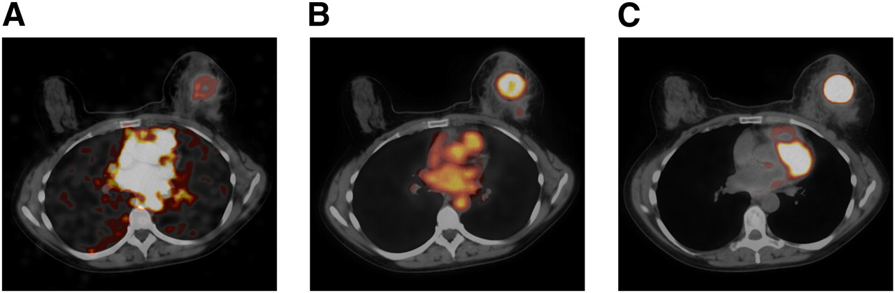
- FIGURE 1.
First-pass dynamic (A), early static (B), and delayed static (C) fused PET/CT images obtained, respectively, 30 s, 8 min, and 90 min after bolus injection of 18F-FDG, in 57-y-old woman with TN invasive ductal carcinoma of left breast.
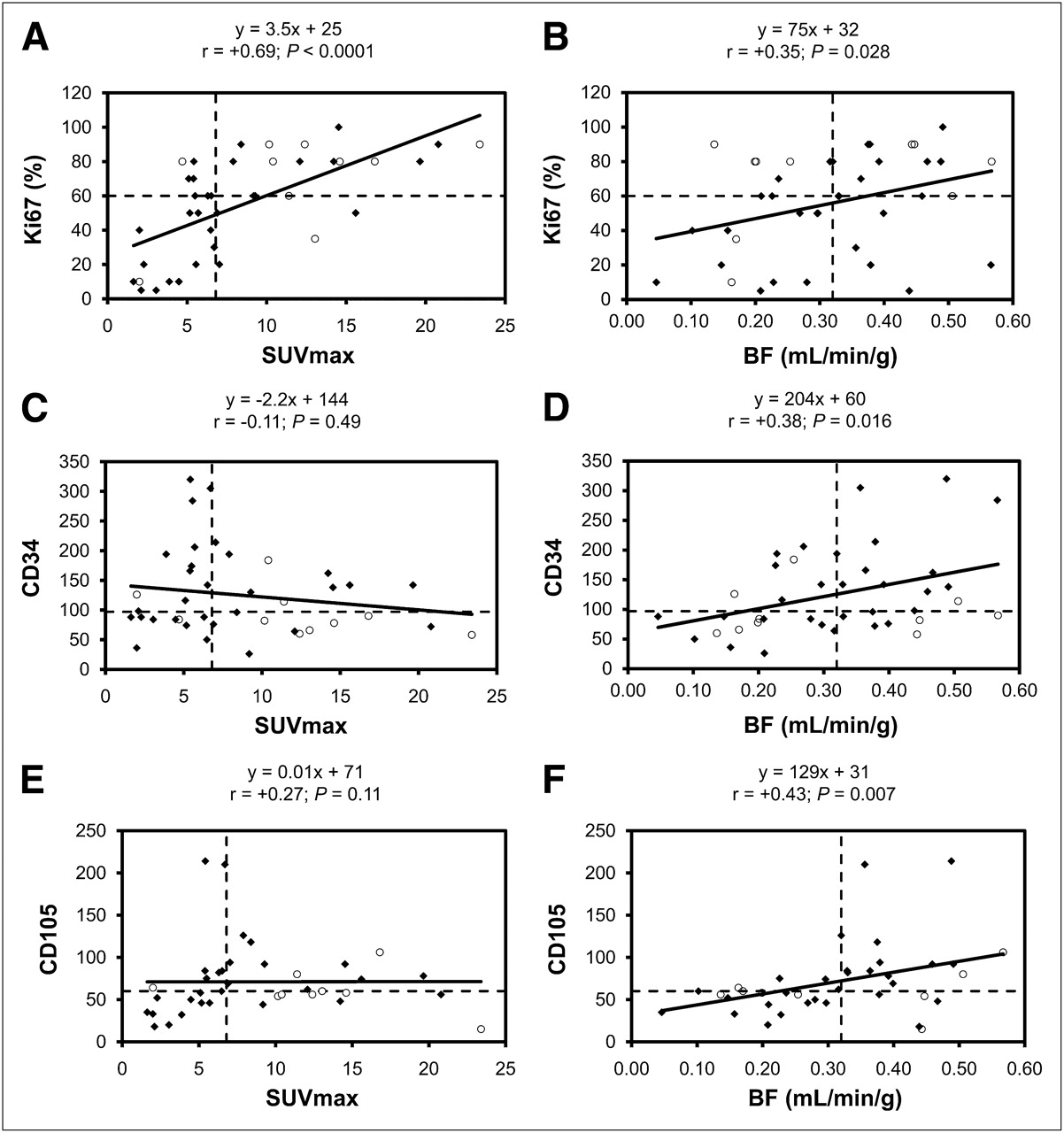
- FIGURE 2.
Spearman rank correlation with regression lines between SUVmax and Ki67 (A), tumor blood flow (BF) and Ki67 (B), SUVmax and CD34 (C), tumor blood flow and CD34 (D), SUVmax and CD105 (E), and tumor blood flow and CD105 (F). Horizontal and vertical dashed lines represent median value for each variable (6.8 for SUVmax, 60% for Ki67, 0.32 mL/min/g for blood flow, 97 for CD34, and 60 for CD105). ○ = TN breast tumors; ◆ = non-TN breast tumors.
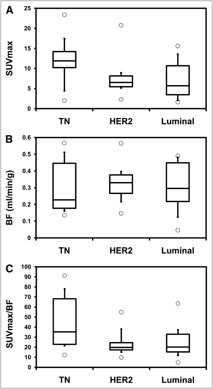
- FIGURE 3.
SUVmax (A), tumor blood flow (BF) (B), and SUVmax-to-blood flow ratio (C) according to subtypes.

Tables
- TABLE 1
Comparison Between SUVmax and Blood Flow in the Patient and Clinical and Tumor Characteristics
SUVmax Blood flow (mL/min/g) Variable Parameter n Median Interquartile range P Median Interquartile range P Menopausal No 22 (55%) 6.7 5.2–9.9 0.48 0.31 0.23–0.38 0.72 Yes 18 (45%) 6.8 5.0–14.5 0.32 0.20–0.43 Tumor size <5 cm 24 (60%) 6.8 5.3–10.2 0.50 0.33 0.21–0.38 0.29 ≥5 cm 16 (40%) 7.9 5.0–13.3 0.31 0.22–0.47 Lymph node involvement No 13 (32%) 5.4 3.9–13.0 0.54 0.28 0.20–0.39 0.40 Yes 27 (68%) 7.9 5.5–11.7 0.33 0.23–0.42 Histologic grade 1/2 26 (65%) 5.6 4.0–8.1 <0.001 0.27 0.20–0.37 0.030 3 14 (35%) 13.3 8.5–16.3 0.39 0.32–0.46 ER − 14 (35%) 10.3 6.8–12.9 0.08 0.29 0.20–0.43 0.86 + 26 (65%) 6.1 4.6–8.9 0.32 0.23–0.40 PR − 19 (48%) 9.2 6.5–12.7 0.021 0.36 0.25–0.42 0.18 + 21 (52%) 5.5 3.1–9.3 0.28 0.20–0.38 HER2 − 25 (63%) 9.3 4.5–13.0 0.60 0.28 0.20–0.45 0.64 + 15 (37%) 6.5 5.5–8.1 0.33 0.27–0.38 TN Non-TN 30 (75%) 6.4 5.1–9.0 0.042 0.32 0.23–0.39 0.82 TN 10 (25%) 11.9 10.2–14.2 0.23 0.18–0.45 Lymphovascular invasion No 6 (15%) 6.9 5.3–11.2 0.65 0.32 0.21–0.38 0.91 Yes 34 (85%) 5.4 3.6–12.1 0.28 0.22–0.41 Ki67 score ≤median 23 (58%) 5.7 3.5–7.0 <0.001 0.28 0.19–0.37 0.09 >median 17 (42%) 12.1 7.9–14.6 0.38 0.25–0.45 CD34 score ≤median 20 (50%) 7.7 4.6–12.6 0.57 0.24 0.17–0.38 0.027 >median 20 (50%) 6.6 5.4–10.7 0.36 0.26–0.46 CD105 score ≤median 20 (50%) 6.1 3.7–12.6 0.31 0.23 0.17–0.32 0.002 >median 20 (50%) 7.5 6.4–11.9 0.37 0.32–0.44 Univariate Multivariate Predictor OR 95% CI P OR 95% CI P Histopathologic grade 3 6.93 1.53–31.4 0.012 — — — TN breast cancer 6.00 1.08–33.3 0.040 — — — Ki67 score (%) 1.06 1.02–1.09 0.002 1.05 1.02–1.09 0.003 OR = odds ratio; CI = confidence interval.
Univariate Multivariate Predictor OR 95% CI P OR 95% CI P Histopathologic grade 3 6.93 1.53–31.4 0.012 9.56 1.60–57.2 0.013 CD34 score 1.01 1.00–1.02 0.046 — — — CD105 score 1.05 1.01–1.09 0.011 1.05 1.01–1.09 0.021 OR = odds ratio; CI = confidence interval.
- TABLE 4
Studies Evaluating Tumor Blood Flow in Patients with Large or Locally Advanced Breast Cancer
Tumor blood flow (mL/min/g) Study No. of patients Tracer Mean ± SD Range Wilson et al. (1992) (34) 20 15O-water 0.30 ± 0.17 0.11–0.77 Mankoff et al. (2002) (35) 37 15O-water 0.32 Zasadny et al. (2003) (19) 9 15O-water 0.15 ± 0.08 0.08–0.29 Tseng et al. (2004) (20) 35 15O-water 0.30 Our study 40 18F-FDG 0.32 ± 0.13 0.05–0.57


Jump to section
Related Articles
Cited By...
- Total-Body Quantitative Parametric Imaging of Early Kinetics of 18F-FDG
- Reply: Semiquantification Limitations: FMTVDM(C)copysr Demonstrates Quantified Tumor Response to Treatment with Both Regional Blood Flow and Metabolic Changes
- Breast Cancer Blood Flow and Metabolism on Dual-Acquisition 18F-FDG PET: Correlation with Tumor Phenotype and Neoadjuvant Chemotherapy Response
- 18F-FDG PET-Derived Tumor Blood Flow Changes After 1 Cycle of Neoadjuvant Chemotherapy Predicts Outcome in Triple-Negative Breast Cancer
- Getting the Most out of 18F-FDG PET Scans: The Predictive Value of 18F-FDG PET-Derived Blood Flow Estimates for Breast Cancer
- Role of Positron Emission Tomography for the Monitoring of Response to Therapy in Breast Cancer