


Article Figures & Data
Figures
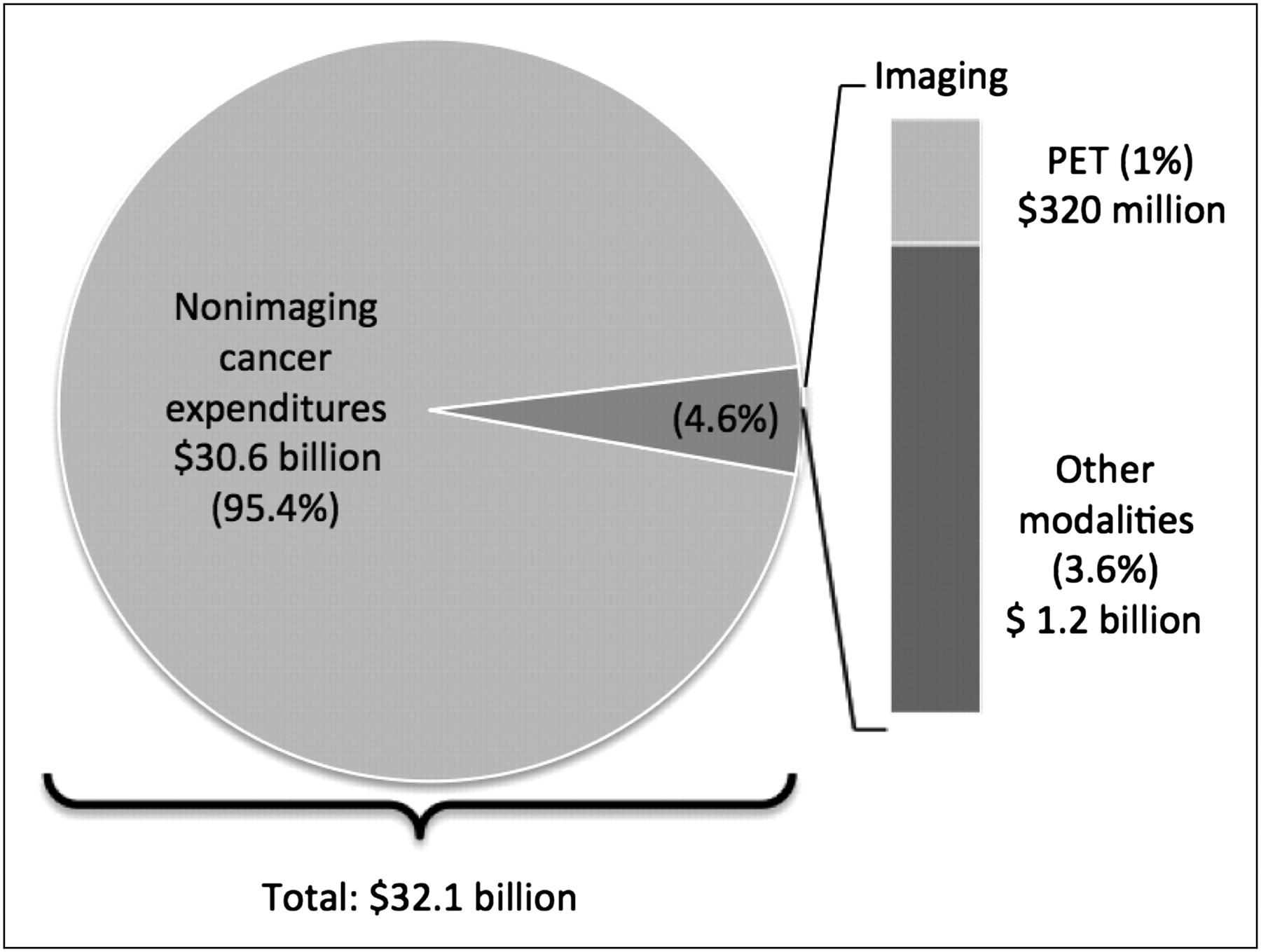
- FIGURE 1.
Estimated contribution of all imaging modalities and PET alone to total Medicare cancer care expenditures in 2006. Estimates are based on data from Dinan et al. (6) and Potetz and DeWilde (15). These data reflect sample from study of Dinan et al. (6) and do not represent HOPPS or non–hospital-based imaging use data.

Tables
Tumor type Initial treatment strategy Subsequent treatment strategy Colorectal Covered Covered Esophagus Covered Covered Head and neck (not thyroid or central nervous system) Covered Covered Lymphoma Covered Covered Non–small cell lung Covered Covered Ovary Covered Covered Brain Covered CED Cervix Covered with exception* Covered Small cell lung Covered CED Soft-tissue sarcoma Covered CED Pancreas Covered CED Testis Covered CED Breast (female and male) Covered with exception† Covered Melanoma Covered with exception‡ Covered Prostate Not covered CED Thyroid Covered Covered with exception or CED§ All other solid tumors Covered CED Myeloma Covered Covered All other cancers not listed CED CED ↵* Cervical cancer nationally not covered for initial diagnosis.
↵† Breast cancer nationally not covered for initial diagnosis or staging of axillary lymph nodes.
↵‡ Melanoma nationally not covered for initial staging of regional lymph nodes.
↵§ Thyroid cancer nationally covered for subsequent treatment strategy for recurrent or residual thyroid cancer of follicular cell origin, previously treated by thyroidectomy and radioiodine ablation, with serum thyroglobulin level of greater than 10 ng/mL, and negative 131I whole-body scan results.
CED = coverage with evidence development.
(Adapted from (55).)


Jump to section
Related Articles
Cited By...
- Is Long-Axial-Field-of-View PET/CT Cost-Effective? An International Health-Economic Analysis
- Palliative care and imaging utilisation for patients with cancer
- Humana and 18F-FDG PET/CT: Another Sequel to the Injustice of Being Judged by the Errors of Others
- Of Sheep and Wolves: Curtailing Coverage for Essential Imaging Tests Based on Flawed Use and Cost Arguments
- The Future of Nuclear Medicine as an Independent Specialty
- Geographic Variation in Postoperative Imaging for Low-Risk Breast Cancer
- Impact of 68Ga-PSMA-11 PET/CT on the Management of Prostate Cancer Patients with Biochemical Recurrence
- Variations in PET/MRI Operations: Results from an International Survey Among 39 Active Sites
- PET and MRI: Is the Whole Greater than the Sum of Its Parts?
- 18F-FDG PET/CT and PET/MRI Perform Equally Well in Cancer: Evidence from Studies on More Than 2,300 Patients
- PET/MR Imaging: A Critical Appraisal
- Nuclear Medicine at a Crossroads
- Generating Evidence for Clinical Benefit of PET/CT in Diagnosing Cancer Patients