
Main menu

  • Home
  • Content
    • Current
    • Ahead of print
    • Past Issues
    • JNM Supplement
    • SNMMI Annual Meeting Abstracts
    • Continuing Education
    • JNM Podcasts
  • Subscriptions
    • Subscribers
    • Institutional and Non-member
    • Rates
    • Journal Claims
    • Corporate & Special Sales
  • Authors
    • Submit to JNM
    • Information for Authors
    • Assignment of Copyright
    • AQARA requirements
  • Info
    • Reviewers
    • Permissions
    • Advertisers
  • About
    • About Us
    • Editorial Board
    • Contact Information
  • More
    • Alerts
    • Feedback
    • Help
    • SNMMI Journals
  • SNMMI
    • JNM
    • JNMT
    • SNMMI Journals
    • SNMMI

User menu

  • Subscribe
  • My alerts
  • Log in
  • My Cart

Search

  • Advanced search
Journal of Nuclear Medicine
  • SNMMI
    • JNM
    • JNMT
    • SNMMI Journals
    • SNMMI
  • Subscribe
  • My alerts
  • Log in
  • My Cart
Journal of Nuclear Medicine

Advanced Search

  • Home
  • Content
    • Current
    • Ahead of print
    • Past Issues
    • JNM Supplement
    • SNMMI Annual Meeting Abstracts
    • Continuing Education
    • JNM Podcasts
  • Subscriptions
    • Subscribers
    • Institutional and Non-member
    • Rates
    • Journal Claims
    • Corporate & Special Sales
  • Authors
    • Submit to JNM
    • Information for Authors
    • Assignment of Copyright
    • AQARA requirements
  • Info
    • Reviewers
    • Permissions
    • Advertisers
  • About
    • About Us
    • Editorial Board
    • Contact Information
  • More
    • Alerts
    • Feedback
    • Help
    • SNMMI Journals
  • View or Listen to JNM Podcast
  • Visit JNM on Facebook
  • Join JNM on LinkedIn
  • Follow JNM on Twitter
  • Subscribe to our RSS feeds
Research ArticleBasic Science Investigations

Brain and Whole-Body Imaging in Rhesus Monkeys of 11C-NOP-1A, a Promising PET Radioligand for Nociceptin/Orphanin FQ Peptide Receptors

Yasuyuki Kimura, Masahiro Fujita, Jinsoo Hong, Talakad G. Lohith, Robert L. Gladding, Sami S. Zoghbi, Johannes A. Tauscher, Nancy Goebl, Karen S. Rash, Zhaogen Chen, Concepcion Pedregal, Vanessa N. Barth, Victor W. Pike and Robert B. Innis
Journal of Nuclear Medicine October 2011, 52 (10) 1638-1645; DOI: https://doi.org/10.2967/jnumed.111.091181
Yasuyuki Kimura
  • Find this author on Google Scholar
  • Find this author on PubMed
  • Search for this author on this site
Masahiro Fujita
  • Find this author on Google Scholar
  • Find this author on PubMed
  • Search for this author on this site
Jinsoo Hong
  • Find this author on Google Scholar
  • Find this author on PubMed
  • Search for this author on this site
Talakad G. Lohith
  • Find this author on Google Scholar
  • Find this author on PubMed
  • Search for this author on this site
Robert L. Gladding
  • Find this author on Google Scholar
  • Find this author on PubMed
  • Search for this author on this site
Sami S. Zoghbi
  • Find this author on Google Scholar
  • Find this author on PubMed
  • Search for this author on this site
Johannes A. Tauscher
  • Find this author on Google Scholar
  • Find this author on PubMed
  • Search for this author on this site
Nancy Goebl
  • Find this author on Google Scholar
  • Find this author on PubMed
  • Search for this author on this site
Karen S. Rash
  • Find this author on Google Scholar
  • Find this author on PubMed
  • Search for this author on this site
Zhaogen Chen
  • Find this author on Google Scholar
  • Find this author on PubMed
  • Search for this author on this site
Concepcion Pedregal
  • Find this author on Google Scholar
  • Find this author on PubMed
  • Search for this author on this site
Vanessa N. Barth
  • Find this author on Google Scholar
  • Find this author on PubMed
  • Search for this author on this site
Victor W. Pike
  • Find this author on Google Scholar
  • Find this author on PubMed
  • Search for this author on this site
Robert B. Innis
  • Find this author on Google Scholar
  • Find this author on PubMed
  • Search for this author on this site
  • Article
  • Figures & Data
  • Supplemental
  • Info & Metrics
  • PDF
Loading

Article Figures & Data

Figures

  • Tables
  • Additional Files
  • FIGURE 1.
    • Download figure
    • Open in new tab
    • Download powerpoint
    FIGURE 1.

    Time–activity curves for source organs after 11C-NOP-1A injection at baseline and after receptor blockade with SB-612111. Symbols represent mean from 3 monkeys and are same for each of 3 pairs of panels (baseline and blocked). Data are decay-corrected to time of injection.
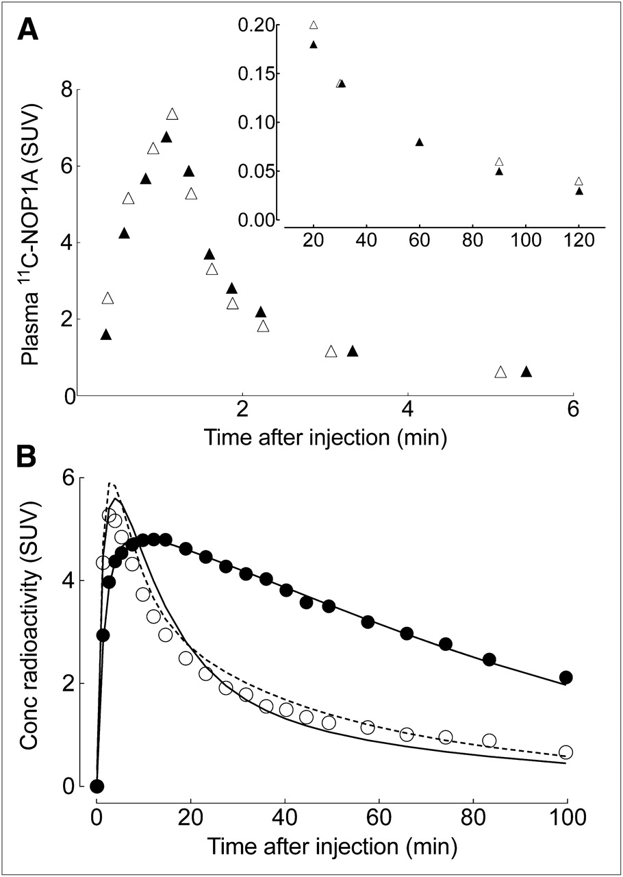
  • FIGURE 2.
    • Download figure
    • Open in new tab
    • Download powerpoint
    FIGURE 2.

    Representative time–activity curves of 11C-NOP-1A in plasma and brain at baseline and after receptor blockade with SB-612111 in single monkey. (A) 11C-NOP-1A concentration, separated from radiometabolites, in arterial plasma is plotted at baseline (▴) and after receptor blockade (▵). Values from 0 to 6 min and from 20 to 100 min are plotted on separate graphs, which differ in range of y-axis. (B) Concentrations of radioactivity in occipital cortex after injection of 11C-NOP-1A are shown at baseline (•) and after receptor blockade (○). Measured brain data were fit with 1-tissue (solid line) and 2-tissue (dotted line)-compartment models. At baseline conditions, 2 fittings overlap almost completely.
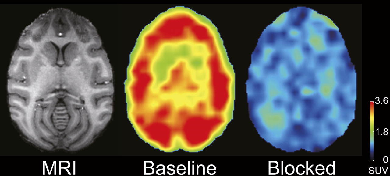
  • FIGURE 3.
    • Download figure
    • Open in new tab
    • Download powerpoint
    FIGURE 3.

    Transaxial PET images of brain uptake at baseline and after receptor blockade in same monkey. PET images from 40 to 80 min after injection of 11C-NOP-1A were summed, and pixel values represent mean concentration of radioactivity (standardized uptake value). Monkey was injected with 222 MBq of 11C-NOP-1A at baseline and 222 MBq after receptor blockade. Radioactivity concentration was expressed as standardized uptake value (SUV), which normalizes for injected activity and body mass. Coregistered MR image shows that PET slice extends through caudate, putamen, and cerebellum.

Tables

  • Figures
  • Additional Files
    • View popup
    TABLE 1.

    Radiation Dosimetry Estimates for 11C-NOP-1A in Standard Reference Man

    Target organRadiation dose (μGy/MBq)
    Adrenals3.4
    Brain5.5
    Breasts1.9
    Gallbladder wall35.6
    Lower large intestine wall2.4
    Small intestine4.1
    Stomach wall2.6
    Upper large intestine wall2.8
    Heart wall12.8
    Kidneys13.5
    Liver13.8
    Lungs11.2
    Muscle2.1
    Ovaries2.5
    Pancreas3.5
    Red marrow5.0
    Osteogenic cells4.5
    Skin1.5
    Spleen11.9
    Testes1.8
    Thymus2.3
    Thyroid1.9
    Urinary bladder wall15.7
    Uterus2.8
    Total body2.8
    Effective dose (μSv/MBq)5.0
    • View popup
    TABLE 2.

    Distribution Volume of 11C-NOP-1A in Monkey Brain

    VT (mL · cm−3)
    BaselineBlocked
    OrganMeanSDMeanSDVS (mL · cm−3), baseline − blockedVS/VT (%)
    Prefrontal cortex18.10.96.62.311.664
    Basal frontal cortex16.61.06.11.710.563
    Parietal cortex19.32.56.82.212.565
    Occipital cortex20.42.26.61.813.767
    Insula19.82.27.51.912.362
    Lateral temporal cortex21.31.07.22.014.266
    Medial temporal cortex16.51.36.61.29.960
    Amygdala21.03.77.31.813.765
    Hippocampus18.13.07.11.511.061
    Anterior cingulate17.71.47.41.810.358
    Posterior cingulate17.61.47.21.810.459
    Caudate15.30.27.21.98.153
    Putamen16.01.37.61.98.553
    Thalamus16.20.78.02.08.251
    Hypothalamus12.61.27.41.85.241
    Cerebellum12.91.16.71.46.248

Additional Files

  • Figures
  • Tables
  • Supplemental Data

    Files in this Data Supplement:

    • Supplemental Data
PreviousNext
Back to top

In this issue

Journal of Nuclear Medicine: 52 (10)
Journal of Nuclear Medicine
Vol. 52, Issue 10
October 1, 2011
  • Table of Contents
  • Table of Contents (PDF)
  • About the Cover
  • Index by author
Print
Download PDF
Article Alerts
Sign In to Email Alerts with your Email Address
Email Article

Thank you for your interest in spreading the word on Journal of Nuclear Medicine.

NOTE: We only request your email address so that the person you are recommending the page to knows that you wanted them to see it, and that it is not junk mail. We do not capture any email address.

Enter multiple addresses on separate lines or separate them with commas.
Brain and Whole-Body Imaging in Rhesus Monkeys of 11C-NOP-1A, a Promising PET Radioligand for Nociceptin/Orphanin FQ Peptide Receptors
(Your Name) has sent you a message from Journal of Nuclear Medicine
(Your Name) thought you would like to see the Journal of Nuclear Medicine web site.
Citation Tools
Brain and Whole-Body Imaging in Rhesus Monkeys of 11C-NOP-1A, a Promising PET Radioligand for Nociceptin/Orphanin FQ Peptide Receptors
Yasuyuki Kimura, Masahiro Fujita, Jinsoo Hong, Talakad G. Lohith, Robert L. Gladding, Sami S. Zoghbi, Johannes A. Tauscher, Nancy Goebl, Karen S. Rash, Zhaogen Chen, Concepcion Pedregal, Vanessa N. Barth, Victor W. Pike, Robert B. Innis
Journal of Nuclear Medicine Oct 2011, 52 (10) 1638-1645; DOI: 10.2967/jnumed.111.091181

Citation Manager Formats

  • BibTeX
  • Bookends
  • EasyBib
  • EndNote (tagged)
  • EndNote 8 (xml)
  • Medlars
  • Mendeley
  • Papers
  • RefWorks Tagged
  • Ref Manager
  • RIS
  • Zotero
Share
Brain and Whole-Body Imaging in Rhesus Monkeys of 11C-NOP-1A, a Promising PET Radioligand for Nociceptin/Orphanin FQ Peptide Receptors
Yasuyuki Kimura, Masahiro Fujita, Jinsoo Hong, Talakad G. Lohith, Robert L. Gladding, Sami S. Zoghbi, Johannes A. Tauscher, Nancy Goebl, Karen S. Rash, Zhaogen Chen, Concepcion Pedregal, Vanessa N. Barth, Victor W. Pike, Robert B. Innis
Journal of Nuclear Medicine Oct 2011, 52 (10) 1638-1645; DOI: 10.2967/jnumed.111.091181
Twitter logo Facebook logo LinkedIn logo Mendeley logo
  • Tweet Widget
  • Facebook Like
  • Google Plus One
Bookmark this article

Jump to section

  • Article
    • Abstract
    • MATERIALS AND METHODS
    • RESULTS
    • DISCUSSION
    • CONCLUSION
    • DISCLOSURE STATEMENT
    • Acknowledgments
    • Footnotes
    • REFERENCES
  • Figures & Data
  • Supplemental
  • Info & Metrics
  • PDF

Related Articles

  • This Month in JNM
  • PubMed
  • Google Scholar

Cited By...

  • Buprenorphine exposure alters the development and migration of interneurons in the cortex
  • Evaluation of a PET Radioligand to Image O-GlcNAcase in Brain and Periphery of Rhesus Monkey and Knock-Out Mouse
  • Occupancy of Nociceptin/Orphanin FQ Peptide Receptors by the Antagonist LY2940094 in Rats and Healthy Human Subjects
  • Brain and Whole-Body Imaging of Nociceptin/Orphanin FQ Peptide Receptor in Humans Using the PET Ligand 11C-NOP-1A
  • Google Scholar

More in this TOC Section

  • Tumor-Specific Binding of Radiolabeled PEGylated GIRLRG Peptide: A Novel Agent for Targeting Cancers
  • PET/MRI of Hypoxic Atherosclerosis Using 64Cu-ATSM in a Rabbit Model
  • Tumor Uptake of Anti-CD20 Fabs Depends on Tumor Perfusion
Show more Basic Science Investigations

Similar Articles

SNMMI

© 2025 SNMMI

Powered by HighWire