


Article Figures & Data
Figures
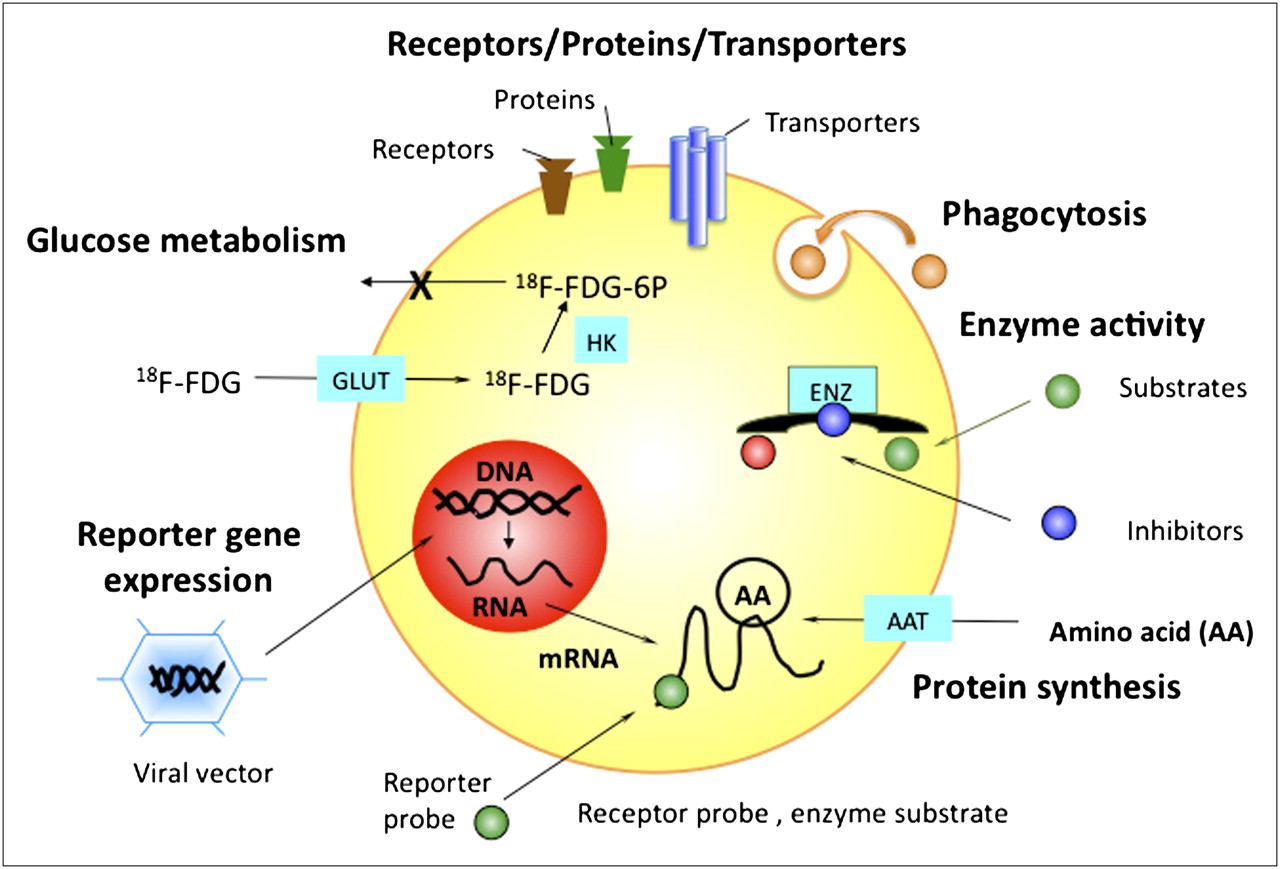
- FIGURE 1.
Examples of cellular targets for molecular imaging agents. AAT = amino acid transporter; ENZ = enzyme; GLUT = glucose transporter; HK = hexokinase; 6P = 6-phosphate.
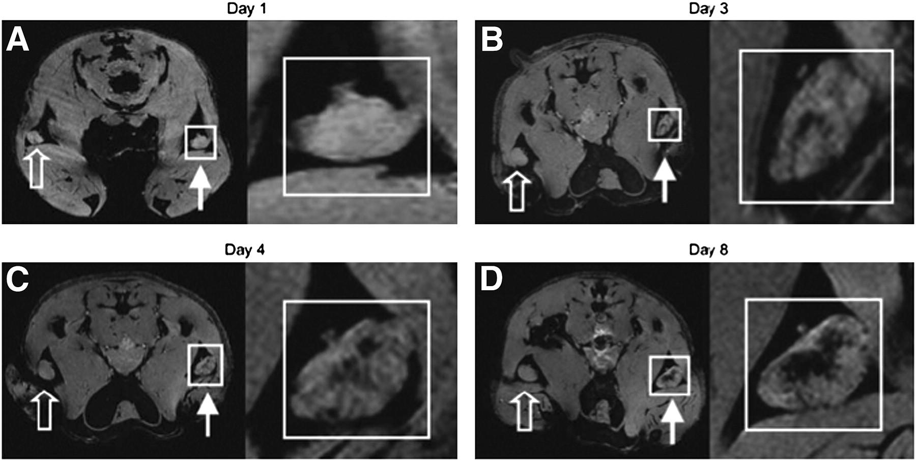
- FIGURE 2.
In vivo MRI monitoring of trafficking of dendritic cells that have taken up SPIOs in vivo after intradermal injection of labeled granulocyte–macrophage colony-stimulating (GM CSF) tumor cell vaccine into footpads of mice. For each day, magnifications of insets in A, B, C, and D are shown at right. Open arrows represent draining popliteal lymph nodes (LNs) for footpads receiving unlabeled GM CSF vaccine. Closed arrows represent draining popliteal LNs for footpads receiving SPIO-labeled GM CSF vaccine. On multigradient-echo images, SPIO-containing LNs have decreased signal intensity. (A) On day 1, popliteal LNs show no evidence of hypointensity. (B) On day 3, decreased signal intensity becomes apparent in LN corresponding to popliteal LN for SPIO-labeled vaccine. (C and D) On day 4 (C) and day 8 (D), respectively, signal decrease persists and then actually increases in popliteal LNs. Images are representative of 3 independent experiments with 5 mice each. (Reprinted with permission of (4).)
- FIGURE 3.
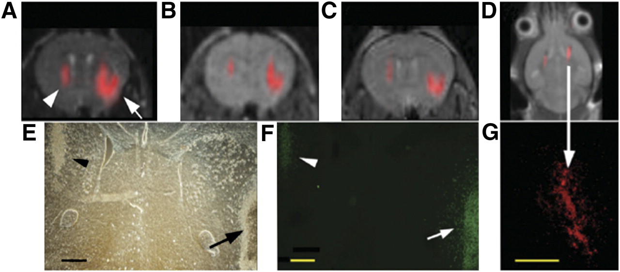
In vivo MRI of transplanted C17.2 neural stem cells, with 19F signal superimposed on 1H MR images. (A–C) MR images at 1 h (A), 3 d (B), and 7 d (C) after injection of 4 × 10⁴ (left hemisphere, arrowhead in A) or 3 × 10⁵ (right hemisphere, arrow in A) cationic perfluoro crown ether (PFCE)–labeled cells. (E and F) Corresponding histopathology determined with phase-contrast microscopy (E) and anti–β-galactosidase immunohistochemistry (F) at day 7 shows that implanted cells remain viable and continue to produce marker enzyme. Arrow in F indicates cells migrating from injection site into brain parenchyma. (D) MR image of different animal at 14 d after injection of 4 × 10⁵ C17.2 cells into both hemispheres shows persistence of 19F signal for 2 wk. (G) Corresponding histopathology shows rhodamine fluorescence from PFCE-labeled cells colocalizing with 19F signal. Bars = 500 μm. (Reprinted with permission of (15).)
- FIGURE 4.
Prosthetic groups for 18F labeling of peptides and proteins. FBA = fluorobenzaldehyde; FBEM = fluorobenzamido–ethylmaleimide; FBzA = fluorobenzoic acid; FPA = fluoropropionic acid; FpyME = fluoropyridinyloxy–propylmaleimide; SFB = succinimidyl fluorobenzoate.
- FIGURE 5.
In vivo PET images of 18F-galacto-RGD uptake (top), 13N-NH3 perfusion (bottom), and their fusion (middle) in transverse views of rat heart without coronary occlusion (sham operation, left) and rat heart with 20 min of coronary occlusion 1 wk after reperfusion (right). Tracer accumulation is visible in chest wall at surgical incision area in both rats (arrowheads), but focal 18F-galacto-RGD uptake in myocardium is observed only after coronary occlusion (arrows). LCA = left anterior descending coronary artery. (Reprinted with permission of (27).)
- FIGURE 6.
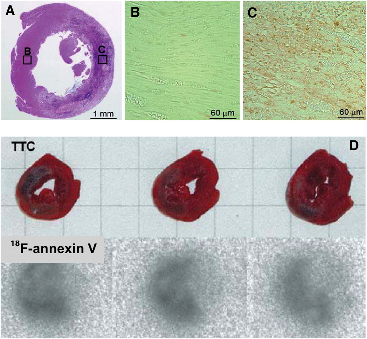
(A–C) Typical results of hematoxylin and eosin staining of left ventricle at 24 h after ischemia (A) and detection of apoptosis in regions shown in boxes B and C (B and C). (D) Typical infarction in left ventricle at 24 h after ischemia (2,3,5-triphenyltetrazolium chloride [TTC] stain) and accumulation of 18F-annexin V. (Reprinted with permission of (28).)
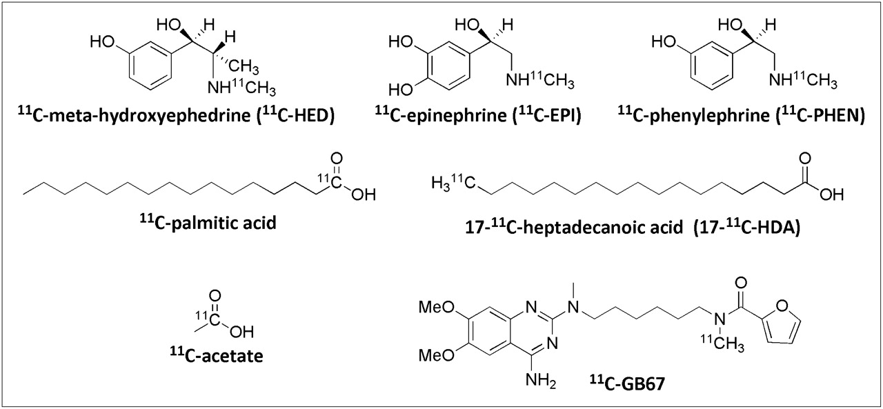
- FIGURE 7.
Selected 11C PET radiotracers for cardiac imaging.
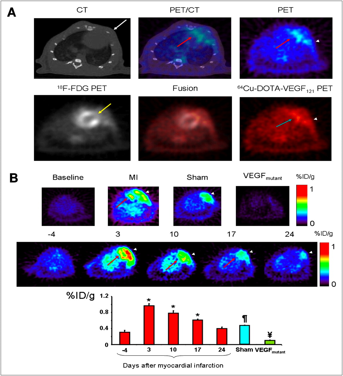
- FIGURE 8.
(A) Myocardial origin of 64Cu-DOTA-VEGF121 PET signal after MI. (Top) Representative coregistered images from micro-CT (left), PET (right), and fused PET/CT (middle) of animal with MI. (Bottom) Representative 64Cu-DOTA-VEGF121 image (left), 18F-FDG image (right), and 64Cu-DOTA-VEGF121–18F-FDG fused image (middle). (B) Time-dependent uptake of 64Cu-DOTA-VEGF121. Arrowhead = area of surgical wound; red arrow = anterolateral myocardium; white arrow = intercostal muscle layer; yellow arrow = ligated coronary artery. *P < 0.05 compared with baseline. ¶P < 0.05 compared with VEGFmutant and 64Cu-DOTA-VEGF121. ¥P < 0.05 compared with sham and 64Cu-DOTA-VEGF121.
- FIGURE 9.
Decay-corrected whole-body coronal small-animal PET images of athymic female nude mice bearing U87MG tumors from 1-h dynamic scan and static scan at 2 h after injection of 68Ga-NOTA-RGD1, 68Ga-NOTA-RGD2, and 68Ga-NOTA-RGD4 (3.7 MBq/mouse). Tumors are indicated by arrowheads.
- FIGURE 10.
Anteroposterior γ-image of mouse with experimental atherosclerotic lesion in left femoral artery (red arrow) and contralateral sham-treated right femoral region (yellow arrow) at 3 h after injection of 99mTc-labeled DTPA-conjugated succinylated polylysine polymer. Injection of polymer followed pretargeting with bispecific antibody Z2D3 F(ab′)2–anti-DTPA F(ab′)2 (44). Lesion in left femoral artery was about 2.5 mg, as determined by immunohistochemical staining.
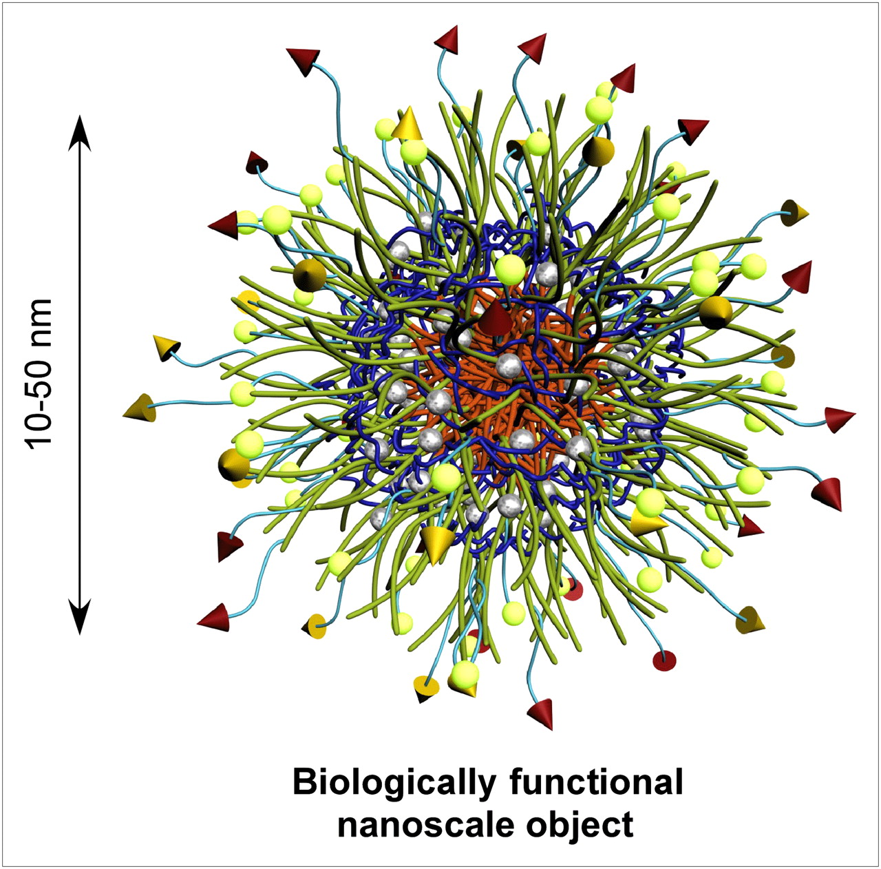
- FIGURE 11.
Schematic illustration of general core–shell morphology of multifunctional spheric nanoparticle.
- FIGURE 12.
Nanomaterials offering potential for high-sensitivity imaging and multimodality imaging relative to singly labeled (☆,Δ,□) targeting ligands (T).
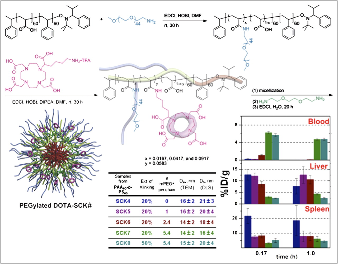
- FIGURE 13.
Pegylated DOTA–shell cross-linked knedel-like nanoparticles (SCKs) originate from amphiphilic block copolymers to which are coupled desired numbers of PEG and DOTA units, so that final assembled nanoparticle has well-defined structure and quantifiable PEG and DOTA levels. Surface coverage by PEG (parameters described in table) alters biodistribution significantly. PEGylated DOTA-SCK# label under nanoparticle structure corresponds to compounds listed in table, and color code in table links nanoparticles to biodistribution bar graphs at right.
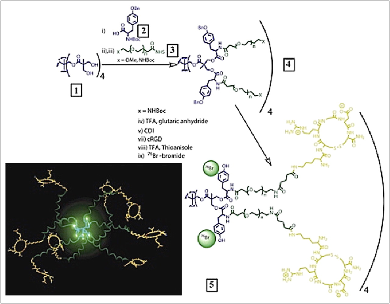
- FIGURE 14.
Preparation of PET nanoprobes targeted to αvβ3-integrin.
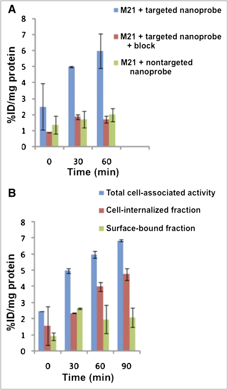
- FIGURE 15.
Cell uptake studies. (A) Percentages of total cell-associated fraction, cell-internalized fraction, and surface-bound fraction for targeted nanoprobe in αvβ3-integrin–positive M21 cells. (B) Percentages of cell-internalized fraction for targeted nanoprobe in absence and presence of block and nontargeted nanoprobe in αvβ3-integrin–positive M21 cells. All values were normalized to protein content per well. Total cell-associated fraction represents sum of cell-internalized fraction and surface-bound fraction. %ID/mg protein = percentage injected or administered dose per milligram of protein.
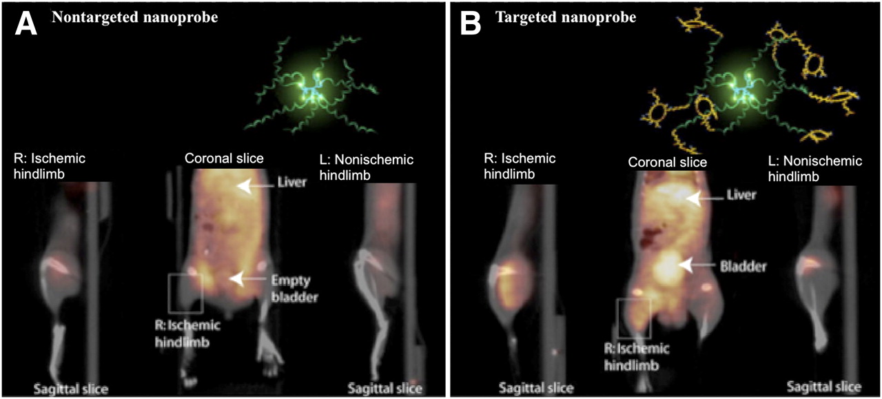
- FIGURE 16.
Noninvasive PET/CT images of angiogenesis induced by hind limb ischemia in murine model. (A) Nontargeted dendritic nanoprobes (bottom, middle). (B) Uptake of αvβ3-integrin–targeted dendritic nanoprobes was higher in ischemic hind limb (left) than in control hind limb (right).


Tables
Radionuclide Reaction Half-life Conventional 15O 14N(d,n)15O 2.04 min 13N 16O(p,n)13N 9.97 min 11C 14N(p,n)11C 20.4 min 18F 18O(p,n)18F 109.8 min Nonconventional 60Cu 60Ni(p,n)60Cu 23.7 min 94mTc 94Mo(p,n)94mTc 52 min 66Ga 66Zn(p,n)66Ga 9.5 h 64Cu 64Ni(p,n)64Cu 12.8 h 86Y 86Sr(p,n)86Y 14.7 h 76Br 76Se(p,n)76Br 16.2 h 89Zr 89Y(p,n)89Zr 78 h 124I 124Te(p,n)124I 4.2 d 68Ga 68Ge/68Ga 68 min 62Cu 62Zn/62Cu 9.74 min


Jump to section
Related Articles
Cited By...
- No citing articles found.