
Main menu

  • Home
  • Content
    • Current
    • Ahead of print
    • Past Issues
    • JNM Supplement
    • SNMMI Annual Meeting Abstracts
    • Continuing Education
    • JNM Podcasts
  • Subscriptions
    • Subscribers
    • Institutional and Non-member
    • Rates
    • Journal Claims
    • Corporate & Special Sales
  • Authors
    • Submit to JNM
    • Information for Authors
    • Assignment of Copyright
    • AQARA requirements
  • Info
    • Reviewers
    • Permissions
    • Advertisers
  • About
    • About Us
    • Editorial Board
    • Contact Information
  • More
    • Alerts
    • Feedback
    • Help
    • SNMMI Journals
  • SNMMI
    • JNM
    • JNMT
    • SNMMI Journals
    • SNMMI

User menu

  • Subscribe
  • My alerts
  • Log in
  • Log out
  • My Cart

Search

  • Advanced search
Journal of Nuclear Medicine
  • SNMMI
    • JNM
    • JNMT
    • SNMMI Journals
    • SNMMI
  • Subscribe
  • My alerts
  • Log in
  • Log out
  • My Cart
Journal of Nuclear Medicine

Advanced Search

  • Home
  • Content
    • Current
    • Ahead of print
    • Past Issues
    • JNM Supplement
    • SNMMI Annual Meeting Abstracts
    • Continuing Education
    • JNM Podcasts
  • Subscriptions
    • Subscribers
    • Institutional and Non-member
    • Rates
    • Journal Claims
    • Corporate & Special Sales
  • Authors
    • Submit to JNM
    • Information for Authors
    • Assignment of Copyright
    • AQARA requirements
  • Info
    • Reviewers
    • Permissions
    • Advertisers
  • About
    • About Us
    • Editorial Board
    • Contact Information
  • More
    • Alerts
    • Feedback
    • Help
    • SNMMI Journals
  • View or Listen to JNM Podcast
  • Visit JNM on Facebook
  • Join JNM on LinkedIn
  • Follow JNM on Twitter
  • Subscribe to our RSS feeds
Research ArticleBasic Science Investigation

Different Modes of Transport for 3H-Thymidine, 3H-FLT, and 3H-FMAU in Proliferating and Nonproliferating Human Tumor Cells

David A. Plotnik, Lindsay E. Emerick, Kenneth A. Krohn, Jashvant D. Unadkat and Jeffrey L. Schwartz
Journal of Nuclear Medicine September 2010, 51 (9) 1464-1471; DOI: https://doi.org/10.2967/jnumed.110.076794
David A. Plotnik
  • Find this author on Google Scholar
  • Find this author on PubMed
  • Search for this author on this site
Lindsay E. Emerick
  • Find this author on Google Scholar
  • Find this author on PubMed
  • Search for this author on this site
Kenneth A. Krohn
  • Find this author on Google Scholar
  • Find this author on PubMed
  • Search for this author on this site
Jashvant D. Unadkat
  • Find this author on Google Scholar
  • Find this author on PubMed
  • Search for this author on this site
Jeffrey L. Schwartz
  • Find this author on Google Scholar
  • Find this author on PubMed
  • Search for this author on this site
  • Article
  • Figures & Data
  • Info & Metrics
  • PDF
Loading

Article Figures & Data

Figures

  • Tables
  • FIGURE 1.
    • Download figure
    • Open in new tab
    • Download powerpoint
    FIGURE 1.

    Time-dependent nucleoside uptake in noncycling, plateau-phase cultures of A549 cells. (A) Intracellular levels of tracer (disintegrations per minute [dpm]/106 cells) in cells incubated in tracer-containing buffer. (B) Intracellular levels of tracer (percentage of activity in cell-equivalent volume of buffer) in cells incubated in tracer-containing buffer. (C) Intracellular levels of tracer (dpm/106 cells) in cells incubated in tracer-containing buffer with 10−4 M NBMPR. (D) Intracellular levels of tracer (percentage of activity in cell-equivalent volume of buffer) in cells incubated in tracer-containing buffer with 10−4 M NBMPR. Results are mean ± SD of at least 3 replicates. ○ = 3H-thymidine; ● = 3H-FLT; ▲ = 3H-FMAU; dotted line = relative activity in buffer.
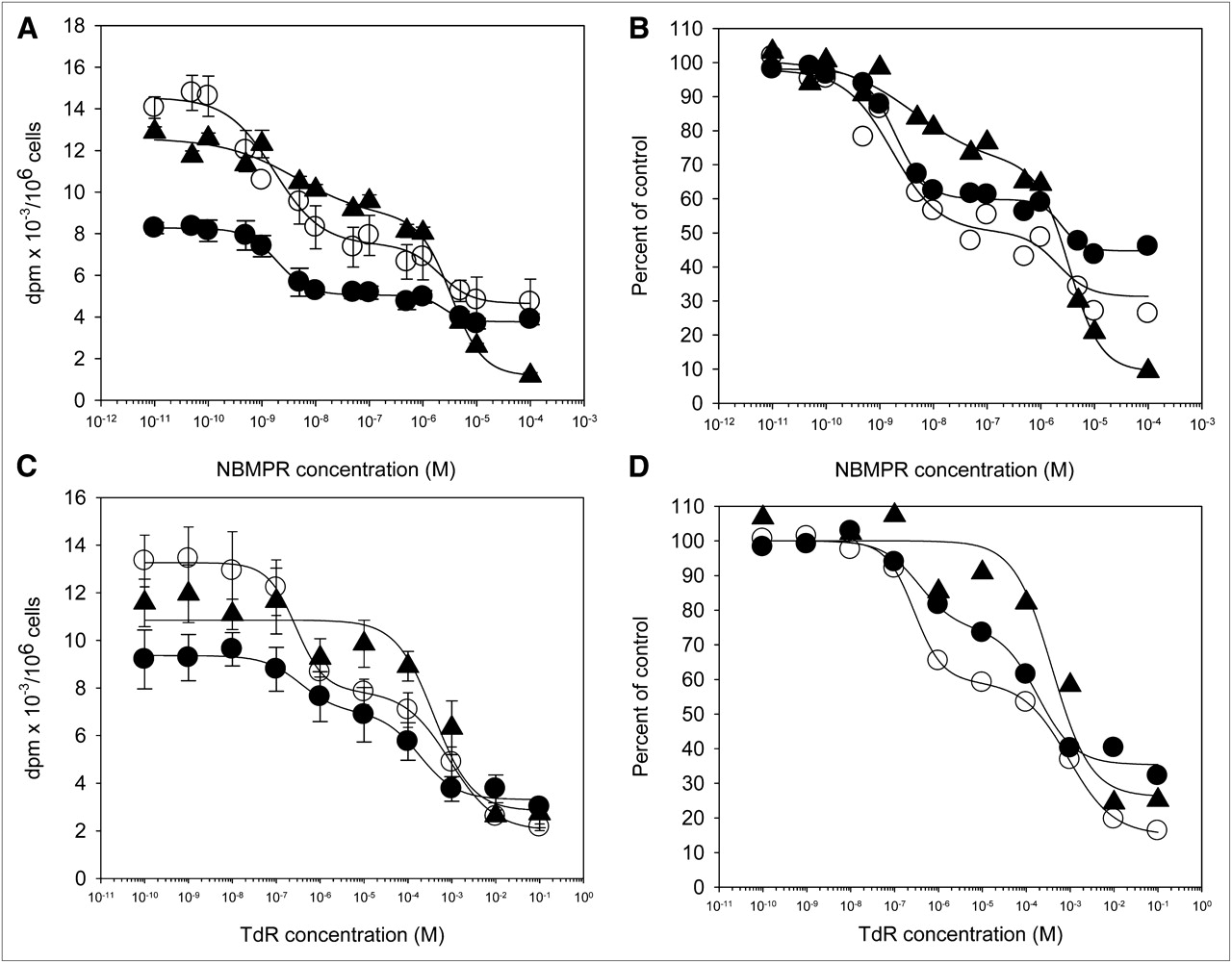
  • FIGURE 2.
    • Download figure
    • Open in new tab
    • Download powerpoint
    FIGURE 2.

    Concentration-dependent inhibition of nucleoside uptake in noncycling, plateau-phase cultures of A549 cells. (A) One-minute nucleoside tracer uptake values (disintegrations per minute [dpm]/106 cells) as function of increasing concentrations of NBMPR. (B) Results normalized to no-inhibitor levels. (C) One-minute nucleoside tracer uptake values (dpm/106 cells) as function of increasing concentrations of carrier-added thymidine. (D) Results normalized to no-carrier-added thymidine. Results are mean ± SD of at least 3 replicates. ○ = 3H-thymidine; ● = 3H-FLT; and ▲ = 3H-FMAU; TdR = thymidine.

  • FIGURE 3.
    • Download figure
    • Open in new tab
    • Download powerpoint
    FIGURE 3.

    Nucleoside efflux from noncycling, plateau-phase cultures of A549 cells. (A) Relationship between nucleoside uptake and efflux in cells preloaded with tracers and then incubated in tracer-free buffer for 15–60 s. (B) NBMPR-sensitive and -resistant nucleoside efflux in cells preloaded with tracers and then incubated in tracer-free buffer for 15–60 s. Results are mean ± SD of at least 3 replicates. ○ = 3H-thymidine; ● = 3H-FLT; ▲ = 3H-FMAU; dpm = disintegrations per minute.

  • FIGURE 4.
    • Download figure
    • Open in new tab
    • Download powerpoint
    FIGURE 4.

    (A) Western blot probed with rabbit anti-hENT1 antibody. Bands from left to right are whole-cell lysates from noncycling cells, whole-cell lysates from exponentially growing cells, membrane preparations from noncycling cells, and membrane preparations from exponentially growing cells. (B) ENT content measured in membrane preparations by 3H-NBMPR binding assay. White bar = noncycling cells; black bar = exponentially growing cells. (C) Tracer uptake after incubation for 60 s in buffer ± 10−4 M NBMPR in plateau-phase, noncycling, and asynchronous exponentially growing A549 cells.

Tables

  • Figures
    • View popup
    TABLE 1.

    Metabolism of 3H-Tracers*

    TimeTracerThymineNucleosideMonophosphateDiphosphateTriphosphateDNA†
    60-s incubation3H-thymidine13.6656.354.0910.8714.760.26
     3H-FLT—95.005.000.000.000.00
     3H-FMAU—100.000.000.000.000.00
    60-min incubation3H-thymidine4.758.514.7314.2962.744.97
     3H-FLT—13.0064.808.0014.000.00
     3H-FMAU—33.2126.379.9128.352.16
    60-min washout‡3H-thymidine0.000.004.7814.1666.3514.72
     3H-FLT—0.0041.1011.4047.500.00
     3H-FMAU—7.7824.4115.7748.581.47
    • ↵* Percentage of total activity in cells.

    • ↵† Activity in DNA.

    • ↵‡ Cells were incubated for 60 min in labeled tracer, then washed and cultured in tracer-free medium for 60 min before analysis.

PreviousNext
Back to top

In this issue

Journal of Nuclear Medicine: 51 (9)
Journal of Nuclear Medicine
Vol. 51, Issue 9
September 1, 2010
  • Table of Contents
  • Table of Contents (PDF)
  • About the Cover
  • Index by author
Print
Download PDF
Article Alerts
Sign In to Email Alerts with your Email Address
Email Article

Thank you for your interest in spreading the word on Journal of Nuclear Medicine.

NOTE: We only request your email address so that the person you are recommending the page to knows that you wanted them to see it, and that it is not junk mail. We do not capture any email address.

Enter multiple addresses on separate lines or separate them with commas.
Different Modes of Transport for 3H-Thymidine, 3H-FLT, and 3H-FMAU in Proliferating and Nonproliferating Human Tumor Cells
(Your Name) has sent you a message from Journal of Nuclear Medicine
(Your Name) thought you would like to see the Journal of Nuclear Medicine web site.
Citation Tools
Different Modes of Transport for 3H-Thymidine, 3H-FLT, and 3H-FMAU in Proliferating and Nonproliferating Human Tumor Cells
David A. Plotnik, Lindsay E. Emerick, Kenneth A. Krohn, Jashvant D. Unadkat, Jeffrey L. Schwartz
Journal of Nuclear Medicine Sep 2010, 51 (9) 1464-1471; DOI: 10.2967/jnumed.110.076794

Citation Manager Formats

  • BibTeX
  • Bookends
  • EasyBib
  • EndNote (tagged)
  • EndNote 8 (xml)
  • Medlars
  • Mendeley
  • Papers
  • RefWorks Tagged
  • Ref Manager
  • RIS
  • Zotero
Share
Different Modes of Transport for 3H-Thymidine, 3H-FLT, and 3H-FMAU in Proliferating and Nonproliferating Human Tumor Cells
David A. Plotnik, Lindsay E. Emerick, Kenneth A. Krohn, Jashvant D. Unadkat, Jeffrey L. Schwartz
Journal of Nuclear Medicine Sep 2010, 51 (9) 1464-1471; DOI: 10.2967/jnumed.110.076794
Twitter logo Facebook logo LinkedIn logo Mendeley logo
  • Tweet Widget
  • Facebook Like
  • Google Plus One
Bookmark this article

Jump to section

  • Article
    • Abstract
    • MATERIALS AND METHODS
    • RESULTS
    • DISCUSSION
    • CONCLUSION
    • Acknowledgments
    • REFERENCES
  • Figures & Data
  • Info & Metrics
  • PDF

Related Articles

  • This Month in JNM
  • PubMed
  • Google Scholar

Cited By...

  • [18F]CFA as a clinically translatable probe for PET imaging of deoxycytidine kinase activity
  • PET Imaging of Proliferation with Pyrimidines
  • Tumor 3'-Deoxy-3'-18F-Fluorothymidine (18F-FLT) Uptake by PET Correlates with Thymidine Kinase 1 Expression: Static and Kinetic Analysis of 18F-FLT PET Studies in Lung Tumors
  • Google Scholar

More in this TOC Section

  • [11C]ZTP-1: An Effective Short-Lived Radioligand for PET of Rat and Monkey Brain Phosphodiesterase Type 4 Subtype B
  • Reduced Renal Uptake of Various Radiopharmaceuticals with Sodium Paraaminohippurate Coadministration in a Rat Model
  • In Vivo Visualization and Quantification of Brain Heat Shock Protein 90 with [11C]HSP990 in Healthy Aging and Neurodegeneration
Show more Basic Science Investigation

Similar Articles

SNMMI

© 2025 SNMMI

Powered by HighWire