
Main menu

  • Home
  • Content
    • Current
    • Ahead of print
    • Past Issues
    • JNM Supplement
    • SNMMI Annual Meeting Abstracts
    • Continuing Education
    • JNM Podcasts
  • Subscriptions
    • Subscribers
    • Institutional and Non-member
    • Rates
    • Journal Claims
    • Corporate & Special Sales
  • Authors
    • Submit to JNM
    • Information for Authors
    • Assignment of Copyright
    • AQARA requirements
  • Info
    • Reviewers
    • Permissions
    • Advertisers
  • About
    • About Us
    • Editorial Board
    • Contact Information
  • More
    • Alerts
    • Feedback
    • Help
    • SNMMI Journals
  • SNMMI
    • JNM
    • JNMT
    • SNMMI Journals
    • SNMMI

User menu

  • Subscribe
  • My alerts
  • Log in
  • My Cart

Search

  • Advanced search
Journal of Nuclear Medicine
  • SNMMI
    • JNM
    • JNMT
    • SNMMI Journals
    • SNMMI
  • Subscribe
  • My alerts
  • Log in
  • My Cart
Journal of Nuclear Medicine

Advanced Search

  • Home
  • Content
    • Current
    • Ahead of print
    • Past Issues
    • JNM Supplement
    • SNMMI Annual Meeting Abstracts
    • Continuing Education
    • JNM Podcasts
  • Subscriptions
    • Subscribers
    • Institutional and Non-member
    • Rates
    • Journal Claims
    • Corporate & Special Sales
  • Authors
    • Submit to JNM
    • Information for Authors
    • Assignment of Copyright
    • AQARA requirements
  • Info
    • Reviewers
    • Permissions
    • Advertisers
  • About
    • About Us
    • Editorial Board
    • Contact Information
  • More
    • Alerts
    • Feedback
    • Help
    • SNMMI Journals
  • View or Listen to JNM Podcast
  • Visit JNM on Facebook
  • Join JNM on LinkedIn
  • Follow JNM on Twitter
  • Subscribe to our RSS feeds
Review ArticleFocus on Molecular Imaging

Tumor pH and Its Measurement

Xiaomeng Zhang, Yuxiang Lin and Robert J. Gillies
Journal of Nuclear Medicine August 2010, 51 (8) 1167-1170; DOI: https://doi.org/10.2967/jnumed.109.068981
Xiaomeng Zhang
  • Find this author on Google Scholar
  • Find this author on PubMed
  • Search for this author on this site
Yuxiang Lin
  • Find this author on Google Scholar
  • Find this author on PubMed
  • Search for this author on this site
Robert J. Gillies
  • Find this author on Google Scholar
  • Find this author on PubMed
  • Search for this author on this site
  • Article
  • Figures & Data
  • Info & Metrics
  • PDF
Loading

Article Figures & Data

Figures

  • FIGURE 1.
    • Download figure
    • Open in new tab
    • Download powerpoint
    FIGURE 1.

    Glucose metabolism in mammalian cells. Afferent blood delivers glucose and oxygen (on hemoglobin) to tissues, where it reaches cells by diffusion. Glucose is taken up by specific transporters, where it is converted first to glucose-6-phosphate by hexokinase and then to pyruvate, generating 2 adenosine triphosphates per glucose. In presence of oxygen, pyruvate is oxidized to HCO3−, generating 36 additional adenosine triphosphates per glucose. In absence of oxygen, pyruvate is reduced to lactate, which is exported from cell. Both processes produce hydrogen ions (H+), which acidify extracellular space. ATP = adenosine triphosphate; HbO2 = oxygenated hemoglobin. (Reprinted with permission of (1).)

  • FIGURE 2.
    • Download figure
    • Open in new tab
    • Download powerpoint
    FIGURE 2.

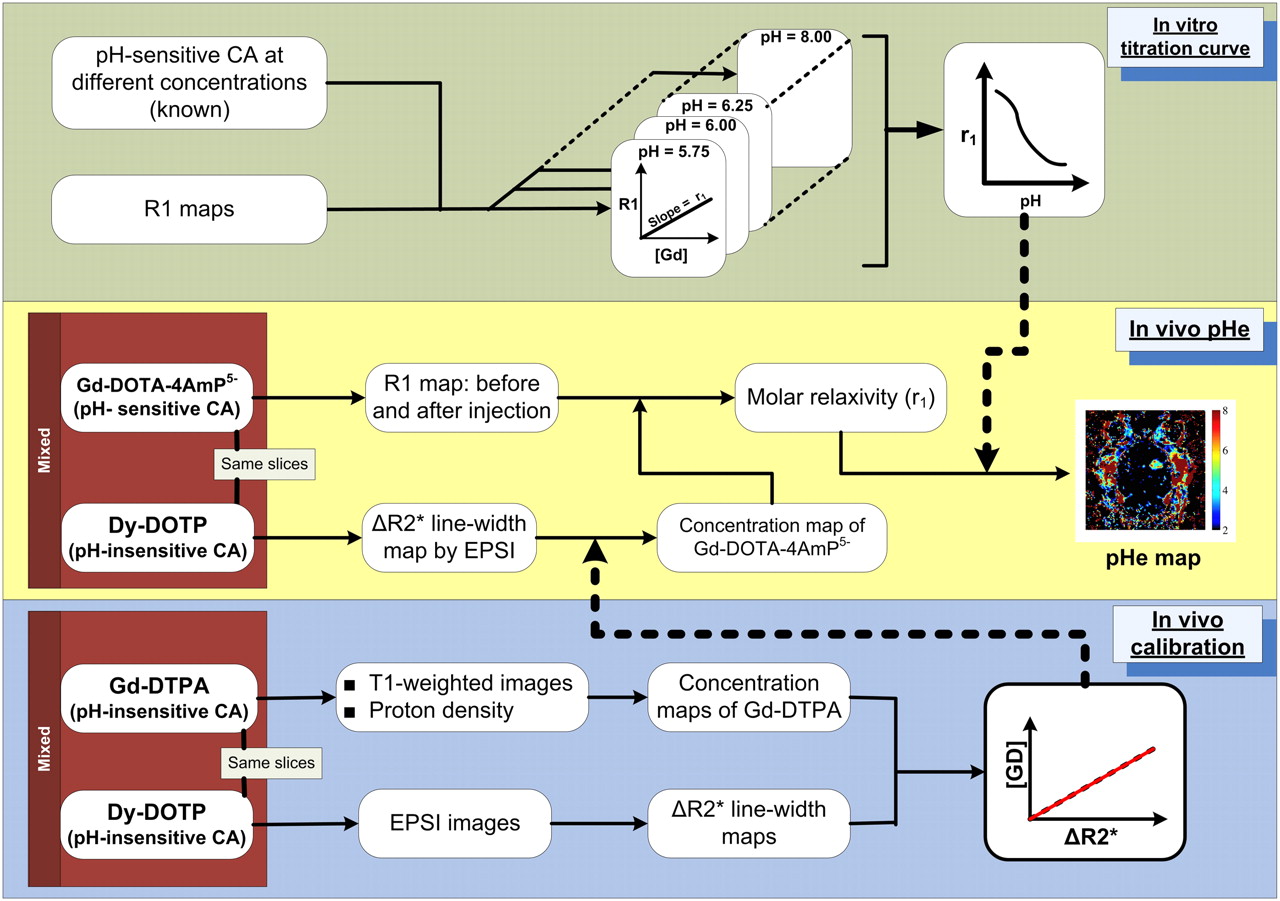
    Schematic overview of single-injection protocol. In vitro calibrations (upper panel) are used to define relationship between molar relaxivity of gadolinium-DOTA-4AmP5− and pH. In vivo calibrations (lower panel) involve coinjection of pH-independent gadolinium-diethylenetriaminepentaacetic acid and dysprosium-DOTP. These data are used to define in vivo relationship between concentration of gadolinium contrast agent and echoplanar spectroscopic imaging–measured line width. In experiment, line width induced by coinjected concentration of dysprosium contrast agent is used to calculate per-pixel concentration of gadolinium-DOTA-4AmP5−, which is then combined with T1 values to calculate molar relaxivity and, hence, pH (9). CA = contrast agent; DOTP = 1,4,7,10-tetraazacyclododecane-N,N′,N′′,N′′′-tetra(methylene phosphonic acid); DTPA = diethylenetriaminepentaacetic acid; EPSI = echoplanar spectroscopic imaging; LW = line width; R1 = spin-lattice relaxation rate (R1 = 1/T1); r1 = molar relaxivity.
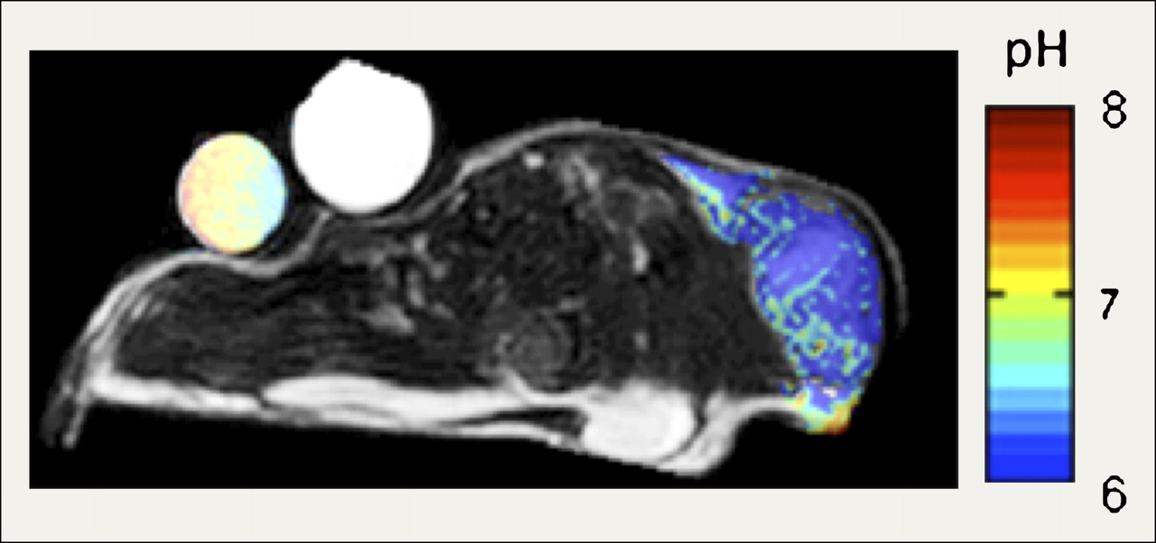
  • FIGURE 3.
    • Download figure
    • Open in new tab
    • Download powerpoint
    FIGURE 3.

    pHe map of mouse MCF-7 breast tumor model. pH was measured by paramagnetic CEST MRI with contrast agent ytterbium-1,4,7,10-tetraazacyclododecane-1,4,7-tetraacetic acid, 10-oaminoanilide. (Data courtesy of M. Pagel (15).)

PreviousNext
Back to top

In this issue

Journal of Nuclear Medicine: 51 (8)
Journal of Nuclear Medicine
Vol. 51, Issue 8
August 1, 2010
  • Table of Contents
  • Table of Contents (PDF)
  • About the Cover
  • Index by author
Print
Download PDF
Article Alerts
Sign In to Email Alerts with your Email Address
Email Article

Thank you for your interest in spreading the word on Journal of Nuclear Medicine.

NOTE: We only request your email address so that the person you are recommending the page to knows that you wanted them to see it, and that it is not junk mail. We do not capture any email address.

Enter multiple addresses on separate lines or separate them with commas.
Tumor pH and Its Measurement
(Your Name) has sent you a message from Journal of Nuclear Medicine
(Your Name) thought you would like to see the Journal of Nuclear Medicine web site.
Citation Tools
Tumor pH and Its Measurement
Xiaomeng Zhang, Yuxiang Lin, Robert J. Gillies
Journal of Nuclear Medicine Aug 2010, 51 (8) 1167-1170; DOI: 10.2967/jnumed.109.068981

Citation Manager Formats

  • BibTeX
  • Bookends
  • EasyBib
  • EndNote (tagged)
  • EndNote 8 (xml)
  • Medlars
  • Mendeley
  • Papers
  • RefWorks Tagged
  • Ref Manager
  • RIS
  • Zotero
Share
Tumor pH and Its Measurement
Xiaomeng Zhang, Yuxiang Lin, Robert J. Gillies
Journal of Nuclear Medicine Aug 2010, 51 (8) 1167-1170; DOI: 10.2967/jnumed.109.068981
Twitter logo Facebook logo LinkedIn logo Mendeley logo
  • Tweet Widget
  • Facebook Like
  • Google Plus One
Bookmark this article

Jump to section

  • Article
    • Abstract
    • PET
    • MR SPECTROSCOPY (MRS) AND MRI
    • OPTICS
    • SUMMARY
    • REFERENCES
  • Figures & Data
  • Info & Metrics
  • PDF

Related Articles

  • This Month in JNM
  • PubMed
  • Google Scholar

Cited By...

  • Non-invasive visualization of pH changes within the tumor-micro-environment by positron emission tomography
  • PH-SENSITIVE NANODROPLETS FOR CONTROLLED DELIVERY OF BERBERINE CHLORIDE
  • Protein kinase inhibitor ceritinib blocks ectonucleotidase CD39 - a promising target for cancer immunotherapy
  • The differential metabolic signature of breast cancer cellular response to olaparib treatment
  • Preclinical Characterization of Relatlimab, a Human LAG-3-Blocking Antibody, Alone or in Combination With Nivolumab
  • Selective Display of a Chemoattractant Agonist on Cancer Cells Activates the Formyl Peptide Receptor 1 on Immune Cells
  • Generating tumor-selective conditionally active biologic anti-CTLA4 antibodies via protein-associated chemical switches
  • INFORM: INFrared-based ORganizational Measurements of tumor and its microenvironment to predict patient survival
  • Breast Tumor-Associated Metalloproteases Restrict Reovirus Oncolysis by Cleaving the {sigma}1 Cell Attachment Protein and Can Be Overcome by Mutation of {sigma}1
  • pH-Dependent Grafting of Cancer Cells with Antigenic Epitopes Promotes Selective Antibody-Mediated Cytotoxicity
  • The Tumor Metabolic Microenvironment: Lessons from Lactate
  • Personalized Tumor Growth Prediction with Multiscale Tumor Modeling
  • Tumor pH and Protein Concentration Contribute to the Signal of Amide Proton Transfer Magnetic Resonance Imaging
  • pH-dependent thermodynamic intermediates of pHLIP membrane insertion determined by solid-state NMR spectroscopy
  • Peptides of pHLIP family for targeted intracellular and extracellular delivery of cargo molecules to tumors
  • In Vivo Imaging of Tumor Metabolism and Acidosis by Combining PET and MRI-CEST pH Imaging
  • Oncogenic p95HER2 regulates Na+-HCO3- cotransporter NBCn1 mRNA stability in breast cancer cells via 3'UTR-dependent processes
  • Probe for the measurement of cell surface pH in vivo and ex vivo
  • Tumour-Endothelial Cell Communications: Important and Indispensable Mediators of Tumour Angiogenesis
  • Targeting Acidity in Pancreatic Adenocarcinoma: Multispectral Optoacoustic Tomography Detects pH-Low Insertion Peptide Probes In Vivo
  • Molecular Mechanism for the Control of Eukaryotic Elongation Factor 2 Kinase by pH: Role in Cancer Cell Survival
  • Cerenkov-Specific Contrast Agents for Detection of pH In Vivo
  • Understanding the pharmacological properties of a metabolic PET tracer in prostate cancer
  • A Comparison of the Imaging Characteristics and Microregional Distribution of 4 Hypoxia PET Tracers
  • pH-(low)-insertion-peptide (pHLIP) translocation of membrane impermeable phalloidin toxin inhibits cancer cell proliferation
  • Google Scholar

More in this TOC Section

  • From Genome to Phenome: Opportunities and Challenges of Molecular Imaging
  • Imaging the Activity of Efflux Transporters at the Blood–Brain Barrier in Neurologic Diseases: Radiotracer Selection Criteria
  • Molecular Imaging of Pulmonary Fibrosis
Show more Focus on Molecular Imaging

Similar Articles

SNMMI

© 2025 SNMMI

Powered by HighWire