


Article Figures & Data
Figures
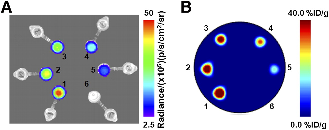
- FIGURE 1.
Phantom images recorded using optical (CLI) imaging (A) and PET (B) of 6 samples of 89Zr activity in water. At time 0 h, the Eppendorf tubes labeled 1–6 corresponded to activity concentrations of 40.3, 32.6, 27.4, 20.4, 13.3, and 0.00 kBq/μL. Optical images were recorded using integration time of 30 s and f/stop of 1.
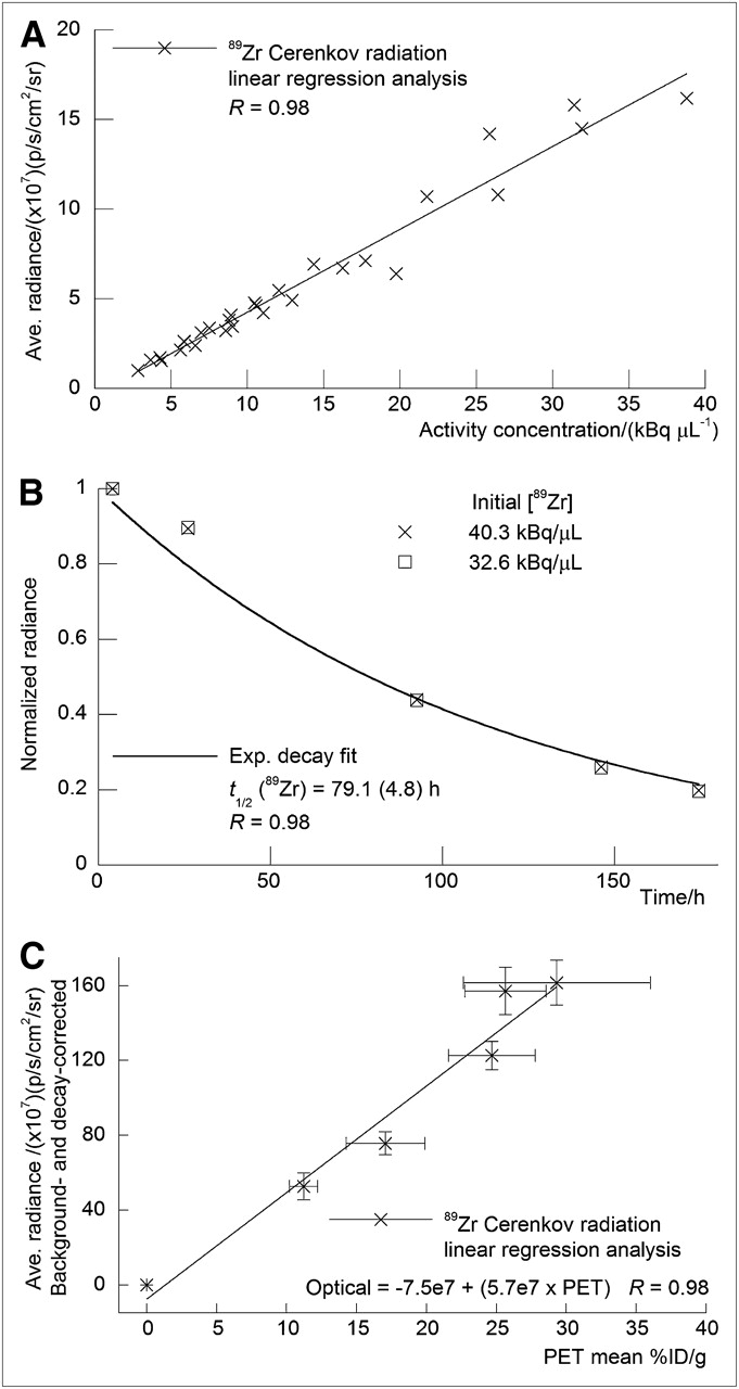
- FIGURE 2.
Quantitative analysis of phantom studies. Positive correlation observed between measured average radiance (background-corrected in units of p/s/cm2/sr) and 89Zr activity concentration (kBq/μL) (A), rate of decay observed in normalized radiance vs. time/h (B), and linear correlation observed between average radiance (background and decay-corrected in units of p/s/cm2/sr) vs. mean PET signal intensity (measured in units of %ID/g, commonly used for quantification of in vivo PET studies) (C). Ave. = average; Exp. = exponential.
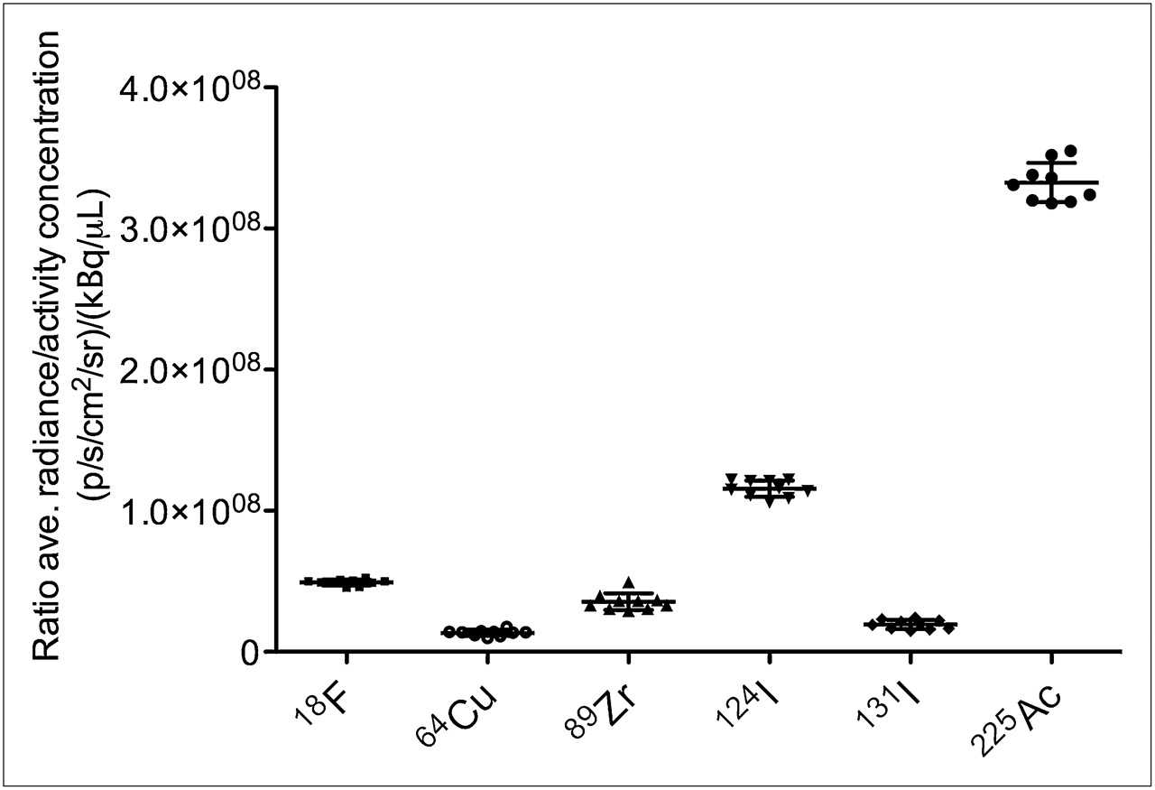
- FIGURE 3.
Plot of ratio of average radiance (p/s/cm2/sr)/activity concentration (μCi/μL) vs. radionuclide. Positron-emitting radionuclides are arranged in order of increasing average β+ kinetic energy (18F: Eβ+ = 249.8 keV [Iβ+ = 100%]; 64Cu: Eβ+ = 278.2 keV [Iβ+ = 17.6%]; 89Zr: Eβ+ = 395.5 keV [Iβ+ = 22.7%]; 124I: Eβ+ = 820 keV [Iβ+ = 22.7%]). For 131I, Eβ− = 181.9 keV (Iβ− 100%). 225Ac decays by 100% α-particle emission with Eα in range of 5,021–5,830 keV (16). Ave. = average.
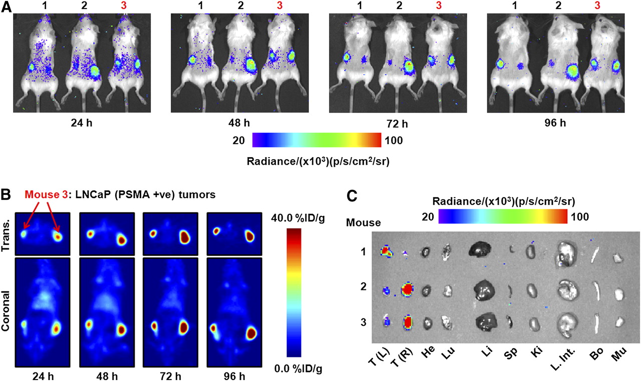
- FIGURE 4.
Temporal images of 89Zr-DFO-J591 uptake (10.9–11.3 MBq [295–305 μCi], 60–62 μg of mAb, in 200 μL of 0.9% sterile saline) recorded in dual subcutaneous LNCaP (PSMA-positive) tumor–bearing severe combined immune deficient mice between 24 and 96 h after administration. (A) Signal observed in optical spectrum from in vivo CLI of 89Zr-DFO-J591 tumor uptake in 3 mice. (B) Corresponding coronal and transverse immuno-PET images recorded for mouse 3. (C) Optical image recorded of organs after acute ex vivo biodistribution at 96 h. Transverse and coronal planar immuno-PET images intersect center of tumors. Upper and lower thresholds of CLI and immuno-PET images in A–C have been adjusted for visual clarity, as indicated by scale bars. Trans. = transverse; +ve = positive; T(L) = left tumor; T(R) = right tumor; He = heart; Lu = lungs; Li = liver; Sp = spleen; Ki = kidneys; L. Int. = large intestine; Bo = bone; Mu = muscle.
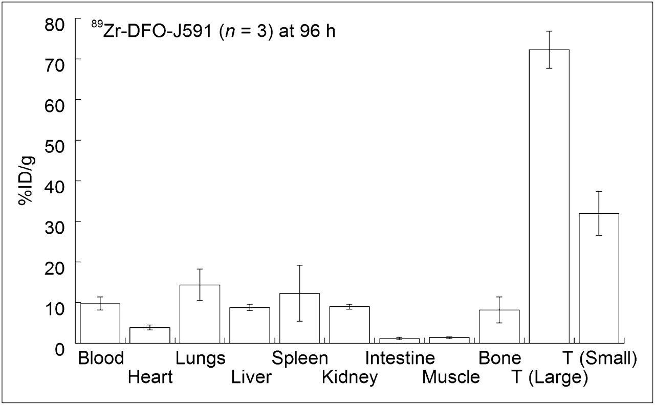
- FIGURE 5.
Bar chart showing selected tissue biodistribution data (%ID/g) for uptake of 89Zr-DFO-J591 in male severe combined immune deficient mice at end of optical and immuno-PET experiments (96 h after injection). T = tumor.
- FIGURE 6.
Time–activity curves showing ROI and volume-of-interest analysis of CLI and immuno-PET images for 89Zr-DFO-J591 uptake in well-established (large) LNCaP tumors. Volume-of-interest analysis of immuno-PET images shows change in 89Zr activity in heart–blood pool and muscle tissue.


Additional Files
Supplemental Data
Files in this Data Supplement:


Jump to section
Related Articles
Cited By...
- Detection of Shortwave-Infrared Cerenkov Luminescence from Medical Isotopes
- Optical Imaging Modalities: Principles and Applications in Preclinical Research and Clinical Settings
- Quo Vadis, Molecular Imaging?
- Leveraging Bioorthogonal Click Chemistry to Improve 225Ac-Radioimmunotherapy of Pancreatic Ductal Adenocarcinoma
- {alpha}-Emitters for Radiotherapy: From Basic Radiochemistry to Clinical Studies--Part 2
- Cerenkov Radiation-Induced Photoimmunotherapy with 18F-FDG
- In Vivo 3-Dimensional Radiopharmaceutical-Excited Fluorescence Tomography
- Cerenkov Luminescence Imaging as a Modality to Evaluate Antibody-Based PET Radiotracers
- Optical Imaging of Ionizing Radiation from Clinical Sources
- Cerenkov Luminescence Imaging for Radiation Dose Calculation of a 90Y-Labeled Gastrin-Releasing Peptide Receptor Antagonist
- Cerenkov-Specific Contrast Agents for Detection of pH In Vivo
- Intratherapeutic Biokinetic Measurements, Dosimetry Parameter Estimates, and Monitoring of Treatment Efficacy Using Cerenkov Luminescence Imaging in Preclinical Radionuclide Therapy
- Cerenkov Luminescence Endoscopy: Improved Molecular Sensitivity with {beta}--Emitting Radiotracers
- In Vivo Localization of 90Y and 177Lu Radioimmunoconjugates Using Cerenkov Luminescence Imaging in a Disseminated Murine Leukemia Model
- Clinical Cerenkov Luminescence Imaging of 18F-FDG
- Designing the Magic Bullet? The Advancement of Immuno-PET into Clinical Use
- Intraoperative Imaging of Tumors Using Cerenkov Luminescence Endoscopy: A Feasibility Experimental Study
- Positron Lymphography: Multimodal, High-Resolution, Dynamic Mapping and Resection of Lymph Nodes After Intradermal Injection of 18F-FDG
- Proof-of-Concept Study of Monitoring Cancer Drug Therapy with Cerenkov Luminescence Imaging
- Harnessing the Power of Radionuclides for Optical Imaging: Cerenkov Luminescence Imaging
- Multimodal Imaging with 18F-FDG PET and Cerenkov Luminescence Imaging After MLN4924 Treatment in a Human Lymphoma Xenograft Model
- Novel insights on imaging sex hormone-dependent tumourigenesis in vivo