


Article Figures & Data
Figures
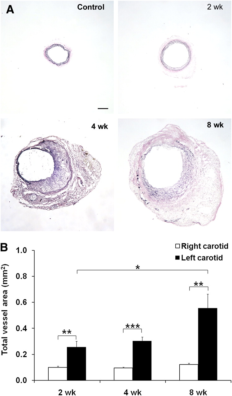
- FIGURE 1.
Carotid aneurysm in apoE−/− mice. (A) Representative examples of elastic van Gieson staining of common carotid artery exposed to NaCl (control) or at 2, 4, and 8 wk after exposure to CaCl2, demonstrating marked enlargement of artery. (B) Morphometric analysis of total vessel area after exposure of left carotid artery to CaCl2. n = 9–13 in each group. *P < 0.05. **P = 0.01. ***P < 0.001. Scale bar = 100 μm.
- FIGURE 2.
MMP expression and activity in aneurysm. (A) Representative examples of MMP-2 and MMP-9 immunofluorescent staining (in red) of carotid arteries at 4 wk, demonstrating diffuse MMP expression in aneurysmal left carotid artery. Elastic membranes are detected by their autofluorescence in green. Nuclei are detected by DAPI in blue. Insets represent staining with control antibodies. Scale bar = 100 μm. (B) Examples of in situ gelatinase zymography of control (right) and aneurysmal (left) carotid arteries at 4 wk. Protease activity is markedly reduced in presence of specific MMP inhibitors (1,10-phenanthroline). Nuclei are detected by DAPI in blue. Scale bar = 20 μm. (C) MMP activity quantified using generic fluorogenic assay at 2, 4, and 8 wk after CaCl2 application. n = 9–13. *P < 0.05. **P < 0.01. AU = arbitrary units; L = lumen.
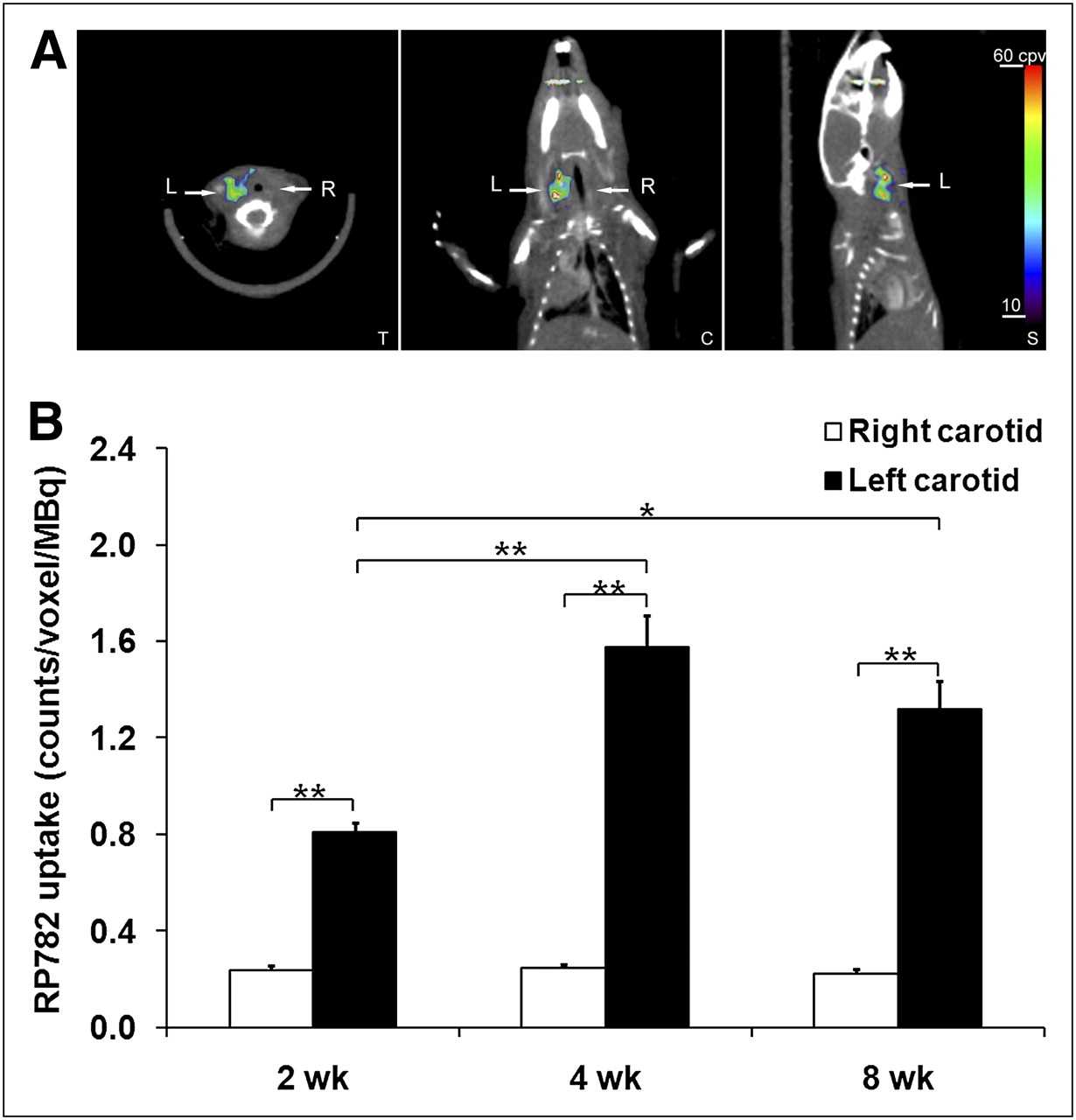
- FIGURE 3.
In vivo imaging of MMP activation in aneurysm. (A) Example of fused micro-SPECT/CT images of mouse at 4 wk after surgery to induce carotid aneurysm. Arrows point to aneurysmal left (L) and control right (R) carotid arteries. (B) Image-derived quantitative analysis of background-corrected RP782 carotid uptake. Background-corrected tracer uptake in left carotid artery peaked at 4 wk after surgery and was significantly higher than uptake in right carotid artery at every time point studied. n = 16–18 in each group. *P = 0.01. **P < 0.001. C = coronal slice; S = sagittal slice; T = transverse slice.
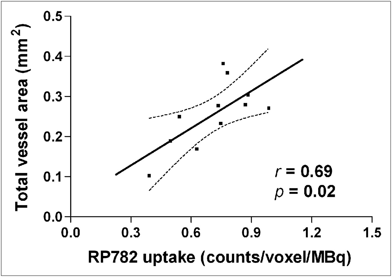
- FIGURE 4.
Ex vivo analysis of MMP tracer uptake in aneurysm. (A) Example of carotid arteries and aorta at 4 wk after aneurysm induction, harvested and photographed after RP782 imaging (left), with corresponding autoradiography (right). Figure represents data from at least 5 animals from each time point. (B) Quantitative analysis of RP782 uptake by γ-well counting. n = 8–9 in each group. *P < 0.001. (C) Correlation between carotid RP782 uptake and MMP activity assessed by quantitative zymography. AU = arbitrary units.
- FIGURE 5.
MMP tracer uptake specificity. (A) Example of RP782 micro-SPECT/CT after administration of 50-fold excess of precursor in mouse 4 wk after surgery to induce carotid aneurysm. Arrows point to aneurysmal left (L) and control right (R) carotid arteries. (B) Image-derived quantitative analysis of RP782 carotid uptake in absence (n = 18) or presence (n = 2) of 50-fold excess of nonlabeled precursor. *P < 0.01. C = coronal slice; S = sagittal slice; T = transverse slice.
- FIGURE 6.
MMP activation and aneurysm size. There is significant correlation between MMP tracer uptake in aneurysmal carotid artery at 2 wk and total vessel area measured at 4 wk in same animal. n = 11. Dotted lines represent 95% confidence interval.


Additional Files
Supplemental Data
Files in this Data Supplement:


Jump to section
Related Articles
Cited By...
- Matrix Metalloproteinases: From Molecular Mechanisms to Physiology, Pathophysiology, and Pharmacology
- Preclinical Evaluation of RYM1, a Matrix Metalloproteinase-Targeted Tracer for Imaging Aneurysm
- Novel Molecular Imaging Approaches to Abdominal Aortic Aneurysm Risk Stratification
- Could Statin Use Be Associated with Reduced Recurrence Rates following Coiling in Ruptured Intracranial Aneurysms?
- Predicting Aortic Aneurysm Expansion by PET
- Multimodality and Molecular Imaging of Matrix Metalloproteinase Activation in Calcific Aortic Valve Disease
- Imaging Vessel Wall Biology to Predict Outcome in Abdominal Aortic Aneurysm
- Molecular Imaging of the Cardiac Extracellular Matrix
- Cardiovascular Molecular Imaging: The Road Ahead
- Integrin-Targeted Imaging of Inflammation in Vascular Remodeling