


Abstract
Despite significant advancements in medical and device-based therapies, cardiovascular disease remains the number one cause of death in the United States. Early detection of atherosclerosis, prevention of myocardial infarction and sudden cardiac death, and modulation of adverse ventricular remodeling still remain elusive goals. Molecular imaging focuses on identifying critical cellular and molecular targets and therefore plays an integral role in understanding these biologic processes in vivo. Because many imaging targets are upregulated before irreversible tissue damage occurs, early detection could ultimately lead to development of novel, preventive therapeutic strategies. This review addresses recent work on radionuclide imaging of cardiovascular inflammation, infection, and infarct healing. We further discuss opportunities provided by multimodality approaches such as PET/MRI and PET/optical imaging.
Despite recent advances in diagnosis, treatment, and guideline-based management, cardiovascular disease remains the number one cause of death in the United States. Consequently, there is a strong impetus for shifting from late-stage diagnosis and in-hospital care to detection of subclinical disease and prevention. Molecular imaging is positioned to play a key role in this transition by addressing challenges such as identification of vulnerable plaque and assessment of postinfarction ventricular remodeling. Our objective in this focused review is to update the reader on the most recent advancements in the field of nuclear cardiovascular imaging of inflammation, infection, and infarct healing. We will then look at the road ahead for opportunities afforded by recent developments in multimodality imaging and radioisotope chemistry. Because of space restrictions, we refer the reader to earlier in-depth reviews (1–3).
INFLAMMATION
Atherosclerosis
Inflammation plays a key role in the pathogenesis of atherosclerotic plaque formation and progression. Key steps involved in the inflammatory cascade include endothelial cell dysfunction, expression of cellular adhesion molecules, lipid retention, monocyte recruitment and differentiation into macrophages, foam cell formation, proteolysis, apoptosis, and angiogenesis. Ultimately, the plaque erodes and ruptures, leading to arterial thrombosis and myocardial infarction or stroke. All of these processes serve as potential targets for the development of molecular imaging probes that detect disease at a time point when intervention can prevent irreversible damage (Fig. 1).
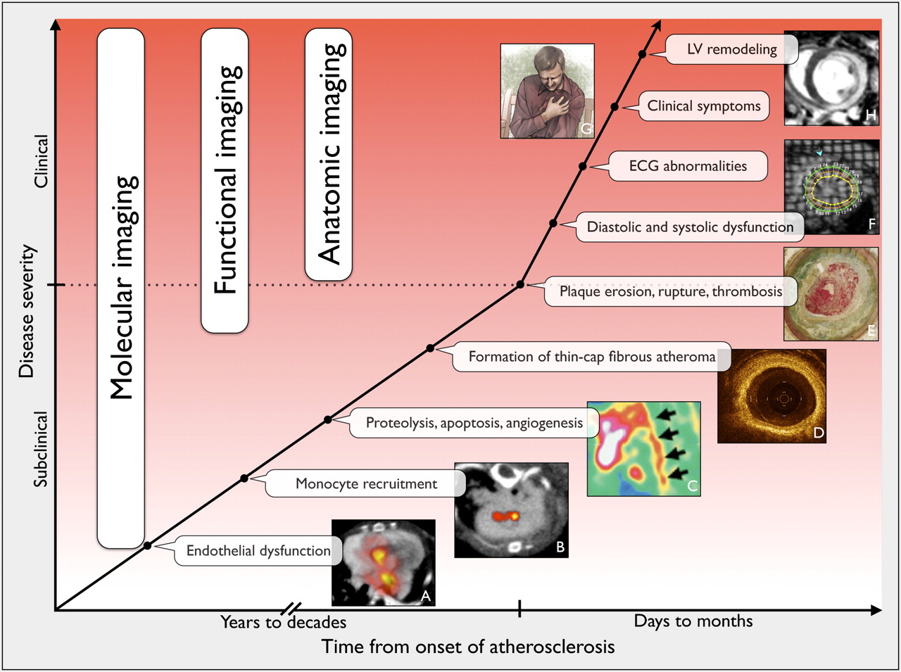
Imaging and ischemic cascade. In this schematic representation of disease progression, plaque rupture and coronary thrombosis represent the inflection point. (A) PET/CT of VCAM-1 expression (4). (B) PET/CT of macrophages (5). (C) SPECT/CT of MMP (7). (D) Intracoronary optical coherence tomography (courtesy of Kevin Croce). (E) Thrombotic occlusion of a coronary artery (reproduced with permission from British Medical Journal publishing group). (F) Tagging MRI (our own data). (G) Angina pectoris (reproduced with permission from American Medical Association). (H) Late gadolinium enhancement MRI showing myocardial infarction (our own data). ECG = electrocardiography; LV = left ventricular.
Expression of adhesion molecules on activated endothelial cells is one of the first steps in the inflammatory cascade and facilitates recruitment of monocytes into plaque. Agents that target adhesion molecules may be particularly suited to detecting early atherosclerotic lesions. We developed an 18F-labeled vascular cell adhesion molecule (VCAM)–1 ligand, 18F-4V, and demonstrated the feasibility of imaging VCAM-1 expression by PET/CT in a murine model of atherosclerosis (4). 18F-4V detected nascent lesions and measured differences in VCAM-1 expression caused by statin treatment (Fig. 2A). Dextranated magnetofluorescent nanoparticles are rapidly ingested by macrophages in atherosclerotic plaques and therefore report on a more downstream target (5). Cross-linked iron oxide particles were labeled with 64Cu, thereby permitting PET and MRI sensing of macrophage content within the plaque (Fig. 2B). Another approach toward macrophage imaging targets proteins on mononuclear phagocytes. PK11195 is a selective ligand for the peripheral benzodiazepine receptor, a translocator protein expressed on activated macrophages. Gaemperli et al. (6) imaged 32 patients with carotid stenosis with the PET agent 11C-PK11195 and found that symptomatic carotid plaques had high target-to-background ratios.
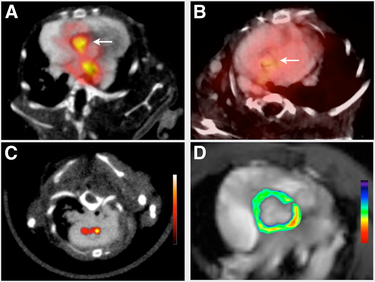
Targeted imaging of atherosclerosis. (A) 18F-4V PET/CT of VCAM-1 expression (4). (B) Higher uptake is seen in ApoE−/− mice than in statin-treated mice. (C) Nanoparticle PET/CT of macrophages (5). (D) MRI with pseudocolored T2-weighted signal intensity, which decreased because of accumulation of iron oxide nanoparticles.
Matrix metalloproteinases (MMP) promote plaque instability. Fujimoto et al. used a radiolabeled (99mTc) MMP inhibitor to noninvasively quantify MMP activity in atherosclerotic lesions in mice (7). The same group also imaged atherosclerotic lesions in rabbits (8) to quantify the effect of dietary modification, as well as statin therapy, on plaque MMP activity. Increasingly, imaging approaches examine multiple biologic processes at the same time to gain broader perspective on disease progression. Haider et al. pursued dual molecular imaging using radiolabeled metalloproteinase inhibitor and the annexin probe AA5 (9). MMP and apoptosis signals correlated with each other (r = 0.6, P < 0.001) and were significantly higher in rabbits on a high-cholesterol diet than in those on a normal diet or treated with a statin.
The length and cost of traditional clinical endpoint studies have created interest in using molecular imaging to study therapeutic efficiency. Fayad's group first optimized 18F-FDG PET protocols (10) and then applied them in the dal-PLAQUE trial. This multicenter clinical trial used 18F-FDG PET/CT and MRI to assess structural and inflammatory indices as clinical endpoints (11). Patients were randomly assigned to placebo or to therapy with a cholesterol ester transfer protein inhibitor. This trial provided interesting links between high-density lipoprotein level, vascular inflammation, and subsequent structural vascular changes.
Arterial Aneurysms
Inflammation has been implicated in aneurysm formation, dilation, and rupture. Although there is only a moderate correlation between aneurysm size and risk of rupture, it is plausible that inflammatory activity in the vessel wall may be more predictive of rupture. We used a macrophage-targeted nanoparticle labeled with 18F for PET/CT in apolipoprotein E double-knockout (ApoE−/−) mice with aneurysms (12) and found that the PET signal was increased in progressing aneurysms (Fig. 3A). Another strategy targeted MMP activity in the vessel wall (13). Razavian et al. examined carotid aneurysms in ApoE−/− mice exposed to calcium chloride using RP782, an 111In-labeled tracer with specific activity for activated MMPs. Focal uptake of RP782 in carotid arteries peaked at 4 wk after aneurysm induction. Once translated, these approaches could enable individualized risk-stratification and development of new decision algorithms based on pathobiology rather than anatomy.

Vascular inflammation, infection, and infarct healing. (A) Macrophage-targeted PET/CT of inflammation in aortic aneurysm with 64Cu-labeled cross-linked iron oxide particles (12). Dotted circle outlines aneurysm in ascending aorta. (B) PET/CT of S. aureus endocarditic vegetation (arrow) in mice with 64Cu-ProT (14). (C) Hybrid PET/MRI of murine postinfarction inflammation with macrophage-targeted 64Cu-labeled nanoparticle (our own data). Infarct area is detected by late gadolinium enhancement on MRI (inset, arrows). ce = contrast-enhanced.
INFECTIVE ENDOCARDITIS
Detection and management of infective endocarditis remain challenging because standard diagnostic criteria, such as fever or new heart murmur, lack sensitivity and specificity. Although helpful, most echocardiographic findings are manifested at late stages of disease and cannot inform on the infective activity in vegetations. Targeted molecular imaging aimed at visualizing bacteria or their products in vegetations may lead to earlier and more reliable diagnosis and can help to find septic emboli. We developed an agent for identifying staphylococcal infections by targeting staphylocoagulase, a virulence factor secreted by Staphylococcus aureus (14). An engineered prothrombin analog detected endocarditis vegetations via noninvasive fluorescence and PET in a mouse model (Fig. 3B). In contrast to blood cultures that detect circulating bacteria, the agent reported on the type and activity of bacteria in vegetations.
MYOCARDIAL INFARCTION AND VENTRICULAR REMODELING
With increased infarct survival, there has been a rise in the number of patients with heart failure. Identifying predictors of adverse ventricular remodeling may help with further risk stratification of patients and open novel therapeutic avenues. Molecular imaging of cell death, inflammatory response, angiogenesis, and fibrosis has pursued a deeper understanding of the remodeling process.
Injury to the myocardium results in an acute inflammatory response dominated by cells of the mononuclear phagocyte system (15). Similar to the strategy used for inflammatory atherosclerosis, 18F-FDG PET was used to assess inflammation after ischemic myocardial injury (16,17). Berr et al. followed ischemia–reperfusion injury with serial PET and MRI in a murine model and found increased 18F-FDG uptake on day 7 after myocardial infarction (16). Although the 18F-FDG signal in acute murine infarcts derives mainly from inflammatory cells (17), the high myocyte uptake of 18F-FDG and the ischemia-induced shift in myocardial metabolism toward glycolysis pose challenges in interpretation, especially if infarct tissue and surviving myocardium are interspersed.
Angiogenesis provides an interesting target for noninvasive molecular imaging of wound healing. Rodriguez-Porcel et al. developed a PET tracer, 64Cu-DOTA–vascular endothelial growth factor 121, to image in vivo VEGF receptor expression in rats with myocardial infarction (18). Serial imaging demonstrated that the VEGF receptor expression level increased significantly after myocardial infarction and remained elevated for 2 wk. RGD (arginine-glycine-aspartate), a binding motif for integrins, serves as >an affinity ligand for αvβ3-integrin–targeted probes. Sadeghi and Bender first studied αvβ3-integrin SPECT in a vascular injury model in ApoE−/− mice with RP748, an 111In-labeled peptidomimetic with affinity for αvβ3-integrin (19). The same group then imaged hypoxia and angiogenesis in parallel using 99mTc-BRU59-21 and 111In-RP748 in rodent and canine models of myocardial infarction (20). These studies demonstrated the prospect of noninvasive tracking of hypoxia-induced integrin activation as a marker of angiogenesis.
Immune cells recruited to the injured myocardium secrete cytokines and proteolytic enzymes that play an important role in the healing process. In particular, MMPs accelerate left ventricular remodeling by digesting extracellular matrix, and probes targeting MMPs can follow these processes after myocardial infarction. Su et al. assessed temporal changes in MMP activation after myocardial infarction with a broad MMP-targeted agent, 111In-RP782, in a murine model (21). More recently, MMP-targeted SPECT/CT followed postinfarct remodeling in a porcine model using 99mTc-labeled RP805 (22). Increased agent retention was seen throughout the heart early after myocardial infarction, and the signal remained elevated for 1 mo.
MULTIMODALITY IMAGING
Multimodality imaging combines individual strengths of different modalities and offers integrated molecular, physiologic, and anatomic information. In addition, by incorporating complementary agents, hybrid imaging permits simultaneous evaluation of several disease pathways. Hybrid PET/MRI may be particularly useful for following molecular and cellular events after myocardial infarction. We labeled a macrophage-targeted nanoparticle with 64Cu to pursue PET/MRI in a mouse 3 d after myocardial infarction. Delayed-enhancement MRI after injection of gadolinium-DTPA delineated the infarcted myocardium, whereas the PET signal reflected inflammatory cells that ingested the nanoparticles (Fig. 3C, unpublished data, 2011). We speculate that this signal could represent a key wound-healing process—removal of necrotic tissue—and may predict outcome. The concept could be expanded further by adding a molecular MRI agent to trace 2 biologic targets simultaneously.
Hybrid PET/optical reporter imaging may also have a role in a staged diagnostic/therapeutic approach. A potential scenario for managing patients with atherosclerosis could use the PET isotope on a macrophage-targeted nanoparticle to localize inflamed plaque and decide whether invasive intervention is necessary. In a second step, the fluorochrome attached to the same nanoparticle could guide local therapy while detected with a fluorescence-sensing intravascular catheter (23).
Finally, multimodal techniques already routinely guide imaging agent development. Especially with larger probes such as nanoparticles or proteins, derivatization with fluorochromes allows initial screening with optical techniques such as fluorescence tomography, fluorescence reflectance imaging, fluorescence histology, and flow cytometry (24). Once a lead preparation is identified, one can then move on to more time-consuming and expensive nuclear imaging, thus accelerating the throughput of the probe discovery process. These approaches are enhanced by bioorthogonal coupling reactions that enable modular attachment of radionuclides and fluorochromes to imaging probes (25). Cycloaddition reactions, often referred to as “click” chemistry, also permit modular labeling of small molecules with commonly used PET isotopes such as 18F.
CONCLUSION
There is a need for early noninvasive detection of atherosclerosis as well as imaging of processes that promote adverse ventricular remodeling. Multimodality molecular imaging has become part of the toolbox in basic science. A major goal for realizing the true impact of imaging on clinical care is to accelerate the movement of imaging probes through the regulatory approval process. The road ahead for molecular imaging is promising, but many miles need to be covered to establish the comparative (cost-) effectiveness of such strategies and to integrate them into clinical decision making.
Acknowledgments
This work was funded in part by grants from the NIH (R01HL095629, R01HL096576, and Translational Program of Excellence in Nanotechnology HHSN268201000044C). We acknowledge fruitful discussions with Drs. Ralph Weissleder and Peter Libby. No other potential conflict of interest relevant to this article was reported.
Footnotes
Published online Apr. 9, 2012.
- © 2012 by the Society of Nuclear Medicine, Inc.
REFERENCES
- Received for publication January 3, 2012.
- Accepted for publication March 14, 2012.


Jump to section
Related Articles
Cited By...
- Leukocytes Link Local and Systemic Inflammation in Ischemic Cardiovascular Disease: An Expanded "Cardiovascular Continuum"
- Scintillating Balloon-Enabled Fiber-Optic System for Radionuclide Imaging of Atherosclerotic Plaques
- Recent Developments in Vascular Biology
- Translational Molecular Imaging: Repurposing an Old Technique to Track Cell Migration Into Human Atheroma
- Dual-Contrast Molecular Imaging Allows Noninvasive Characterization of Myocardial Ischemia/Reperfusion Injury After Coronary Vessel Occlusion in Mice by Magnetic Resonance Imaging
- Imaging and Nanomedicine in Inflammatory Atherosclerosis
- Preclinical and Translational PET/MR Imaging
- Positron Emission Tomography/Computed Tomographic and Magnetic Resonance Imaging in a Murine Model of Progressive Atherosclerosis Using 64Cu-Labeled Glycoprotein VI-Fc
- PET Imaging of Chemokine Receptors in Vascular Injury-Accelerated Atherosclerosis
- Monocyte and Macrophage Heterogeneity in the Heart