
Main menu

  • Home
  • Content
    • Current
    • Ahead of print
    • Past Issues
    • JNM Supplement
    • SNMMI Annual Meeting Abstracts
    • Continuing Education
    • JNM Podcasts
  • Subscriptions
    • Subscribers
    • Institutional and Non-member
    • Rates
    • Journal Claims
    • Corporate & Special Sales
  • Authors
    • Submit to JNM
    • Information for Authors
    • Assignment of Copyright
    • AQARA requirements
  • Info
    • Reviewers
    • Permissions
    • Advertisers
  • About
    • About Us
    • Editorial Board
    • Contact Information
  • More
    • Alerts
    • Feedback
    • Help
    • SNMMI Journals
  • SNMMI
    • JNM
    • JNMT
    • SNMMI Journals
    • SNMMI

User menu

  • Subscribe
  • My alerts
  • Log in
  • My Cart

Search

  • Advanced search
Journal of Nuclear Medicine
  • SNMMI
    • JNM
    • JNMT
    • SNMMI Journals
    • SNMMI
  • Subscribe
  • My alerts
  • Log in
  • My Cart
Journal of Nuclear Medicine

Advanced Search

  • Home
  • Content
    • Current
    • Ahead of print
    • Past Issues
    • JNM Supplement
    • SNMMI Annual Meeting Abstracts
    • Continuing Education
    • JNM Podcasts
  • Subscriptions
    • Subscribers
    • Institutional and Non-member
    • Rates
    • Journal Claims
    • Corporate & Special Sales
  • Authors
    • Submit to JNM
    • Information for Authors
    • Assignment of Copyright
    • AQARA requirements
  • Info
    • Reviewers
    • Permissions
    • Advertisers
  • About
    • About Us
    • Editorial Board
    • Contact Information
  • More
    • Alerts
    • Feedback
    • Help
    • SNMMI Journals
  • View or Listen to JNM Podcast
  • Visit JNM on Facebook
  • Join JNM on LinkedIn
  • Follow JNM on Twitter
  • Subscribe to our RSS feeds
OtherBASIC SCIENCE INVESTIGATIONS

Novel PET Probes Specific for Deoxycytidine Kinase

Chengyi J. Shu, Dean O. Campbell, Jason T. Lee, Andrew Q. Tran, Jordan C. Wengrod, Owen N. Witte, Michael E. Phelps, Nagichettiar Satyamurthy, Johannes Czernin and Caius G. Radu
Journal of Nuclear Medicine July 2010, 51 (7) 1092-1098; DOI: https://doi.org/10.2967/jnumed.109.073361
Chengyi J. Shu
  • Find this author on Google Scholar
  • Find this author on PubMed
  • Search for this author on this site
Dean O. Campbell
  • Find this author on Google Scholar
  • Find this author on PubMed
  • Search for this author on this site
Jason T. Lee
  • Find this author on Google Scholar
  • Find this author on PubMed
  • Search for this author on this site
Andrew Q. Tran
  • Find this author on Google Scholar
  • Find this author on PubMed
  • Search for this author on this site
Jordan C. Wengrod
  • Find this author on Google Scholar
  • Find this author on PubMed
  • Search for this author on this site
Owen N. Witte
  • Find this author on Google Scholar
  • Find this author on PubMed
  • Search for this author on this site
Michael E. Phelps
  • Find this author on Google Scholar
  • Find this author on PubMed
  • Search for this author on this site
Nagichettiar Satyamurthy
  • Find this author on Google Scholar
  • Find this author on PubMed
  • Search for this author on this site
Johannes Czernin
  • Find this author on Google Scholar
  • Find this author on PubMed
  • Search for this author on this site
Caius G. Radu
  • Find this author on Google Scholar
  • Find this author on PubMed
  • Search for this author on this site
  • Article
  • Figures & Data
  • Supplemental
  • Info & Metrics
  • PDF
Loading

Article Figures & Data

Figures

  • Tables
  • Additional Files
  • FIGURE 1. 
    • Download figure
    • Open in new tab
    • Download powerpoint
    FIGURE 1. 

    In vivo metabolite analysis of 18F-FAC. (A) Chemical structures of 18F-FAC and 18F-FAU and schematic showing extracellular and intracellular routes by which 18F-FAC can be deaminated to 18F-FAU. (B) Percentage of total detected radioactivity in plasma that is attributable to 18F-FAC over time as determined by HLPC analysis. Analysis is performed after intravenous injection of 18F-FAC in mice. 5′-NT = 5′nucleotidase; DCTD = deoxycytidylate deaminase (catabolic enzymes are shown in red fonts); MP = monophosphate.

  • FIGURE 2. 
    • Download figure
    • Open in new tab
    • Download powerpoint
    FIGURE 2. 

    18F-FAC and 18F-clofarabine (18F-CA) small-animal PET/CT scans of C57BL/6J mice (images are representative of pattern observed in 3 mice scanned with each probe). %ID/g = percentage injected dose per gram of tissue; Bl = urinary bladder; BF = brown fat; BM = bone marrow; GI = gastrointestinal tract; K = kidney; L = liver; S = spleen; Thy = thymus.
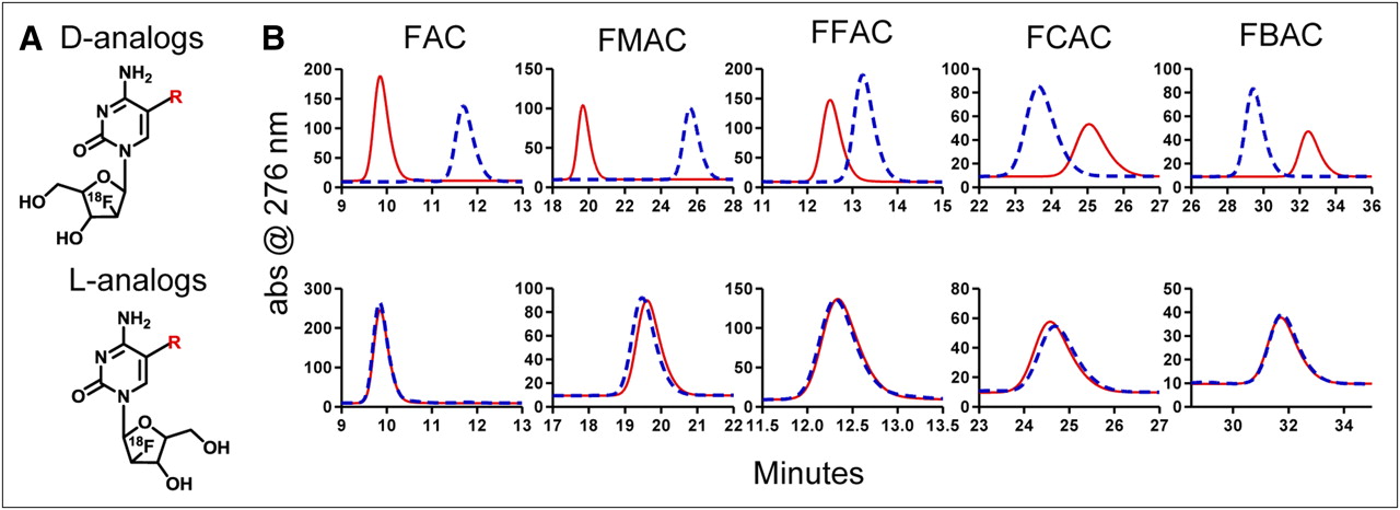
  • FIGURE 3. 
    • Download figure
    • Open in new tab
    • Download powerpoint
    FIGURE 3. 

    Candidate dCK-specific PET probes that resist deamination. (A) Chemical structures of deoxycytidine analogs amenable to 18F labeling. (B) In vitro deamination assay. After incubation at 37°C for 1 h in presence (blue dashed traces) or absence (red solid traces) of recombinant purified CDA, candidate probes were analyzed on HPLC. d-analogs are shown in top row, and l-analogs are shown in bottom row. abs = absorbance. R = H (FAC); R = CH3 (FMAC); R = F (FFAC); R = Cl (FCAC); R = Br (FBAC).
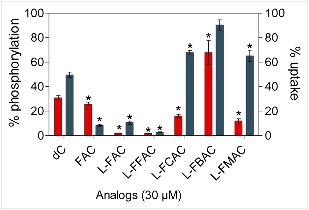
  • FIGURE 4. 
    • Download figure
    • Open in new tab
    • Download powerpoint
    FIGURE 4. 

    Analyses of affinity of l-analogs for dCK. l-analogs were tested for their ability to competitively inhibit phosphorylation (red bars; left axis) and uptake (blue bars; right axis) of tritium-labeled deoxycytidine (3H-dC) using dCK-expressing L1210 cells. Results represent 2 independent experiments. P values are calculated relative to water (n = 3). *P < 0.05. dC = deoxycytidine.
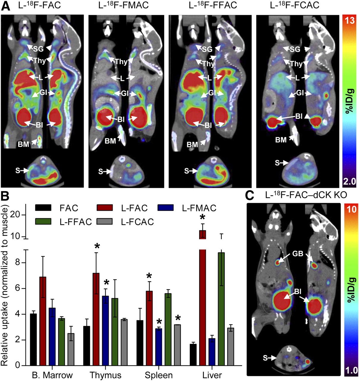
  • FIGURE 5. 
    • Download figure
    • Open in new tab
    • Download powerpoint
    FIGURE 5. 

    Biodistribution of 18F-labeled unnatural nucleosides in mice. (A) Small-animal PET/CT images of C57BL/6J mice. (B) Quantification of PET data. Probe uptake was normalized to muscle background (absolute uptake values are shown in Supplemental Fig. 6). (C) l-18F-FAC small-animal PET/CT scan of dCK knockout mouse. P values were calculated relative to FAC for each specific tissue. *P < 0.05; n = 5 (18F-FAC), n = 3 (l-18F-FAC), n = 3 (l-18F-FMAC), n = 2 (l-18F-FFAC), and n = 2 (l-18F-FCAC). %ID/g = percentage injected dose per gram of tissue; Bl = urinary bladder; B. Marrow = bone marrow; BM = bone marrow; GI = gastrointestinal tract; KO = knockout; L = liver; S = spleen; SG = salivary gland; Thy = thymus.
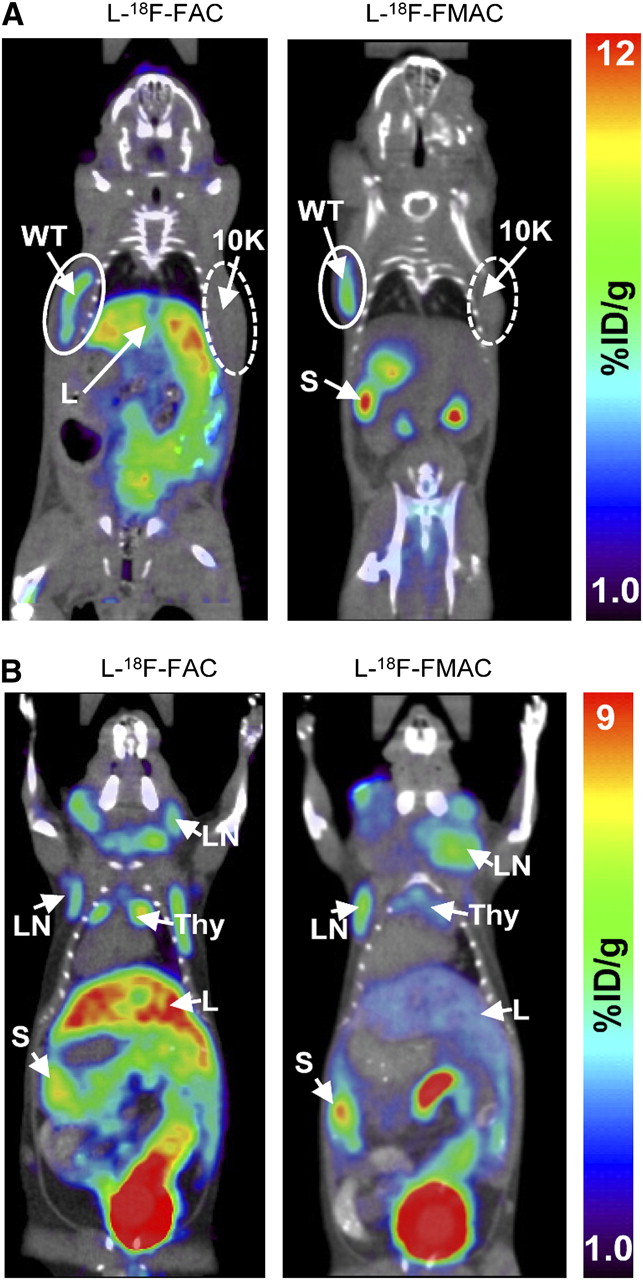
  • FIGURE 6. 
    • Download figure
    • Open in new tab
    • Download powerpoint
    FIGURE 6. 

    l-18F-FAC and l-18F-FMAC small-animal PET/CT images of malignant and autoimmune lymphoproliferative disorders. (A) l-18F-FAC and l-18F-FMAC small-animal PET/CT of L1210 lymphoma tumors. L1210 parental cell line (WT, solid-lined circle) and dCK-deficient variant L1210-10K (10K, dash-lined circle) were injected subcutaneously under the left and right shoulders of the mouse, respectively. Only the parental cell line accumulated both probes. (B) l-18F-FAC and l-18F-FMAC small-animal PET/CT of autoimmune B6.MRL-Faslpr/J mice. Both probes detected cervical, axillary, and brachial lymphadenopathy in these mice. %ID/g = percentage injected dose per gram of tissue; L = liver; LN = lymph nodes; S = spleen; Thy = thymus; WT = wild-type. Number of mice per probe ≥ 3.

Tables

  • Figures
  • Additional Files
    • View popup
    TABLE 1

    Kinetic Parameters for FAC and Its Analogs with l-Chirality

    ProbeKM (μM)Relative kcatRelative kcat/KM
    FAC0.76 ± 0.151.01.0
    l-FMAC1.02 ± 0.151.00.96
    l-FCAC0.61 ± 0.090.961.20
    l-FBAC6.54 ± 0.992.700.31
    • kcat and specificity constant (kcat/KM) were determined using purified recombinant human dCK, and values are given relative to FAC.

Additional Files

  • Figures
  • Tables
  • Supplemental Data

    Files in this Data Supplement:

    • Supplemental Materials
PreviousNext
Back to top

In this issue

Journal of Nuclear Medicine: 51 (7)
Journal of Nuclear Medicine
Vol. 51, Issue 7
July 2010
  • Table of Contents
  • Table of Contents (PDF)
  • About the Cover
  • Index by author
Print
Download PDF
Article Alerts
Sign In to Email Alerts with your Email Address
Email Article

Thank you for your interest in spreading the word on Journal of Nuclear Medicine.

NOTE: We only request your email address so that the person you are recommending the page to knows that you wanted them to see it, and that it is not junk mail. We do not capture any email address.

Enter multiple addresses on separate lines or separate them with commas.
Novel PET Probes Specific for Deoxycytidine Kinase
(Your Name) has sent you a message from Journal of Nuclear Medicine
(Your Name) thought you would like to see the Journal of Nuclear Medicine web site.
Citation Tools
Novel PET Probes Specific for Deoxycytidine Kinase
Chengyi J. Shu, Dean O. Campbell, Jason T. Lee, Andrew Q. Tran, Jordan C. Wengrod, Owen N. Witte, Michael E. Phelps, Nagichettiar Satyamurthy, Johannes Czernin, Caius G. Radu
Journal of Nuclear Medicine Jul 2010, 51 (7) 1092-1098; DOI: 10.2967/jnumed.109.073361

Citation Manager Formats

  • BibTeX
  • Bookends
  • EasyBib
  • EndNote (tagged)
  • EndNote 8 (xml)
  • Medlars
  • Mendeley
  • Papers
  • RefWorks Tagged
  • Ref Manager
  • RIS
  • Zotero
Share
Novel PET Probes Specific for Deoxycytidine Kinase
Chengyi J. Shu, Dean O. Campbell, Jason T. Lee, Andrew Q. Tran, Jordan C. Wengrod, Owen N. Witte, Michael E. Phelps, Nagichettiar Satyamurthy, Johannes Czernin, Caius G. Radu
Journal of Nuclear Medicine Jul 2010, 51 (7) 1092-1098; DOI: 10.2967/jnumed.109.073361
Twitter logo Facebook logo LinkedIn logo Mendeley logo
  • Tweet Widget
  • Facebook Like
  • Google Plus One
Bookmark this article

Jump to section

  • Article
    • Abstract
    • MATERIALS AND METHODS
    • RESULTS
    • DISCUSSION
    • CONCLUSION
    • Acknowledgments
    • Footnotes
    • References
  • Figures & Data
  • Supplemental
  • Info & Metrics
  • PDF

Related Articles

  • This Month in JNM
  • Erratum
  • PubMed
  • Google Scholar

Cited By...

  • STING-driven interferon signaling triggers metabolic alterations in pancreas cancer cells visualized by [18F]FLT PET imaging
  • Predicting Gemcitabine Delivery by 18F-FAC PET in Murine Models of Pancreatic Cancer
  • 18F-FAC PET Visualizes Brain-Infiltrating Leukocytes in a Mouse Model of Multiple Sclerosis
  • Imaging of Activated T Cells as an Early Predictor of Immune Response to Anti-PD-1 Therapy
  • 18F-FAC PET Selectively Images Liver-Infiltrating CD4 and CD8 T Cells in a Mouse Model of Autoimmune Hepatitis
  • Production of diverse PET probes with limited resources: 24 18F-labeled compounds prepared with a single radiosynthesizer
  • Detection of immune responses after immunotherapy in glioblastoma using PET and MRI
  • A PET Imaging Strategy to Visualize Activated T Cells in Acute Graft-versus-Host Disease Elicited by Allogenic Hematopoietic Cell Transplant
  • Human Biodistribution and Radiation Dosimetry of 18F-Clofarabine, a PET Probe Targeting the Deoxyribonucleoside Salvage Pathway
  • [18F]CFA as a clinically translatable probe for PET imaging of deoxycytidine kinase activity
  • Co-targeting of convergent nucleotide biosynthetic pathways for leukemia eradication
  • Stratification of Nucleoside Analog Chemotherapy Using 1-(2'-Deoxy-2'-18F-Fluoro-{beta}-D-Arabinofuranosyl)Cytosine and 1-(2'-Deoxy-2'-18F-Fluoro-{beta}-L-Arabinofuranosyl)-5-Methylcytosine PET
  • Structure-guided Engineering of Human Thymidine Kinase 2 as a Positron Emission Tomography Reporter Gene for Enhanced Phosphorylation of Non-natural Thymidine Analog Reporter Probe
  • Google Scholar

More in this TOC Section

  • Counting Rate Characteristics and Image Distortion in Preclinical PET Imaging During Radiopharmaceutical Therapy
  • Design and Fabrication of Kidney Phantoms for Internal Radiation Dosimetry Using 3D Printing Technology
  • Synthesis and Biologic Evaluation of Novel 18F-Labeled Probes Targeting Prostate-Specific Membrane Antigen for PET of Prostate Cancer
Show more Basic Science Investigations

Similar Articles

SNMMI

© 2025 SNMMI

Powered by HighWire