


Article Figures & Data
Figures
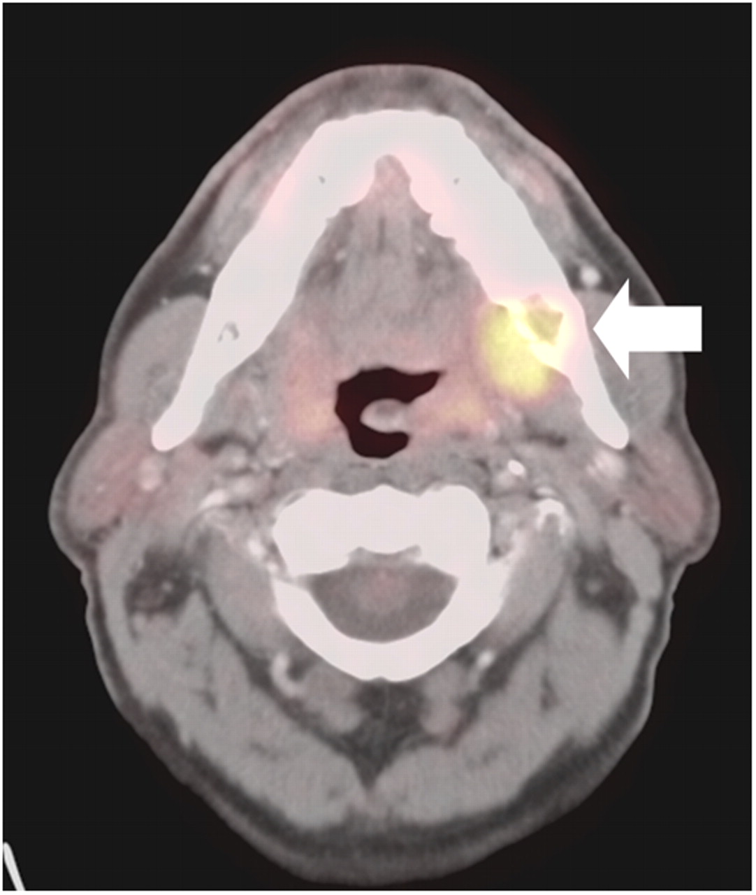
- FIGURE 1.
Site of infection or inflammation may mimic primary tumor by PET/CT. HN PET/CT shows false-positive lesion due to peridontal abscess (solid arrow). Primary tumor was actually small skin lesion that was missed by all imaging modalities.
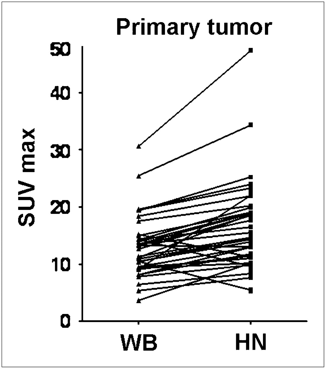
- FIGURE 2.
Change in SUVmax from WB to HN protocol for primary tumor.
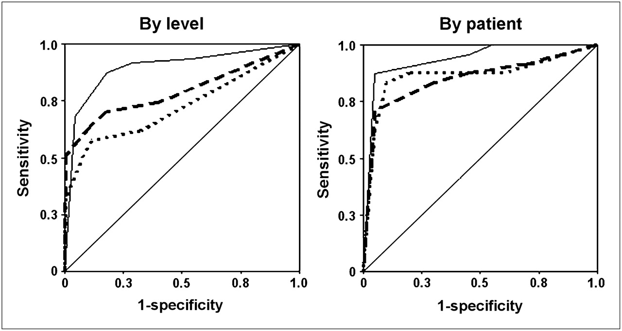
- FIGURE 3.
ROC analyses of CECT, WB PET/CT, and HN PET/CT protocols in detection of nodal metastases by level and by patient. Dotted line = CECT; dashed line = WB; solid line = HN.
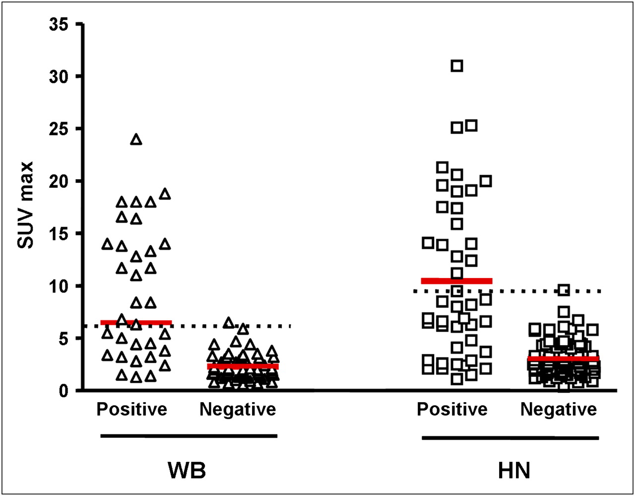
- FIGURE 4.
SUVmax in WB and HN protocols for positive and negative nodes. Dotted black line is placed just above highest SUVmax for negative nodes. Red line shows median of SUVmax for each group.
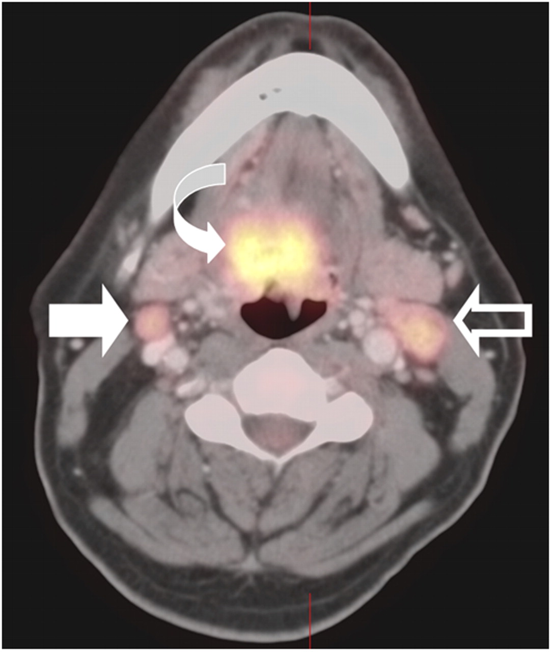
- FIGURE 5.
PET/CT had difficulty in distinguishing reactive from malignant nodes. HN PET/CT protocol shows primary tumor (curved arrow), true-positive left level II node (open arrow), and false-positive right level II node (solid arrow). Both lymph nodes were similar in metabolic activity.
- FIGURE 6.
Change in SUVmax from WB PET/CT protocol (90 min after injection) to HN PET/CT protocol (150 min after injection) for positive and negative nodes.
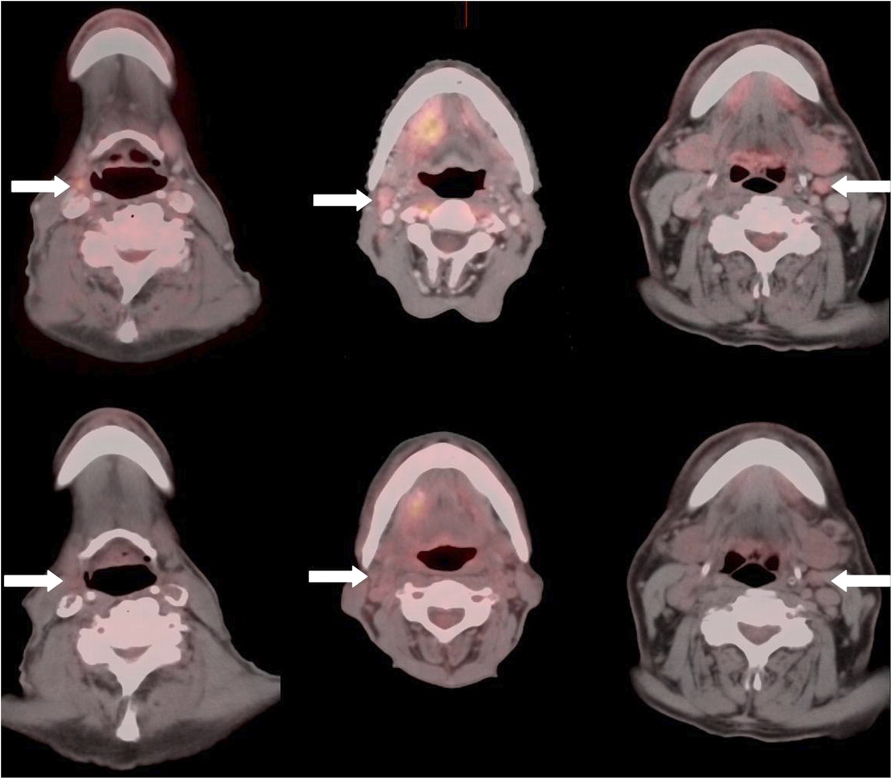
- FIGURE 7.
Three examples in which HN PET/CT protocol (upper panel) detected small (<15 mm) histologically proven positive lymph nodes that were missed by standard WB protocol (lower panel). Arrows indicate discordant nodes.


Tables
- TABLE 1
Acquisition and Processing Parameters for WB PET/CT and High-Resolution, Contrast-Enhanced HN PET/CT (Siemens Biograph PET/CT)
Parameter WB PET/CT HN PET/CT Technique CT kVp, 120; mA, 240 kVp, 120; mA, 200 CT collimation (mm) 1.5 0.75 Bed time (min) 6 12 Matrix (PET image) 168 256, with 1.5 zoom Pixel size (mm) 4.16 1.82 Filter FWHM (mm) 5 (7 if >78.75 kg) 2.0 Iteration 4 6 Subset 8 14 Intravenous contrast No Yes Interval from injection (min) 90 150 Bed Cradle Flat Head position In head holder In head holder (chin up) FWHM = full width at half maximum.
Primary tumor sites Number of patients Total (%) Oropharyngeal 29 66 Hypopharyngeal 1 2 Laryngeal 4 9 Skin 4 9 Unknown primary site 6 14 Total 44 100 CECT feature Sensitivity (%) Specificity (%) PPV (%) NPV (%) Accuracy (%) Size (enlarged) 63 89 66 88 79 Shape (round) 83 66 45 92 73 Contrast enhancement (any) 83 68 47 92 74 Heterogeneous enhancement 63 96 84 89 83 Absence of fatty hilum 97 21 29 95 51 Necrosis 53 98 90 86 81 Extracapsular spread 43 98 88 84 77 Asymmetry 100 21 30 100 52 Overall CT appearance 57 88 63 86 81 Total number of nodal levels evaluated by CECT alone was 77. Prevalence of malignant nodal levels was 25.27%. PPV = positive predictive value; NPV = negative predictive value.
- TABLE 5
ROC Analysis for Detection of Nodal Metastases by CECT, WB PET/CT, and HN PET/CT Protocols
Parameter By level By patient Area under the curve CECT 0.722 0.863 WB protocol 0.790 0.850 HN protocol 0.896 0.936 95% confidence interval CECT 0.623–0.820 0.741–0.984 WB protocol 0.699–0.881 0.730–0.970 HN protocol 0.836–0.956 0.860–1.000 Comparison of CT vs. WB PET/CT Difference between areas 0.069 0.0125 Significance level P = 0.099 P = 0.0811 Comparison of CT vs. HN PET/CT Difference between areas 0.174 0.074 Significance level P < 0.001 P = 0.152 Comparison of WB vs HN PET/CT Difference between areas 0.106 0.086 Significance level P < 0.002 P = 0.059


Jump to section
Related Articles
Cited By...
- Effect of Time-of-Flight Technique on the Diagnostic Performance of 18F-FDG PET/CT for Assessment of Lymph Node Metastases in Head and Neck Squamous Cell Carcinoma
- Negative Predictive Value of Surveillance PET/CT in Head and Neck Squamous Cell Cancer
- Positron Emission Tomography With [18F]Fluorodeoxyglucose Improves Staging and Patient Management in Patients With Head and Neck Squamous Cell Carcinoma: A Multicenter Prospective Study