


Article Figures & Data
Figures
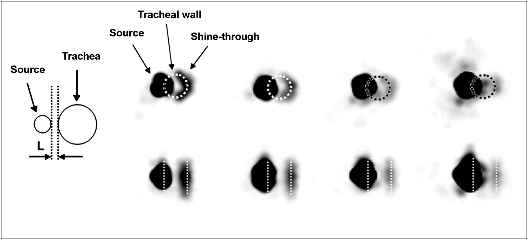
- FIGURE 1.
Schematic drawing of phantom source–trachea configuration in transverse plane and illustration of shine-through as function of stopping-layer thickness for 1-mL syringe. Images from left to right are of stopping-layer thickness of 0.3, 1.85, 2.85, and 3.85 mm. Top row shows transverse slices; bottom row, coronal slices. Position of trachea, indicated by circle and straight lines, was taken from CT images. Upper-level window settings for images were, from left to right, 1.2%, 0.54%, 0.39%, and 0.36% of image maximum.
- FIGURE 2.
Relative magnitude of shine-through in phantom study for different source volumes as function of stopping-layer thickness.
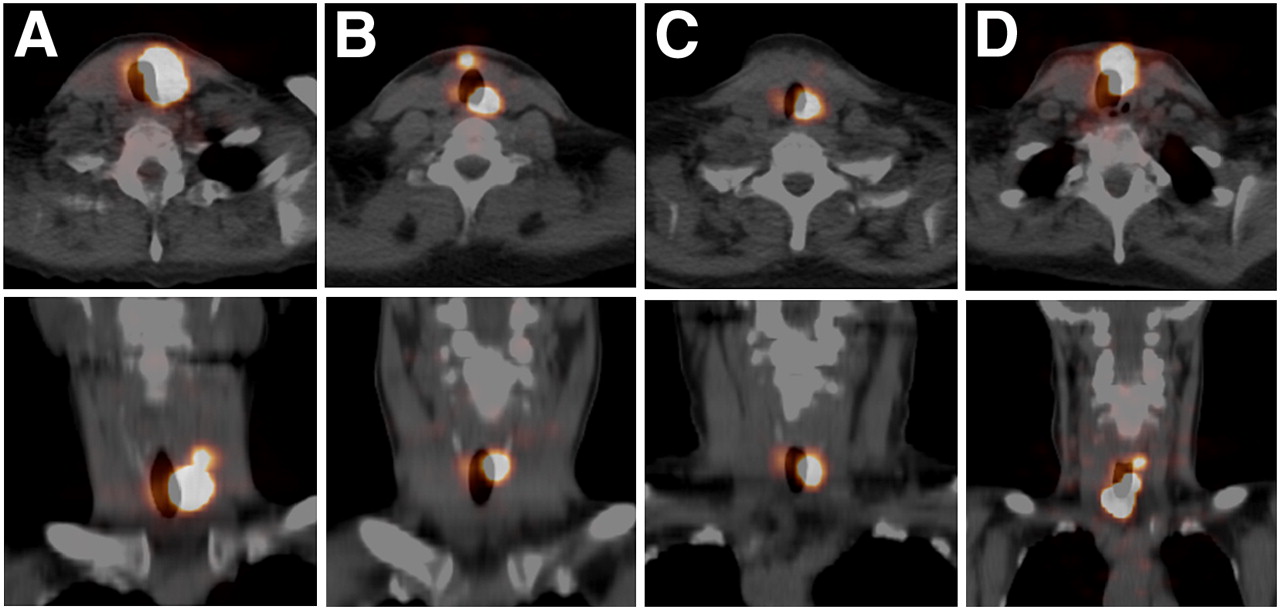
- FIGURE 3.
Shine-through in 4 patient studies at 96 h after oral intake of 20–25 MBq of 124I. Top row shows transaxial PET/CT images; bottom row, coronal images. A–D correspond to patient codes in Table 1. Top level of viewing window was 29%, 4%, 24%, and 2% of image maximum.
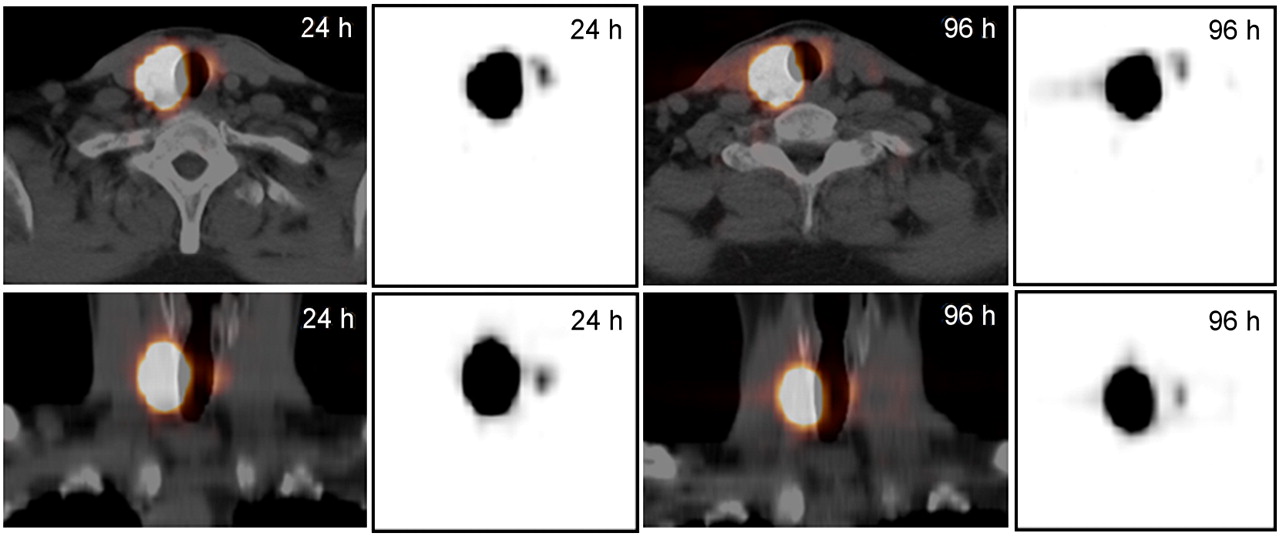
- FIGURE 4.
Shine-through in fifth patient (patient E) at 24 and 96 h after oral intake of 124I. Top row shows transaxial PET/CT images; bottom row, coronal images. Gray-scale PET-only images are shown for comparison with those of phantom in Figure 1.


Tables
Patient code Lesion uptake at 24 h (%) Shine-through at 24 h (%) Shine-through at 96 h (%) A 0.10 14.0 9.2 B 2.53 1.9 1.9 C 0.19 6.7 7.4 D 3.27 0.8 0.9 E 0.30 0.7 0.7


