


Article Figures & Data
Figures
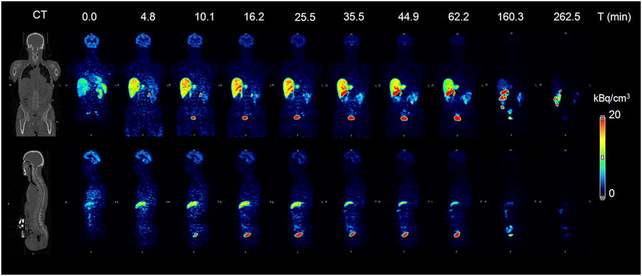
- FIGURE 1.
Whole-body time–activity distribution of 18F-GE067 in subject 1 (Table 1), with representative coronal and sagittal slices as indicated on CT view on left. PET image color intensities are expressed as activation concentration (kBq/cm3). Upper row indicates start (min) of whole-body scan.
- FIGURE 2.
Brain uptake distribution of 18F-GE067 in healthy 64-y-old male subject (top), compared with 68-y-old male AD patient (bottom). Transverse, sagittal, and coronal sections indicate absence of specific gray matter uptake of 18F-GE067 and aspecific uptake in white matter, pons, and thalamus. Images represent standardized uptake value ratios (SUVR) to cerebellar cortex, between 85 and 105 min after injection, obtained from phase I clinical study.
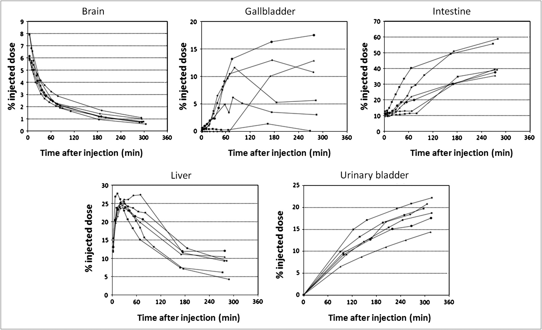
- FIGURE 3.
Mean activity in brain, gallbladder, intestine, liver, and urinary bladder as fraction of total-body activity, at all time points for 6 subjects.


Tables
- TABLE 1
Subject Data, Net Injected Activity of 18F-GE067, and Individual Effective Dose Estimates
Subject no. Sex Age (y) Height (m) Mass (kg) Body mass index (kg/m2) Injected dose (MBq) Individual effective dose (μSv/MBq) 1 M 74 165 63.0 23.1 95.8 30.7 2 M 51 172 69.5 23.5 97.2 33.3 3 F 71 153 58.0 24.8 139.1 37.5 4 M 73 160 71.0 27.8 144.0 29.4 5 M 56 188 81.0 22.9 146.5 37.4 6 M 63 174 80.0 26.4 105.1 34.5 Mean ± SD 64.7 ± 8.8 168.7 ± 11.2 70.4 ± 8.3 24.8 ± 1.8 121.3 ± 22.2 33.8 ± 3.36 - TABLE 2
Radiation Absorbed Dose Estimates (OLINDA) Based on ICRP 30 (13) Gastrointestinal Tract Model
Organ Dose estimate (mGy/MBq) Coefficient of variation (%) Adrenals 1.46E-02 ± 2.35E−03 16.1 Brain 1.19E-02 ± 1.18E−03 9.9 Breasts 5.59E-03 ± 1.05E−03 18.8 Gallbladder wall 2.87E-01 ± 1.64E−01 57.1 LLI wall 8.15E-02 ± 1.51E−02 18.5 Small intestine 1.55E-01 ± 6.19E−02 39.9 Stomach wall 1.52E-02 ± 2.84E−03 18.7 ULI wall 1.73E-01 ± 7.22E−02 41.7 Heart wall 1.21E-02 ± 2.44E−03 20.2 Kidneys 4.01E-02 ± 5.77E−03 14.4 Liver 6.39E-02 ± 1.12E−02 17.5 Lungs 1.67E-02 ± 1.63E−03 9.8 Muscle 9.7E-03 ± 1.48E−03 15.3 Ovaries 3.31E-02 ± 8.59E−03 26.0 Pancreas 1.72E-02 ± 2.91E−03 16.9 Red marrow 1.53E-02 ± 1.63E−03 10.7 Osteogenic cells 1.27E-02 ± 2.15E−03 16.9 Skin 5.54E-03 ± 9.01E−04 16.3 Spleen 1.59E-02 ± 3.06E−03 19.2 Testes 5.30E-03 ± 2.61E−03 49.2 Thymus 6.25E-03 ± 1.28E−03 20.5 Thyroid 6.56E-03 ± 1.96E−03 29.9 Urinary bladder 6.16E-02 ± 6.61E−03 10.7 Uterus 2.72E-02 ± 6.75E−03 24.8 Total body 1.36E-02 ± 2.03E−03 14.9 Effective dose (mSv/MBq) 0.0338 ± 0.0034 10.1 Data are mean ± SD.
Supplemental Data
Files in this Data Supplement:
In this issue


Jump to section
Related Articles
Cited By...
- Association of Enlarged Perivascular Spaces and Measures of Small Vessel and Alzheimer Disease
- An Overview of PET Radiochemistry, Part 1: The Covalent Labels 18F, 11C, and 13N
- Whole-Body Biodistribution and Dosimetry of the Dopamine Transporter Radioligand 18F-FE-PE2I in Human Subjects
- Increased midlife triglycerides predict brain {beta}-amyloid and tau pathology 20 years later
- SNMMI Procedure Standard/EANM Practice Guideline for Amyloid PET Imaging of the Brain 1.0
- Myo-inositol changes precede amyloid pathology and relate to APOE genotype in Alzheimer disease
- Performance Characteristics of Amyloid PET with Florbetapir F 18 in Patients with Alzheimer's Disease and Cognitively Normal Subjects
- 18F-ML-10, a PET Tracer for Apoptosis: First Human Study
- In Vivo Imaging of Amyloid Deposition in Alzheimer Disease Using the Radioligand 18F-AV-45 (Flobetapir F 18)
- Phase 1 Study of the Pittsburgh Compound B Derivative 18F-Flutemetamol in Healthy Volunteers and Patients with Probable Alzheimer Disease