


Article Figures & Data
Figures
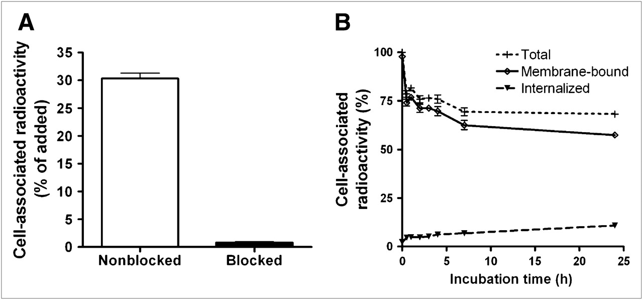
- FIGURE 1.
(A) In vitro specificity analysis. One group of culture dishes with SKOV-3 cells was pretreated with saturating amounts of nonlabeled His6-ZHER2:342 before incubation with 99mTc-ZHER2:2395-C. Cell-associated radioactivity was calculated as percentage of total added radioactivity. (B) Cell-associated radioactivity as function of time after interrupted incubation of SKOV-3 cells with 99mTc-ZHER2:2395-C. Cell-associated radioactivity at time zero after interrupted incubation was considered to be 100%. Radioactivity that was removed from cells by treatment with 4 M urea solution in 0.2 M glycine buffer (pH 2.0) was considered to be membrane bound, and remainder was considered to be internalized. Data are presented as mean ± SD (n = 3). Error bars may not be visible because they are smaller than the symbols.
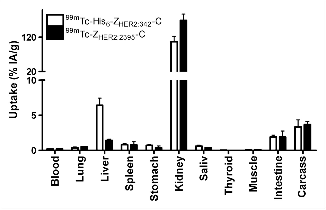
- FIGURE 2.
Biodistributions of radiolabeled Affibody molecules 99mTc-H6-ZHER2:342-C, containing His tag, and 99mTc-ZHER2:2395-C, lacking His tag, in NMRI (normal) mice 4 h after injection. Data are expressed as %IA per whole sample for gastrointestinal tract and contents, thyroid, and carcass and as %IA/g for other tissues and are presented as mean ± SD for 4 animals. Saliv = salivary gland.
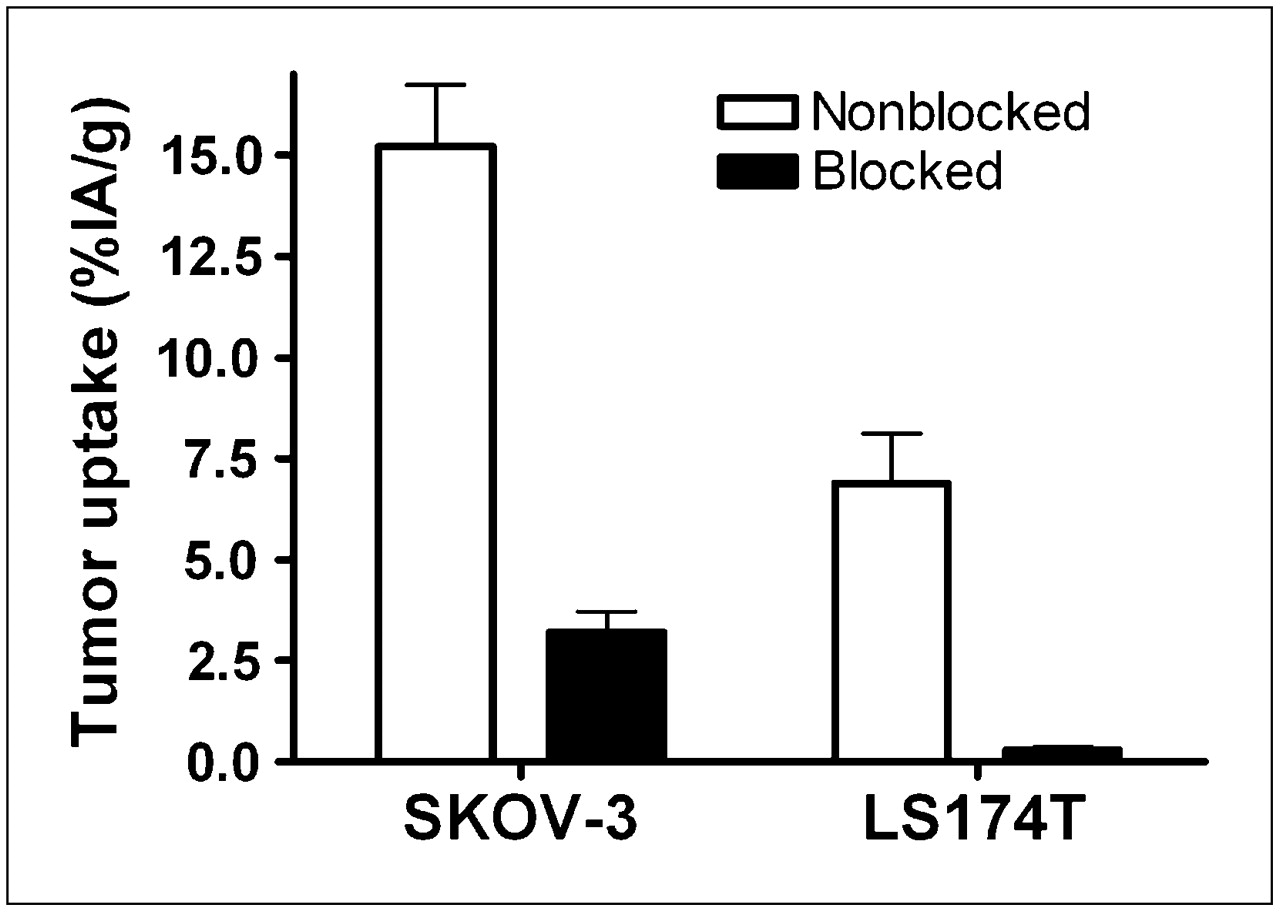
- FIGURE 3.
Specificity of 99mTc-ZHER2:2395-C tumor uptake in SKOV-3 and LS174T xenografts 4 h after injection. To saturate HER2 in tumors, one group of animals for each tumor model was preinjected with 600 μg of nonlabeled His6-ZHER2:342 45 min before injection of radiolabeled conjugate (designated as blocked). All animals were injected with 1 μg of labeled Affibody molecule. Data are expressed as %IA/g and are presented as mean ± SD for 4 animals.
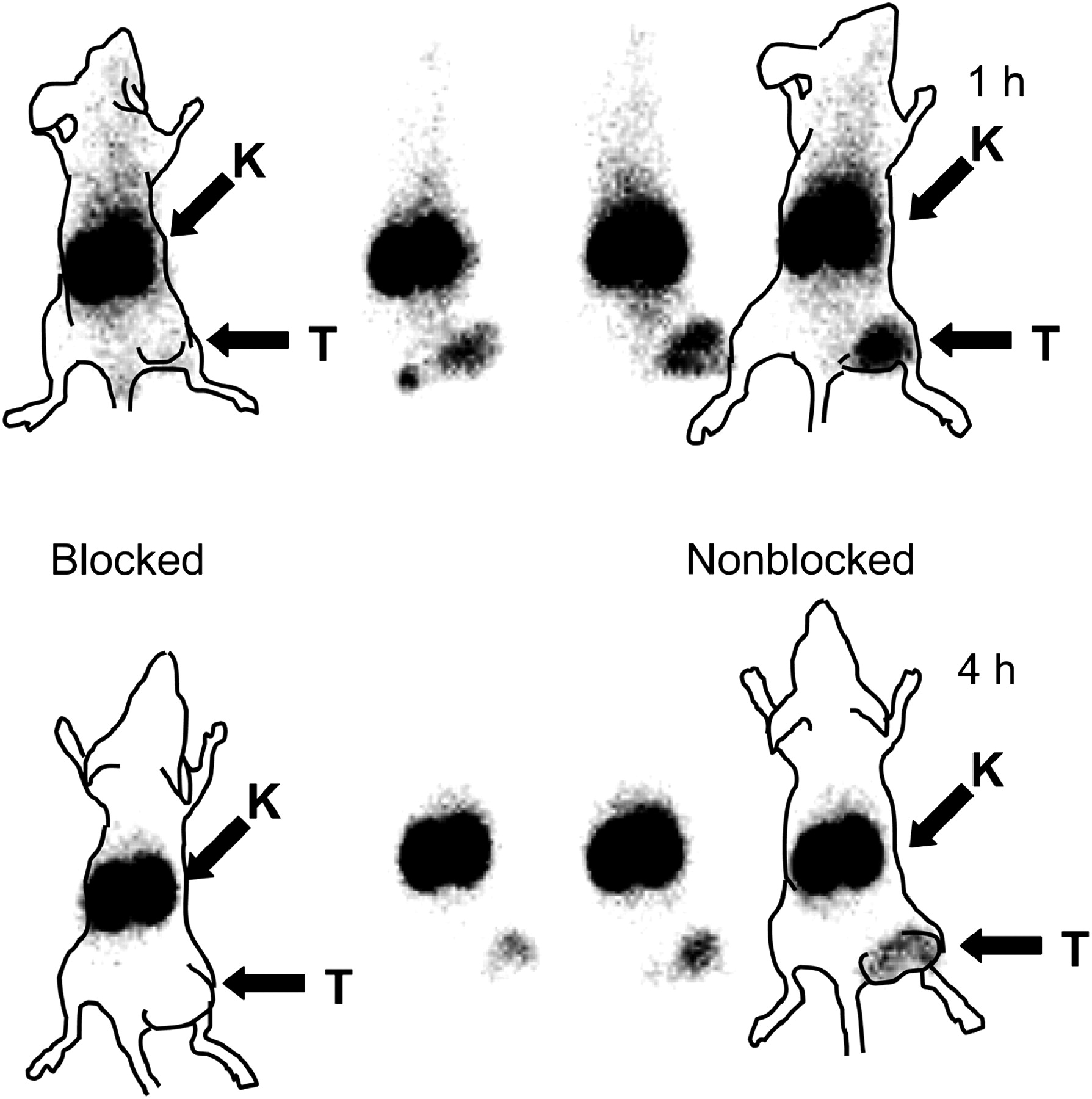
- FIGURE 4.
Imaging of HER2 expression in LS174T xenografts in BALB/c nu/nu mice with 99mTc-ZHER2:2395-C. In blocking experiment, HER2 was saturated by preinjection of 600 μg of nonlabeled Affibody molecule. Planar γ-camera images were acquired 1 and 4 h after administration of 99mTc-ZHER2:2395-C. Tumors (right hind leg) were clearly visualized without blocking but were not seen in blocking experiment, indicating specific HER2 binding to 99mTc-ZHER2:2395-C. Animal contours were derived from digital photographs and superimposed over γ-camera images to facilitate interpretation. K = kidney; T = tumor.
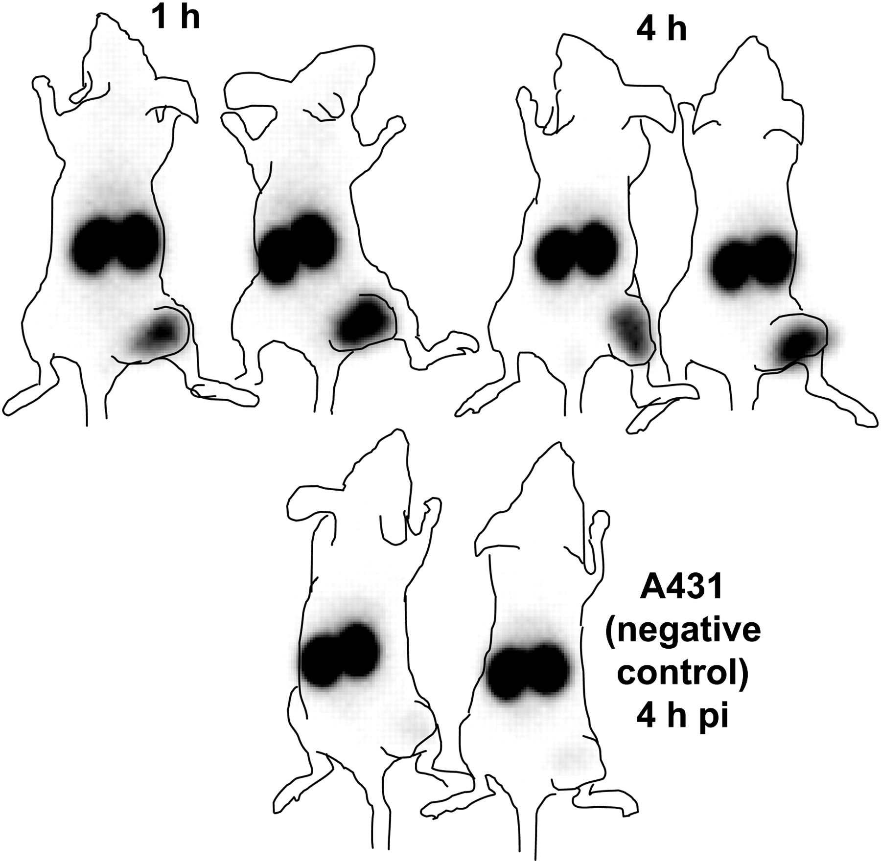
- FIGURE 5.
Imaging of HER2 expression in SKOV-3 and A431 xenografts in BALB/c nu/nu mice with 99mTc-ZHER2:2395-C. A431 tumors were used as negative controls because they are considered to be non–HER2-expressing tumors. Planar γ-camera images were acquired 1 and 4 h after administration of 99mTc-ZHER2:2395-C in mice with SKOV-3 xenografts and 4 h after administration in mice with A431 xenografts. Tumors (right hind leg) were clearly visualized in SKOV-3 xenografts but were not seen in negative controls, indicating specific HER2 binding to 99mTc-ZHER2:2395-C. Animal contours were derived from digital photographs and superimposed over γ-camera images to facilitate interpretation. pi = after injection.


Tables
Uptake (%IA/g) at: Tissue 1 h 2 h 4 h Blood 7 ± 2.0 2.7 ± 0.5 1.3 ± 0.4 Heart 3.0 ± 0.5 1.5 ± 0.4 0.7 ± 0.3 Lung 7 ± 1.9 1.8 ± 0.6 1.5 ± 0.3 Liver 19 ± 1.9 17 ± 3.6 19 ± 5 Spleen 2.0 ± 1.0 2.1 ± 0.2 2.0 ± 0.2 Pancreas 0.7 ± 0.2 0.5 ± 0.1 0.40 ± 0.04 Kidney 63 ± 12 89 ± 10 73 ± 9 Stomach 1.4 ± 0.6 1.1 ± 0.1 0.6 ± 0.1 Tumor 6 ± 1.9 8.3 ± 1.6 8.9 ± 3.1 Skin 2.8 ± 0.5 1.3 ± 0.4 1.8 ± 0.5 Muscle 0.4 ± 0.1 0.20 ± 0.03 0.14 ± 0.03 Bone 1.1 ± 0.2 1.3 ± 0.3 0.7 ± 0.2 Data are presented as mean ± SD for 3 animals.
- TABLE 2
Uptake of 99mTc-ZHER2:2395-C in BALB/c nu/nu Mice Bearing Xenografts of LS174T or SKOV-3 Tumors
Uptake* in: LS174T at: SKOV-3 at: Tissue 0.5 h 1 h 4 h 6 h 4 h Blood 2.4 ± 0.2 1.0 ± 0.2 0.08 ± 0.02 0.05 ± 0.02 0.13 ± 0.04 Lung 3.1 ± 0.5 2.0 ± 0.5 0.39 ± 0.07 0.5 ± 0.3 0.45 ± 0.07 Liver 1.7 ± 0.3 2.0 ± 0.4 1.5 ± 0.2 1.7 ± 0.3 1.6 ± 0.1 Spleen 1.3 ± 0.2 1.5 ± 1.3 0.40 ± 0.06 0.38 ± 0.06 0.5 ± 0.2 Stomach 1.9 ± 0.4 1.3 ± 0.4 0.37 ± 0.05 0.36 ± 0.03 0.44 ± 0.08 Kidney 147 ± 20 184 ± 18 191 ± 15 157 ± 18 143 ± 20 Salivary gland 1.1 ± 0.2 0.7 ± 0.2 0.28 ± 0.05 0.28 ± 0.03 0.35 ± 0.07 Thyroid 0.040 ± 0.002 0.026 ± 0.003 0.006 ± 0.001 0.007 ± 0.004 0.005 ± 0.001 Tumor 7.2 ± 2.3 8.7 ± 1.7 6.9 ± 2.5† 6.6 ± 0.8 15 ± 3† Muscle 0.55 ± 0.08 0.36 ± 0.06 0.2 ± 0.1 0.07 ± 0.01 0.09 ± 0.01 Intestine 2.5 ± 0.4 1.8 ± 0.5 1.5 ± 0.3 2.2 ± 0.5 2.1 ± 0.3 Carcass 153 ± 2.5 9.4 ± 1.1 3.7 ± 0.5 2.8 ± 0.2 3.9 ± 0.4 ↵* Uptake is reported as %IA/g for all tissues except gastrointestinal tract and its contents, thyroid, and carcass, for which uptake is reported as %IA per whole sample.
↵† Significant difference (P < 0.05) between LS174T xenografts (moderate level of HER2 expression) and SKOV-3 xenografts (high level of HER2 expression).
Data are presented as mean ± SD for 4 animals.
- TABLE 3
Tumor-to-Organ Ratios for 99mTc-ZHER2:2395-C in BALB/c nu/nu Mice Bearing Xenografts of LS174T or SKOV-3 Tumors
Tumor-to-organ ratio LS174T at: SKOV-3 at: Organ 0.5 h 1 h 4 h 6 h 4 h Blood 3.1 ± 1.2 9.1 ± 2.2 88 ± 24 129 ± 35 121 ± 24 Lung 2.3 ± 0.6 4.3 ± 0.5 17 ± 6 15 ± 7 34 ± 5 Liver 4.3 ± 1.2 4.4 ± 0.6 4.5 ± 1.4 4.0 ± 0.4 9.3 ± 1.5 Spleen 5.8 ± 1.9 7.8 ± 3.9 17 ± 4 17 ± 1 33 ± 7 Stomach 3.9 ± 1.3 6.7 ± 1.6 19 ± 6 19 ± 3 35 ± 7 Kidney 0.05 ± 0.01 0.05 ± 0.01 0.04 ± 0.01 0.042 ± 0.002 0.11 ± 0.02 Salivary gland 6.4 ± 1.9 12.0 ± 2.6 26 ± 16 24 ± 4 44 ± 9 Muscle 13 ± 5 24 ± 6 60 ± 38 91 ± 22 172 ± 24 Data are presented as mean ± SD for 4 animals.
Supplemental Data
Files in this Data Supplement:
In this issue


Jump to section
Related Articles
Cited By...
- Preclinical and clinical applications of specific molecular imaging for HER2-positive breast cancer
- 188Re-ZHER2:V2, a Promising Affibody-Based Targeting Agent Against HER2-Expressing Tumors: Preclinical Assessment
- HER2-Positive Tumors Imaged Within 1 Hour Using a Site-Specifically 11C-Labeled Sel-Tagged Affibody Molecule
- Molecular Design and Optimization of 99mTc-Labeled Recombinant Affibody Molecules Improves Their Biodistribution and Imaging Properties
- Targeting of HER2-Expressing Tumors Using 111In-ABY-025, a Second-Generation Affibody Molecule with a Fundamentally Reengineered Scaffold
- Targeting Prostate Cancer Cells In Vivo Using a Rapidly Internalizing Novel Human Single-Chain Antibody Fragment