


Article Figures & Data
Figures
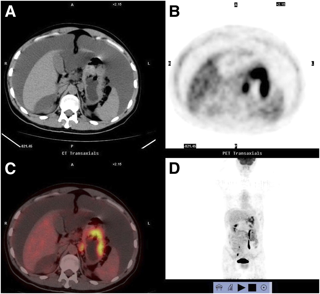
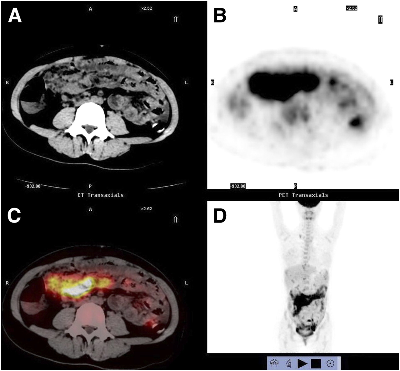
- FIGURE 1.
Images of 25-y-old man who presented with ascites for 1 mo: axial CT (A), axial PET (B), axial fused PET/CT (C), and 3-dimensional PET (D). Cytology of ascitic fluid was positive for malignancy, but noninvasive examinations could not detect primary cause of malignant ascites. PET/CT images showed high uptake in gastric area (SUVmax of 8.0, maximal diameter of 6.6 cm). Abdominal cavity metastasis showed smudging sign. After PET/CT examination, gastroscopy was repeated and biopsy confirmed malignant gastric lesion.
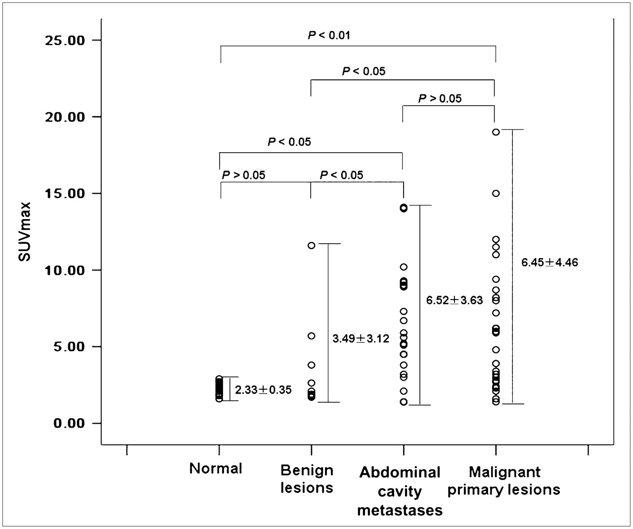
- FIGURE 2.
Comparison of SUVmax in peritoneum of healthy volunteers (normal), benign ascites lesions, abdominal cavity metastases, and malignant primary lesions.
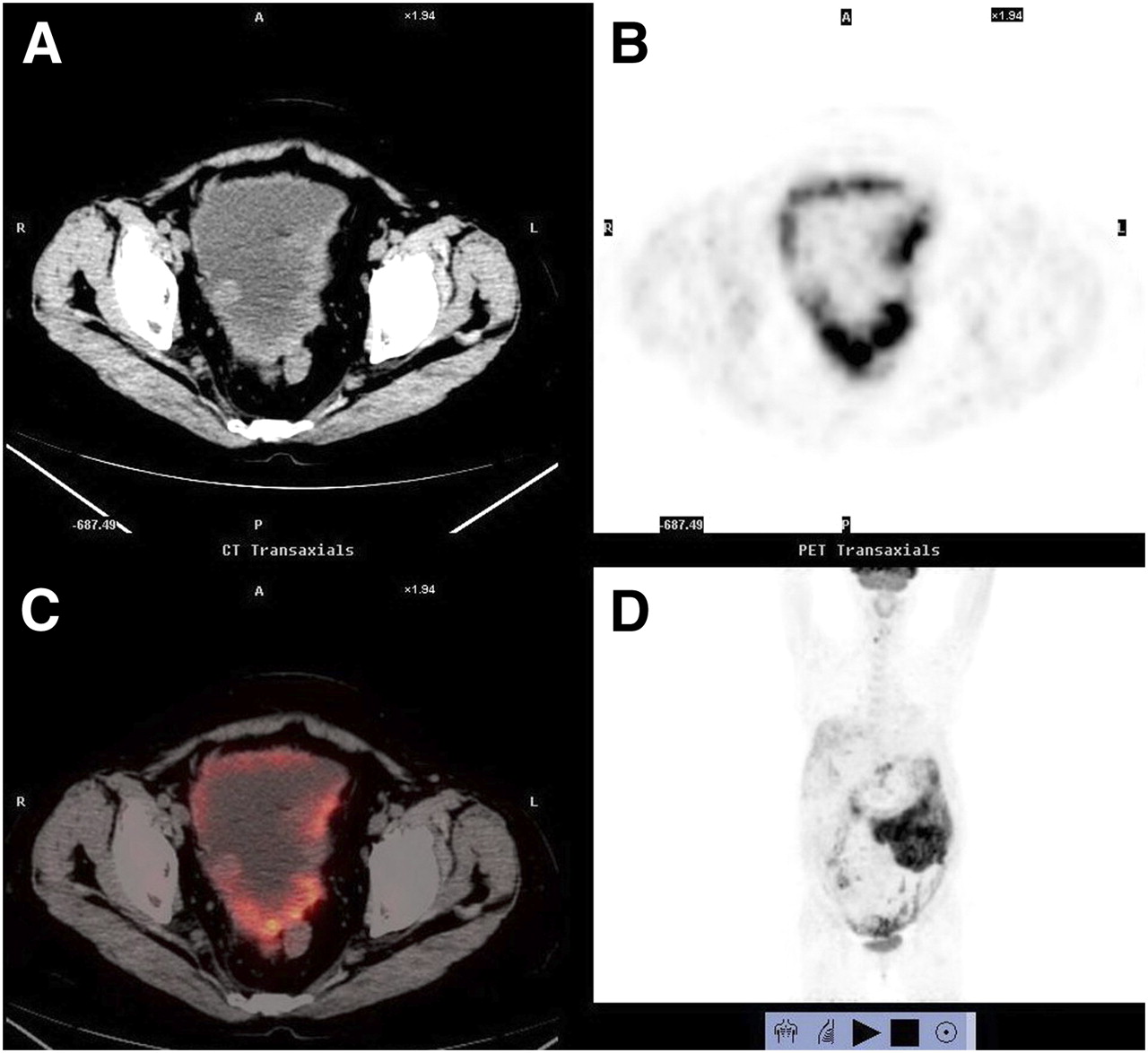
- FIGURE 3.
PET/CT images of 65-y-old woman with ascites for 1 mo: axial CT (A), axial PET (B), axial fused PET/CT (C), and 3-dimensional PET (D). Patient had serum CA12-5 of 210 and carcinoembryonic antigen of 10.5. Cytology of ascitic fluid was positive for malignancy, but noninvasive examinations could not detect primary cause. PET/CT failed to find primary cancer but showed diffuse nodular-shadow signs and omental-caking sign. High uptake (SUVmax of 14.1, maximal diameter of 13.1 cm) in abdominal cavity helped to confirm ascites to be malignant. Laparotomy and pathology confirmed that primary cancer and metastases in abdominal cavity were poorly differentiated right ovarian adenocarcinoma.
- FIGURE 4.
PET/CT images of false-positive case, 32-y-old woman who presented with ascites for 1 mo: axial CT (A), axial PET (B), axial fused PET/CT (C), and 3-dimensional PET (D). Patient had serum CA12-5 of 1,579, normal carcinoembryonic antigen, normal CA19-9, and history of fever and night sweats 3 wk previously. Cytology of ascitic fluid and purified protein derivatives test were negative for malignancy. PET/CT showed diffuse high uptake in abdominal cavity (SUVmax of 11.6, maximal diameter of 17.4 cm) and omental-caking sign, mimicking malignant lesions. Laparoscopy at another hospital confirmed that patient had peritoneal tuberculosis, and she recovered after antituberculosis treatment.
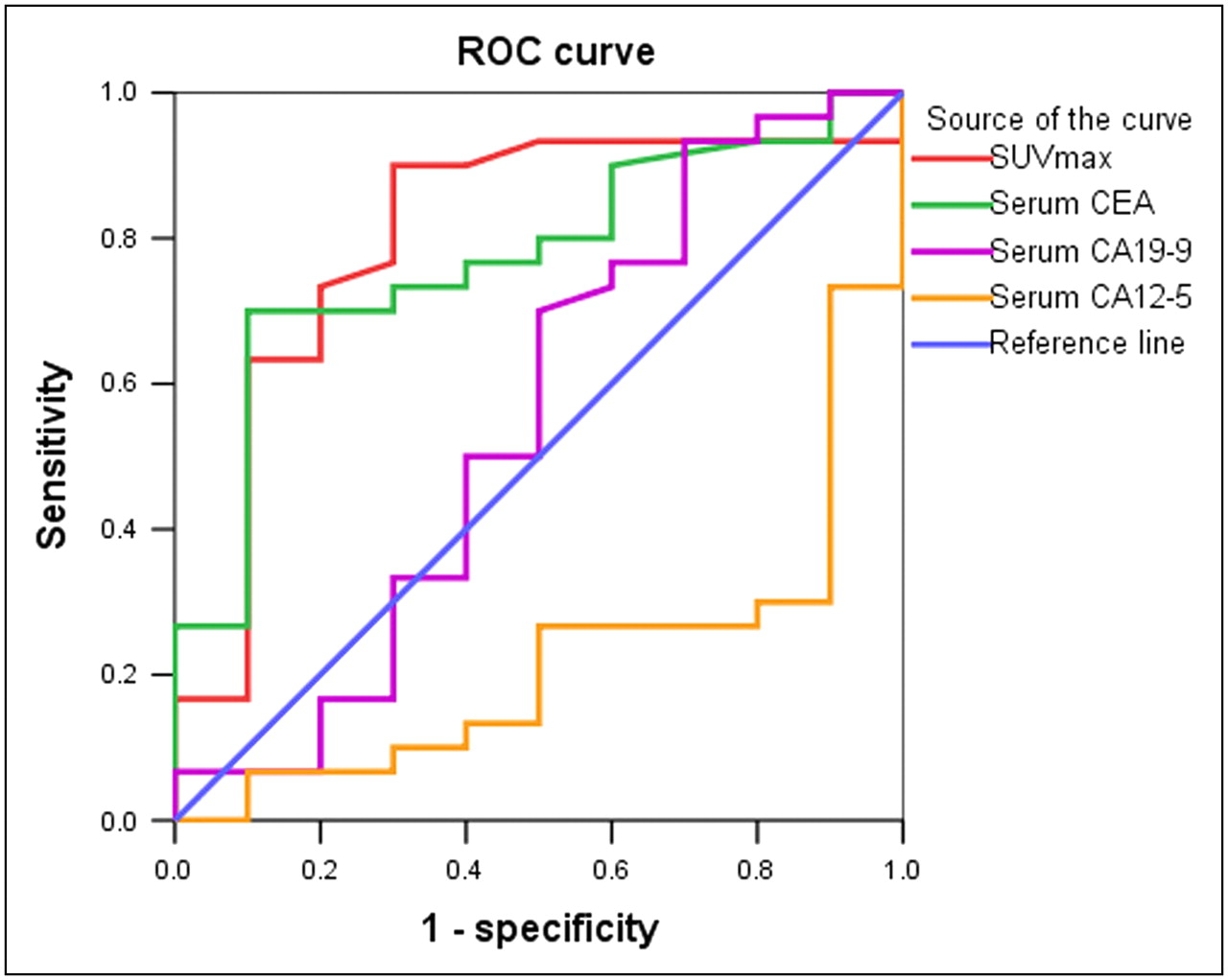
- FIGURE 5.
ROC curve of SUVmax, serum carcinoembryonic antigen (CEA), CA19-9, and CA12-5 in differential diagnosis of ascites.


Tables
Modality TP FN TN FP Sensitivity (%) Specificity (%) PPV (%) NPV (%) Accuracy (%) CT 11 19 8 2 36.7 80 84.6 29.6 47.5 PET/CT 19 11 7 3 63.3 70 86.4 38.9 65 P 0.039* 0.606 0.886 0.519 0.115 ↵* Statistically significant at 0.05 level.
TP = true-positive; FN = false-negative; TN = true-negative; FP = false-positive; NPV = negative predictive value; PPV = positive predictive value.
Lesions of all sizes in 40 patients were included. Statistical analysis used χ2 test.
Modality TP FN TN FP Sensitivity (%) Specificity (%) PPV (%) NPV (%) Accuracy (%) CT 6 16 16 2 27.3 88.7 75 50 55 PET/CT 19 3 15 3 86.4 83.3 86.4 83.3 85 P 0.000* 0.63 0.46 0.02* 0.003* ↵* Statistically significant at 0.05 level.
TP = true-positive; FN = false-negative; TN = true-negative; FP = false-positive; NPV = negative predictive value; PPV = positive predictive value.
Lesions of all sizes in 40 patients were included. Statistical analysis used χ2 test.


