
Main menu

  • Home
  • Content
    • Current
    • Ahead of print
    • Past Issues
    • JNM Supplement
    • SNMMI Annual Meeting Abstracts
    • Continuing Education
    • JNM Podcasts
  • Subscriptions
    • Subscribers
    • Institutional and Non-member
    • Rates
    • Journal Claims
    • Corporate & Special Sales
  • Authors
    • Submit to JNM
    • Information for Authors
    • Assignment of Copyright
    • AQARA requirements
  • Info
    • Reviewers
    • Permissions
    • Advertisers
  • About
    • About Us
    • Editorial Board
    • Contact Information
  • More
    • Alerts
    • Feedback
    • Help
    • SNMMI Journals
  • SNMMI
    • JNM
    • JNMT
    • SNMMI Journals
    • SNMMI

User menu

  • Subscribe
  • My alerts
  • Log in
  • My Cart

Search

  • Advanced search
Journal of Nuclear Medicine
  • SNMMI
    • JNM
    • JNMT
    • SNMMI Journals
    • SNMMI
  • Subscribe
  • My alerts
  • Log in
  • My Cart
Journal of Nuclear Medicine

Advanced Search

  • Home
  • Content
    • Current
    • Ahead of print
    • Past Issues
    • JNM Supplement
    • SNMMI Annual Meeting Abstracts
    • Continuing Education
    • JNM Podcasts
  • Subscriptions
    • Subscribers
    • Institutional and Non-member
    • Rates
    • Journal Claims
    • Corporate & Special Sales
  • Authors
    • Submit to JNM
    • Information for Authors
    • Assignment of Copyright
    • AQARA requirements
  • Info
    • Reviewers
    • Permissions
    • Advertisers
  • About
    • About Us
    • Editorial Board
    • Contact Information
  • More
    • Alerts
    • Feedback
    • Help
    • SNMMI Journals
  • View or Listen to JNM Podcast
  • Visit JNM on Facebook
  • Join JNM on LinkedIn
  • Follow JNM on Twitter
  • Subscribe to our RSS feeds
Research ArticleCLINICAL INVESTIGATIONS

Quantification of Tryptophan Transport and Metabolism in Lung Tumors Using PET

Csaba Juhász, Otto Muzik, Xin Lu, M. Salik Jahania, Ayman O. Soubani, Majid Khalaf, Fangyu Peng, Thomas J. Mangner, Pulak K. Chakraborty and Diane C. Chugani
Journal of Nuclear Medicine March 2009, 50 (3) 356-363; DOI: https://doi.org/10.2967/jnumed.108.058776
Csaba Juhász
  • Find this author on Google Scholar
  • Find this author on PubMed
  • Search for this author on this site
Otto Muzik
  • Find this author on Google Scholar
  • Find this author on PubMed
  • Search for this author on this site
Xin Lu
  • Find this author on Google Scholar
  • Find this author on PubMed
  • Search for this author on this site
M. Salik Jahania
  • Find this author on Google Scholar
  • Find this author on PubMed
  • Search for this author on this site
Ayman O. Soubani
  • Find this author on Google Scholar
  • Find this author on PubMed
  • Search for this author on this site
Majid Khalaf
  • Find this author on Google Scholar
  • Find this author on PubMed
  • Search for this author on this site
Fangyu Peng
  • Find this author on Google Scholar
  • Find this author on PubMed
  • Search for this author on this site
Thomas J. Mangner
  • Find this author on Google Scholar
  • Find this author on PubMed
  • Search for this author on this site
Pulak K. Chakraborty
  • Find this author on Google Scholar
  • Find this author on PubMed
  • Search for this author on this site
Diane C. Chugani
  • Find this author on Google Scholar
  • Find this author on PubMed
  • Search for this author on this site
  • Article
  • Figures & Data
  • Info & Metrics
  • PDF
Loading

Article Figures & Data

Figures

  • Tables
  • FIGURE 1. 
    • Download figure
    • Open in new tab
    • Download powerpoint
    FIGURE 1. 

    Three-compartmental model for AMT kinetics in lung tumor, using first-order rate constants (17). Transport rate constant K1 and outflow rate constant k2 describe exchange of AMT between vascular space (CP) and cell cytoplasm, in which it comprises free compartment (Cf). Irreversible enzymatic conversion of AMT to its metabolites (in metabolic pool, Cm) is characterized by metabolic rate constant k3. Efflux of AMT metabolites from metabolic compartment can be represented by rate constant k4. Although, theoretically, AMT metabolites could move from Cm to both Cf and CP, applied kinetic modeling included efflux of tracer metabolites from entire tissue compartment. Dotted arrow represents k4, which was poorly identifiable in full model and eventually set to zero; thus, final analysis included only 3 kinetic rate constants (K1, k2, and k3).

  • FIGURE 2. 
    • Download figure
    • Open in new tab
    • Download powerpoint
    FIGURE 2. 

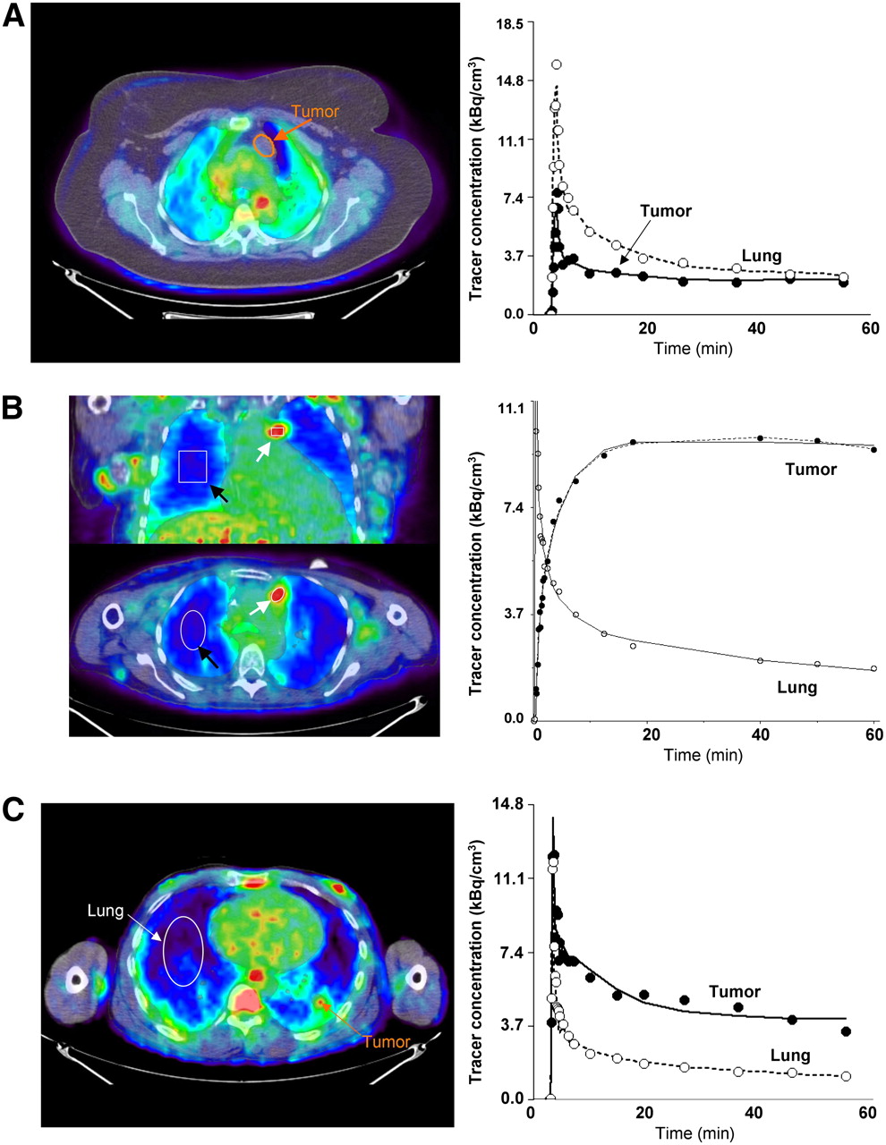
    (A) Images of AMT tracer uptake between 40 and 60 min after injection (left) in patient with benign cyst (patient 1, Table 2). There is low accumulation of tracer in cyst, with activity being lower than normal lung tissue at all time points. (B) Images of AMT tracer uptake between 40 and 60 min after injection (left) in patient with NSCLC with multiple local metastases (patient 6, Table 2). Images show high accumulation in tumor tissue (2-NSCLC, Table 2) and excellent contrast between tumor and lung tissue. ROIs were defined for tumor nodules (white arrows) and lung tissue (black arrows). Corresponding tumor time–activity curve (right) indicates rapid initial uptake of AMT, followed by slight decrease at late time points. Curve fit applying reduced compartmental model (k4 = 0) is shown as solid line, and full compartmental model is displayed as broken line. In addition, 2-compartmental model fit is shown for lung tissue. (C) Representative image of AMT tracer uptake between 40 and 60 min after injection (left) in patient (patient 10, 2-NSCLC, Table 2). Two tumor sites were identified in this patient on basis of CT and 18F-FDG, which both were believed to represent NSCLC. First tumor (1-NSCLC, Table 2) showed time–activity curves similar to Figure 2B and high 18F-FDG SUV. In contrast, second site (orange arrow) showed relatively low uptake. Inspection of corresponding time–activity curve showed high initial uptake, with subsequent plateau followed by tracer washout. Neither full nor reduced (shown) 3-compartmental model fit data well; however, parameter identifiability was much improved using reduced model (CN 5.3 vs. 21.8).

  • FIGURE 3. 
    • Download figure
    • Open in new tab
    • Download powerpoint
    FIGURE 3. 

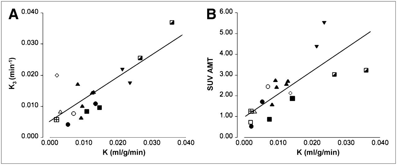
    Correlation between unidirectional uptake rate (K) derived from reduced compartmental model (k4 = 0) and either k3 parameter (A) or SUV for AMT (B). Tumors obtained from different patients are marked by different symbols. k3 parameter was highly correlated with K, indicating that metabolic conversion is dominant factor in AMT tracer uptake in tumors. Moreover, K was also significantly correlated with SUV for AMT, suggesting that this semiquantitative value can provide estimate of magnitude of metabolic conversion.

  • FIGURE 4. 
    • Download figure
    • Open in new tab
    • Download powerpoint
    FIGURE 4. 

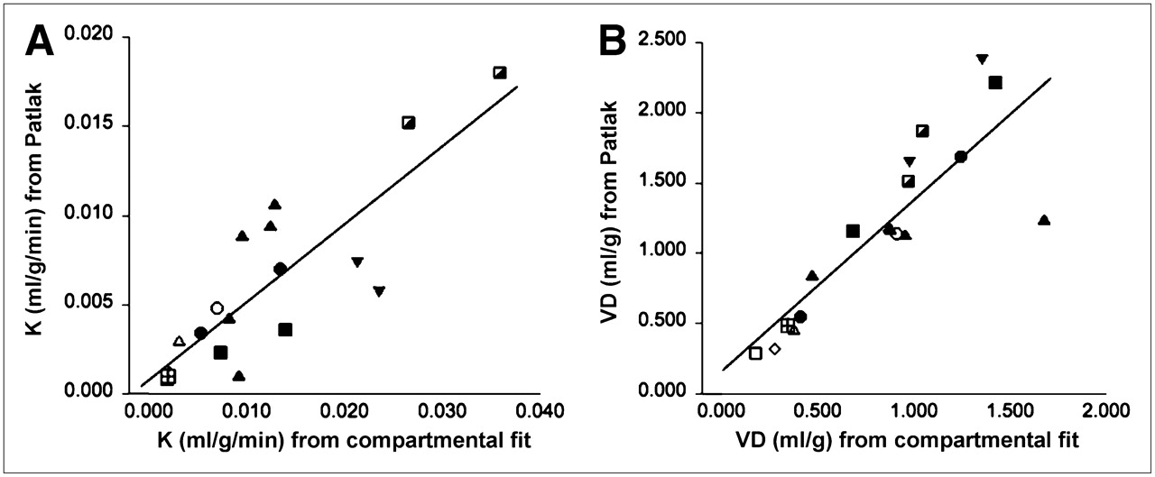
    (A) Correlation of K values derived from reduced compartmental fit and Patlak graphical analysis. r was determined as 0.83, indicating an excellent correlation between these 2 measures. Tumors obtained from different patients are marked by different symbols. (B) Excellent correlation was also determined between VD determined using compartmental fit and Patlak graphical analysis.

Tables

  • Figures
    • View popup
    TABLE 1

    Patient Data and SUVs Measured in Each Lesion

    Time between 18F-FDG and AMT PETSUV
    Patient no.SexAge (y)DiagnosisPrevious therapy (before AMT)Lesion no.18F-FDGAMT
    1F54Thymic cystNone18F-FDG PET not done1—0.5
    2M39HamartomaNone54 d (18F-FDG later)10.90.7
    3M66Metastasis (rectal cancer)None11 d12.81.3
    4M58NSCLC (scc)Chemotherapy (2 wk)*1 mo115.72.5
    5F58NSCLC (pd)Radiotherapy + chemotherapy (>6 mo)†1 mo18.05.5
    26.84.4
    6F56NSCLC (lcc)Radiotherapy + chemotherapy (>6 mo)†7 d15.52.4
    22.82.7
    332.6
    42.71.6
    52.72.7
    7M79NSCLC (acc)None6 d12.81.2
    8M49NSCLC (scc)None11 d17.32.1
    23.31.7
    9F47NSCLC (acc)None18F-FDG PET not done1—3.2
    2—3.0
    10M46NSCLC (pd)None28 d14.71.9
    21.60.8
    • ↵* Therapy started after 18F-FDG PET but 2 wk before AMT PET scan.

    • ↵† Therapy was finished more than 6 mo before AMT PET scan.

    • scc = squamous cell carcinoma; pd = poorly differentiated carcinoma; lcc = large cell carcinoma; acc = adenocarcinoma.

    • Tumors are consecutively numbered in patients with multiple lesions.

    • View popup
    TABLE 2

    Individual Parameter Estimates Derived from Reduced Compartmental Model

    Patient no.LesionK1 (mL/g/min)k2 (min−1)k3 (min−1)K (mL/g/min)Patlak slope
    1Thymic cyst0.0120.0920.0190.00210.0017
    2Hamartoma0.0980.2920.0060.00190.0016
    3Metastasis0.0980.2790.0060.00200.0007
    4NSCLC*0.1700.1790.0080.00700.0051
    51-NSCLC0.2870.1950.0170.02360.0108
    2-NSCLC0.1870.1700.0220.02130.0074
    61-NSCLC0.0730.0670.010.00960.0088
    2-NSCLC0.1280.1310.0150.01290.0107
    3-NSCLC0.1040.1050.0140.01250.0095
    4-NSCLC0.0640.1170.0170.00820.0043
    5-NSCLC0.0630.0360.0060.00920.0035
    7NSCLC†0.0820.2100.0080.00310.0030
    81-NSCLC0.1760.1310.0110.01340.0074
    2-NSCLC*0.2750.2110.0040.00530.0058
    91-NSCLC0.2420.2130.0370.03590.0180
    2-NSCLC0.1840.1510.0260.02660.0152
    101-NSCLC0.1140.0700.0110.01450.0078
    2-NSCLC*0.1510.2010.010.00750.0034
    Total MeanNSCLC0.1530.1460.0140.01400.0081
    Total SD0.0730.0580.0080.00910.0043
    Mean for unaffected lungs0.0360.208———
    SD for unaffected lungs0.0140.047———
    • ↵* High initial uptake, subsequent tracer washout.

    • ↵† Small tumor size (volume, ∼0.8 cm3), possible image distortions because of partial-volume effect.

PreviousNext
Back to top

In this issue

Journal of Nuclear Medicine: 50 (3)
Journal of Nuclear Medicine
Vol. 50, Issue 3
March 2009
  • Table of Contents
  • Table of Contents (PDF)
  • About the Cover
  • Index by author
Print
Download PDF
Article Alerts
Sign In to Email Alerts with your Email Address
Email Article

Thank you for your interest in spreading the word on Journal of Nuclear Medicine.

NOTE: We only request your email address so that the person you are recommending the page to knows that you wanted them to see it, and that it is not junk mail. We do not capture any email address.

Enter multiple addresses on separate lines or separate them with commas.
Quantification of Tryptophan Transport and Metabolism in Lung Tumors Using PET
(Your Name) has sent you a message from Journal of Nuclear Medicine
(Your Name) thought you would like to see the Journal of Nuclear Medicine web site.
Citation Tools
Quantification of Tryptophan Transport and Metabolism in Lung Tumors Using PET
Csaba Juhász, Otto Muzik, Xin Lu, M. Salik Jahania, Ayman O. Soubani, Majid Khalaf, Fangyu Peng, Thomas J. Mangner, Pulak K. Chakraborty, Diane C. Chugani
Journal of Nuclear Medicine Mar 2009, 50 (3) 356-363; DOI: 10.2967/jnumed.108.058776

Citation Manager Formats

  • BibTeX
  • Bookends
  • EasyBib
  • EndNote (tagged)
  • EndNote 8 (xml)
  • Medlars
  • Mendeley
  • Papers
  • RefWorks Tagged
  • Ref Manager
  • RIS
  • Zotero
Share
Quantification of Tryptophan Transport and Metabolism in Lung Tumors Using PET
Csaba Juhász, Otto Muzik, Xin Lu, M. Salik Jahania, Ayman O. Soubani, Majid Khalaf, Fangyu Peng, Thomas J. Mangner, Pulak K. Chakraborty, Diane C. Chugani
Journal of Nuclear Medicine Mar 2009, 50 (3) 356-363; DOI: 10.2967/jnumed.108.058776
Twitter logo Facebook logo LinkedIn logo Mendeley logo
  • Tweet Widget
  • Facebook Like
  • Google Plus One
Bookmark this article

Jump to section

  • Article
    • Abstract
    • MATERIALS AND METHODS
    • RESULTS
    • DISCUSSION
    • CONCLUSION
    • Acknowledgments
    • Footnotes
    • References
  • Figures & Data
  • Info & Metrics
  • PDF

Related Articles

  • This Month in JNM
  • PubMed
  • Google Scholar

Cited By...

  • The Kynurenine Pathway: A Primary Resistance Mechanism in Patients with Glioblastoma
  • Assessment of Tryptophan Uptake and Kinetics Using 1-(2-18F-Fluoroethyl)-L-Tryptophan and {alpha}-11C-Methyl-L-Tryptophan PET Imaging in Mice Implanted with Patient-Derived Brain Tumor Xenografts
  • Google Scholar

More in this TOC Section

  • Cardiac Presynaptic Sympathetic Nervous Function Evaluated by Cardiac PET in Patients with Chronotropic Incompetence Without Heart Failure
  • Validation and Evaluation of a Vendor-Provided Head Motion Correction Algorithm on the uMI Panorama PET/CT System
  • Efficacy and Safety of 124I-MIBG Dosimetry-Guided High-Activity 131I-MIBG Therapy of Advanced Pheochromocytoma or Neuroblastoma
Show more Clinical Investigations

Similar Articles

SNMMI

© 2025 SNMMI

Powered by HighWire