


Article Figures & Data
Figures
- FIGURE 1.
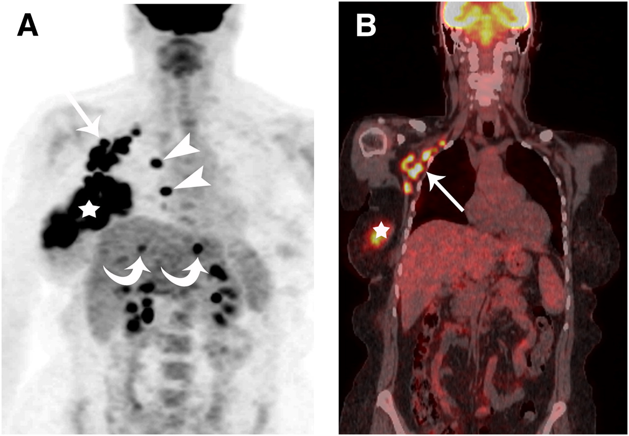
A 52-y-old woman with biopsy-proven right IBC. (A) Maximum-intensity-projection reconstruction of CT-attenuation-corrected PET image shows global hypermetabolic uptake in right breast (star), right subpectoral nodes (arrow), and right internal mammary nodes (arrowheads) and bilobar liver metastases (curved arrows). (B) Coronal PET/CT shows right subpectoral (arrow) and right breast (star) uptake.
- FIGURE 2.
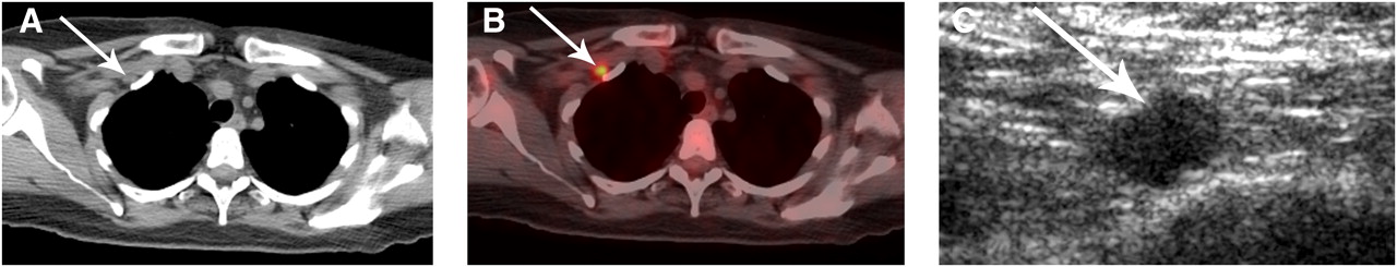
A 53-y-old woman with biopsy-proven right IBC. (A and B) Axial thoracic CT (A) and PET/CT (B) show solitary hypermetabolic, 1-cm medial subpectoral lymph node (arrow). (C) Transverse ultrasound shows abnormal solid, irregular, hypoechoic lymph node in medial subpectoral region (arrow). Ultrasound-guided fine-needle aspiration confirmed metastasis.
- FIGURE 3.
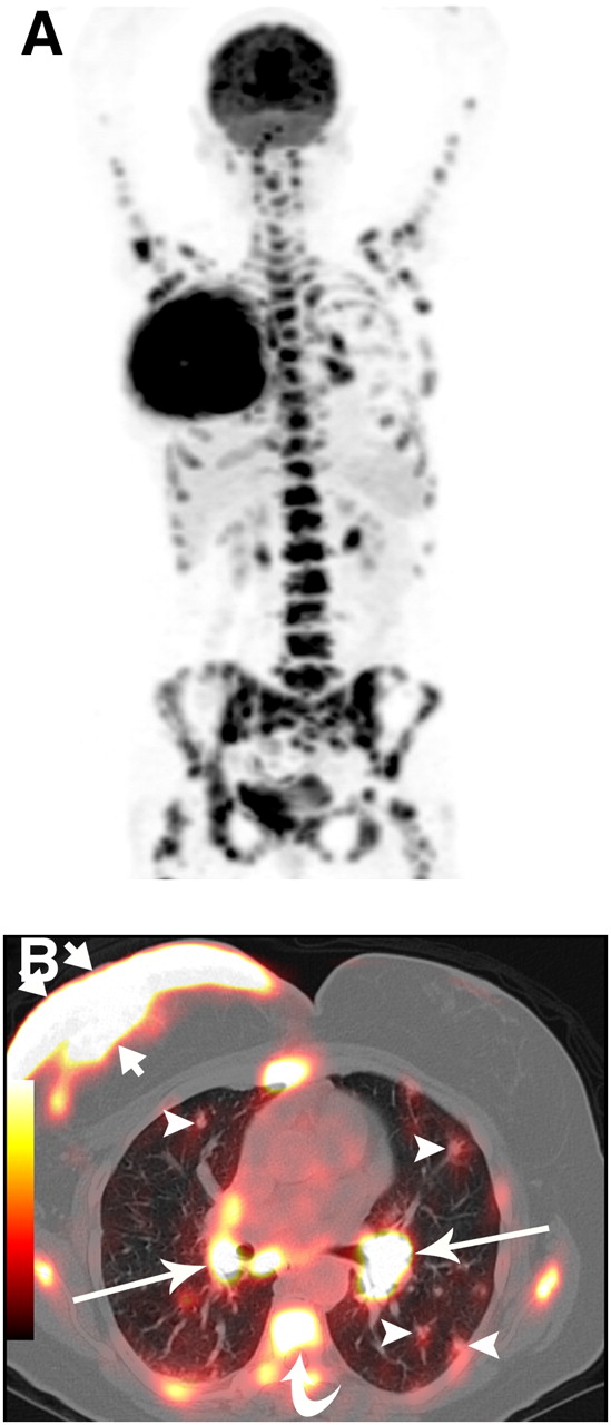
A 52-y-old woman with biopsy-proven right IBC. (A) Maximum-intensity-projection reconstruction of CT-attenuation-corrected PET image shows multiple areas of 18F-FDG uptake consistent with extensive metastasis. (B) Axial PET/CT shows 18F-FDG uptake in right breast, with associated diffuse skin thickening (short arrows), and uptake in bilateral hilar nodes (long arrows), axial skeleton (curved arrow), and pulmonary nodules (arrowheads).
- FIGURE 4.
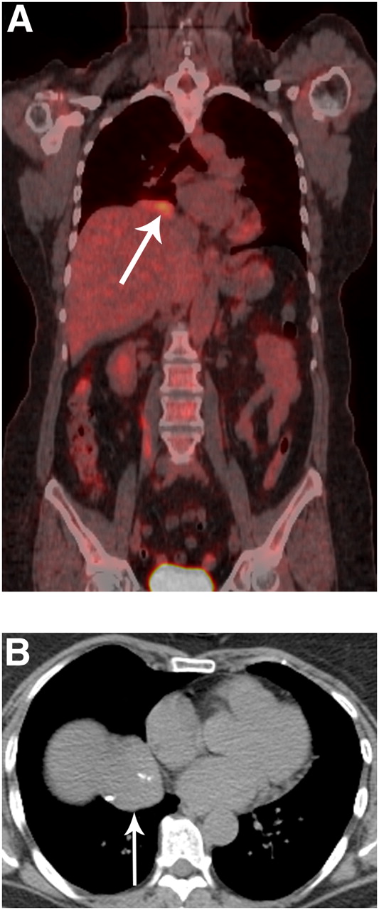
False-positive cardiophrenic angle mass in 59-y-old woman with biopsy-proven right IBC. (A and B) Coronal PET/CT (A) and axial CT (B) show 4-cm 18F-FDG–avid mass (maximum SUV, 5.2; arrows), with associated calcifications at cardiophrenic angle. Biopsy revealed reactive lymphoid tissue.


Tables
Correlative imaging Site Patients with uptake Mean size* (cm) Mean SUV* Biopsy Total Biopsy of 1 nodal site† Distant metastases, no biopsy‡ False-negative False-positive Axillary 38 (90%) 2.5 (0.7–4.6) 11.7 (2.5–36) 29 (71%)§ 9 (22%) 6 (15%) 3 (7%) 1 0 Subpectoral 19 (44%) 1.3 (0.7–2.5) 11 (2.5–34) 4 (10%) 15 (34%) 8 (20%) 7 (14%) 1 0 Supraclavicular 10 (15%) 1.4 (0.7–2.0) 6.8 (3.7–12.6) 5 (12%)‖ 5 (12%) 2 (5%) 3 (7%) 4 0 Internal mammary 9 (22%) 1.2 (0.8–1.6) 7.8 (3.7–13.7) 0 9 (22%) 6 (15%) 3 (7%) 0 0 ↵* Range is in parentheses.
↵† These patients had abnormal regional lymph nodes at multiple sites, all of which were regarded as metastatic after biopsy confirmed that 1 site—axillary, subpectoral, or supraclavicular—was metastatic.
↵‡ In these patients, no biopsy was performed in view of distant metastases.
↵§ One patient had positive biopsy findings and negative PET/CT findings.
↵‖ Four patients had positive biopsy findings and negative PET/CT findings.
- TABLE 2
Correlation Between PET/CT Findings of Distant Metastases and the Findings of Biopsy and Other Imaging Modalities in 20 Patients with IBC
PET/CT findings Chest Abdomen Contralateral LNs Patient no. Musculoskeletal Pulmonary Pleural Mediastinal Liver LNs Axillary Subpectoral Internal mammary Supraclavicular Biopsy Correlative imaging 1* + + Pos (supraclavicular) CT, FU 2† + 0 MRI 3 + + 0 CT, WBS 4 + + + + 0 FU 5† + Pos (supraclavicular) US, FU 6 + + Pos (axillary) CT, FU 7 − (FN) Pos (axillary) US, CT 8 + 0 CT 9* + Pos (chest wall) US, CT 10* + + + + 0 CT, WBS, FU 11† + 0 CT 12 + − (FN) + + 0 CT, WBS, FU 13 − (FN) + + + Pos (pleural) CT 14* + + + + + 0 CT, WBS 15† + 0 MRI 16† + + + + (FP) Neg (axillary) CT, MRI 17 + − (FN) + + + 0 CT, WBS, FU 18 + + Pos (axillary) CT, US 19† + 0 CT 20† + 0 FU ↵* Patients who presented with clinical features suggestive of metastases.
↵† Patients who did not have clinical suspicion or imaging evidence of metastases before PET/CT.
LNs = lymph nodes; + = positive on PET/CT; Pos = positive; FU = follow up with PET/CT; US = ultrasound; − = negative on PET/CT; FN = false-negative; FP = false-positive; Neg = negative.
Site of metastasis Patients (n) Patients with uptake (n) Mean SUV* Patients with biopsy confirmation (n) Patients with correlative imaging and follow-up (n) False-positive False-negative Bone 9 9 13.4 (5.8–24.6) 0 9 0 0 Liver 6 6 9 (4.9–12.7) 0 6 0 0 Abdominal nodes 3 3 8.3 (2.5–10.2) 0 3 0 0 Pulmonary 4 2 12.4 (10.3–14.4) 0 4 0 2 Pleural 2 1 2.5 2 0 0 1 Mediastinal lymph nodes 10 11 8.8 (3.6–17.3) 1 10 1 0 Soft tissue 1 1 4.3 1 0 0 0 Contralateral lymph nodes 7 7 4 Axillary 5 5 4.4 (3.3–7.5) 2 4 1 1 Subpectoral 2 2 4.5 (4.4–4.5) 0 2 0 0 Supraclavicular 3 3 6.5 (4.2–9.2) 2 1 0 0 Internal mammary 2 2 5.5 (5.1–6.2) 0 2 0 0 ↵* Range is in parentheses.


Jump to section
Related Articles
Cited By...
- Assessing 18F-FDG Uptake in the Sentinel Lymph Node in Breast Cancer
- 18F-FDG PET/CT for Staging and Restaging of Breast Cancer
- Retrospective Analysis of 18F-FDG PET/CT for Staging Asymptomatic Breast Cancer Patients Younger Than 40 Years
- 18F-FDG PET/CT in Staging Patients with Locally Advanced or Inflammatory Breast Cancer: Comparison to Conventional Staging
- Inflammatory Breast Cancer: What We Know and What We Need to Learn
- American Society of Clinical Oncology Identifies Five Key Opportunities to Improve Care and Reduce Costs: The Top Five List for Oncology
- The Yield of 18F-FDG PET/CT in Patients with Clinical Stage IIA, IIB, or IIIA Breast Cancer: A Prospective Study
- FDG-PET/CT Compared with Conventional Imaging in the Detection of Distant Metastases of Primary Breast Cancer
- Initial Staging Impact of Fluorodeoxyglucose Positron Emission Tomography/Computed Tomography in Locally Advanced Breast Cancer
- Invasive Breast Cancer
- Inflammatory Breast Cancer