
Main menu

  • Home
  • Content
    • Current
    • Ahead of print
    • Past Issues
    • JNM Supplement
    • SNMMI Annual Meeting Abstracts
    • Continuing Education
    • JNM Podcasts
  • Subscriptions
    • Subscribers
    • Institutional and Non-member
    • Rates
    • Journal Claims
    • Corporate & Special Sales
  • Authors
    • Submit to JNM
    • Information for Authors
    • Assignment of Copyright
    • AQARA requirements
  • Info
    • Reviewers
    • Permissions
    • Advertisers
  • About
    • About Us
    • Editorial Board
    • Contact Information
  • More
    • Alerts
    • Feedback
    • Help
    • SNMMI Journals
  • SNMMI
    • JNM
    • JNMT
    • SNMMI Journals
    • SNMMI

User menu

  • Subscribe
  • My alerts
  • Log in
  • Log out
  • My Cart

Search

  • Advanced search
Journal of Nuclear Medicine
  • SNMMI
    • JNM
    • JNMT
    • SNMMI Journals
    • SNMMI
  • Subscribe
  • My alerts
  • Log in
  • Log out
  • My Cart
Journal of Nuclear Medicine

Advanced Search

  • Home
  • Content
    • Current
    • Ahead of print
    • Past Issues
    • JNM Supplement
    • SNMMI Annual Meeting Abstracts
    • Continuing Education
    • JNM Podcasts
  • Subscriptions
    • Subscribers
    • Institutional and Non-member
    • Rates
    • Journal Claims
    • Corporate & Special Sales
  • Authors
    • Submit to JNM
    • Information for Authors
    • Assignment of Copyright
    • AQARA requirements
  • Info
    • Reviewers
    • Permissions
    • Advertisers
  • About
    • About Us
    • Editorial Board
    • Contact Information
  • More
    • Alerts
    • Feedback
    • Help
    • SNMMI Journals
  • View or Listen to JNM Podcast
  • Visit JNM on Facebook
  • Join JNM on LinkedIn
  • Follow JNM on Twitter
  • Subscribe to our RSS feeds
Research ArticleCLINICAL INVESTIGATIONS

Functional Images Reflect Aggressiveness of Endometrial Carcinoma: Estrogen Receptor Expression Combined with 18F-FDG PET

Tetsuya Tsujikawa, Yoshio Yoshida, Takashi Kudo, Yasushi Kiyono, Tetsuji Kurokawa, Masato Kobayashi, Tatsuro Tsuchida, Yasuhisa Fujibayashi, Fumikazu Kotsuji and Hidehiko Okazawa
Journal of Nuclear Medicine October 2009, 50 (10) 1598-1604; DOI: https://doi.org/10.2967/jnumed.108.060145
Tetsuya Tsujikawa
  • Find this author on Google Scholar
  • Find this author on PubMed
  • Search for this author on this site
Yoshio Yoshida
  • Find this author on Google Scholar
  • Find this author on PubMed
  • Search for this author on this site
Takashi Kudo
  • Find this author on Google Scholar
  • Find this author on PubMed
  • Search for this author on this site
Yasushi Kiyono
  • Find this author on Google Scholar
  • Find this author on PubMed
  • Search for this author on this site
Tetsuji Kurokawa
  • Find this author on Google Scholar
  • Find this author on PubMed
  • Search for this author on this site
Masato Kobayashi
  • Find this author on Google Scholar
  • Find this author on PubMed
  • Search for this author on this site
Tatsuro Tsuchida
  • Find this author on Google Scholar
  • Find this author on PubMed
  • Search for this author on this site
Yasuhisa Fujibayashi
  • Find this author on Google Scholar
  • Find this author on PubMed
  • Search for this author on this site
Fumikazu Kotsuji
  • Find this author on Google Scholar
  • Find this author on PubMed
  • Search for this author on this site
Hidehiko Okazawa
  • Find this author on Google Scholar
  • Find this author on PubMed
  • Search for this author on this site
  • Article
  • Figures & Data
  • Info & Metrics
  • PDF
Loading

Article Figures & Data

Figures

  • Tables
  • FIGURE 1. 
    • Download figure
    • Open in new tab
    • Download powerpoint
    FIGURE 1. 

    Box-and-whisker plots of uptake of each tracer among 3 groups. Horizontal bars inside boxes indicate median values. Error bars indicate farthest points that are not outliers. Significant differences in 18F-FDG accumulation were observed between hyperplasia and high-risk (P < 0.0001) or low-risk (P < 0.005) endometrial carcinoma. 18F-FDG accumulation was not significantly different between high-risk and low-risk carcinoma, although 18F-FES PET showed significant difference between these 2 groups (P < 0.05). High-R = high-risk carcinoma; low-R = low-risk carcinoma; NS = not significant.
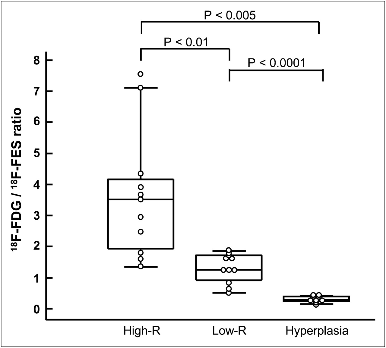
  • FIGURE 2. 
    • Download figure
    • Open in new tab
    • Download powerpoint
    FIGURE 2. 

    Box-and-whisker plots of 18F-FDG–to–18F-FES SUV ratio among 3 groups. Significant differences were observed between high-risk and low-risk carcinoma (P < 0.01), between high-risk carcinoma and hyperplasia (P < 0.005), and between low-risk carcinoma and hyperplasia (P < 0.0001). High-R = high-risk carcinoma; low-R = low-risk carcinoma.
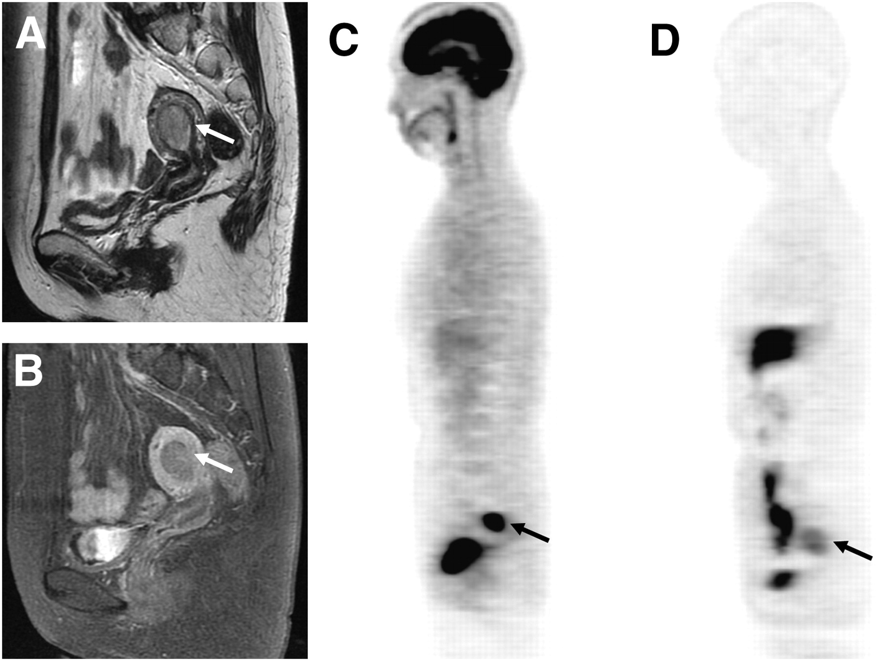
  • FIGURE 3. 
    • Download figure
    • Open in new tab
    • Download powerpoint
    FIGURE 3. 

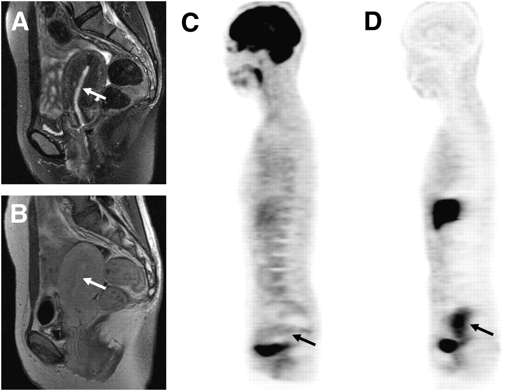
    T2-weighted (A), contrast-enhanced T1-weighted MR (B), 18F-FDG (C), and 18F-FES PET (D) images of 60-y-old woman with endometrial carcinoma (arrows). PET images showed intense 18F-FDG uptake (SUV, 11.6) and moderate 18F-FES uptake (SUV, 4.7) in tumor. Preoperative staging by MRI suggested no myometrial invasion, whereas PET findings with an 18F-FDG–to–18F-FES ratio of 2.5 indicated high-risk carcinoma. Postoperative histopathologic result was high-risk carcinoma with FIGO stage Ic and histologic grade 2.
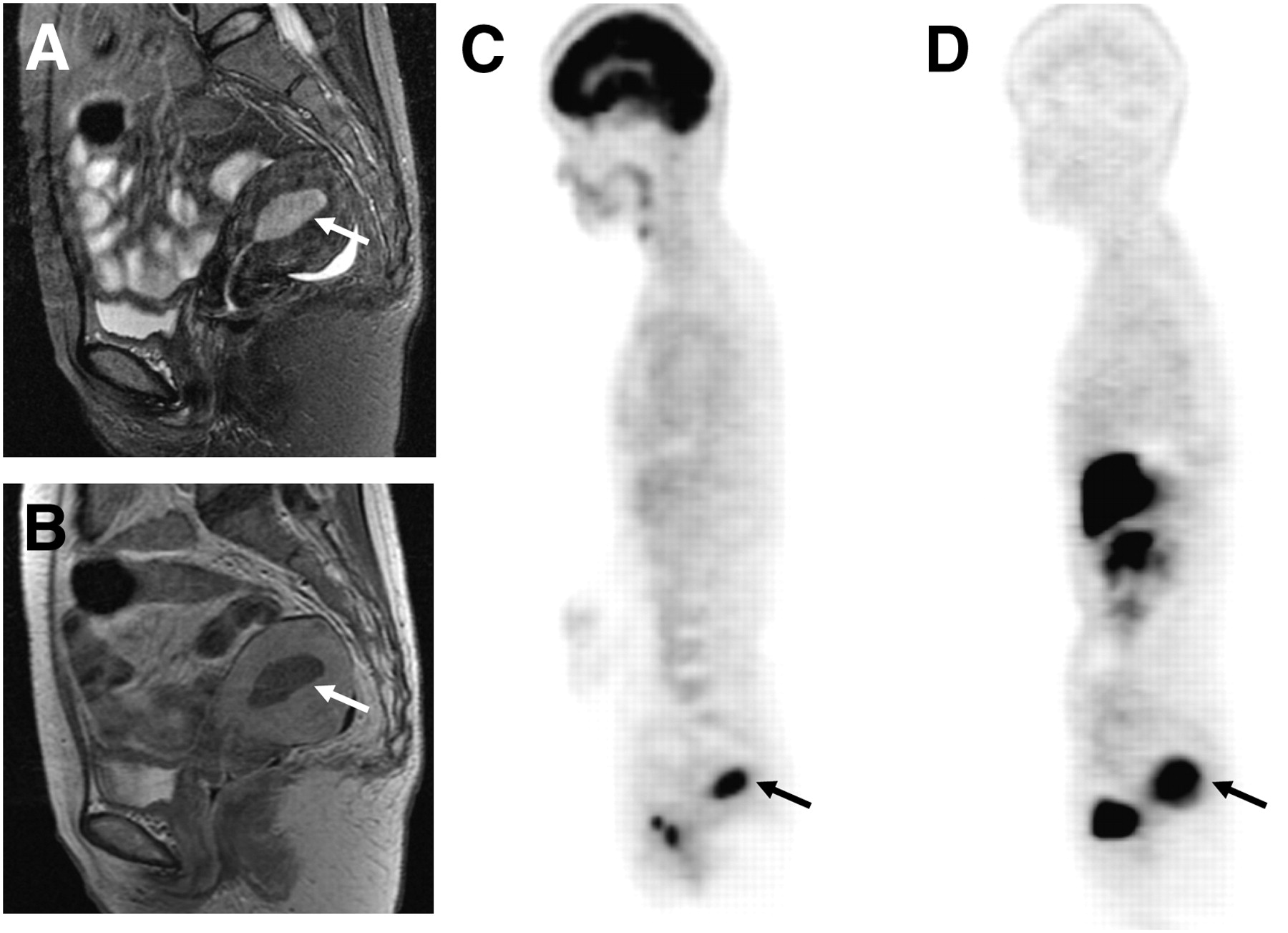
  • FIGURE 4. 
    • Download figure
    • Open in new tab
    • Download powerpoint
    FIGURE 4. 

    Sagittal T2-weighted (A), contrast-enhanced T1-weighted MR (B), 18F-FDG (C), and 18F-FES PET (D) images of 50-y-old woman with low-risk endometrial carcinoma (FIGO stage Ib and histologic grade 1, arrows). PET images showed intense accumulation for both tracers in endometrial lesion. Region of 18F-FES uptake (SUV, 8.1) appeared larger than that of 18F-FDG (SUV, 10.2) because of 18F-FES avidity in myometrial inner layer. 18F-FDG–to–18F-FES ratio was 1.3.

  • FIGURE 5. 
    • Download figure
    • Open in new tab
    • Download powerpoint
    FIGURE 5. 

    A 39-y-old woman who had endometrial hyperplasia with relatively low intensity on sagittal T2-weighted (A) and contrast-enhanced T1-weighted (B) MR images (arrows). PET images showed low 18F-FDG (SUV, 1.4) (C) and high 18F-FES (SUV, 5.9) (D) accumulation in endometrial lesion of uterus. 18F-FDG–to–18F-FES ratio was 0.2.

Tables

  • Figures
    • View popup
    TABLE 1

    Patient Characteristics

    CharacteristicValue
    Total patients (n)31
    Mean age (y)56 ± 15
    Age range (y)30–84
    Menopausal status (n)
     Premenopausal13
     Postmenopausal18
    Histopathologic type (n)
     Endometrial adenocarcinoma22
     Endometrial hyperplasia9
    Stage of carcinoma (n)
     Ia or Ib14
     Ic3
     II2
     III3
    Differentiation grade (n)
     G112
     G29
     G31
    Tumor size (n)
     <1 cm14
     1–2 cm8
     2 cm <9
    High-risk carcinoma (n)11
    Low-risk carcinoma (n)11
    • Carcinoma of stage Ib or lower with grade G2 or G3 was included in high-risk group.

    • View popup
    TABLE 2

    Regional SUVs and 18F-FDG–to–18F-FES SUV Ratio

    ClassPatients18F-FDG SUV18F-FES SUV18F-FDG–to–18F-FES ratio
    High-risk carcinoma (stage ≥ Ic or grade ≥ 2)119.9 ± 2.6* (5.4–14.8)3.4 ± 1.6 (1.3–6.3)3.6 ± 2.1 (1.3–7.4)
    Low-risk carcinoma (stage ≤ Ib and grade 1)117.1 ± 3.7 (2.4–12.5)5.3 ± 1.8 (2.1–8.1)1.3 ± 0.5 (0.5–1.8)
    Endometrial hyperplasia91.5 ± 0.3 (1.1–1.9)5.6 ± 2.4† (2.7–11.0)0.3 ± 0.1 (0.2–0.4)
    • ↵* P < 0.0001.

    • ↵† P < 0.005 significantly higher between the 2 PET tracers (paired t test).

    • Values are mean ± SD. Statistical significance for group comparison is given in Figures 1 and 2. Data in parentheses are ranges.

    • View popup
    TABLE 3

    Diagnostic Accuracy in Differentiating High- and Low-Risk Carcinoma

    SensitivitySpecificityPPVNPVAccuracy
    Parametern95% CI (%)n95% CI (%)n95% CI (%)n95% CI (%)n95% CI (%)
    18F-FDG–to–18F-FES ratio (2.0)*8/11 (73%)39–9411/11 (100%)76–1008/8 (100%)69–10011/14 (79%)49–9519/22 (86%‡)65–97
    18F-FES SUV (4.4)*9/11 (82%)48–978/11 (73%)39–949/12 (75%)43–958/10 (80%)44–9717/22 (77%)55–92
    18F-FDG SUV (4.0)*11/11 (100%)76–1004/11 (36%)11–6911/18 (61%)36–834/4 (100%)47–10015/22 (68%‡)45–86
    MRI†4/8 (50%)16–8413/14 (93%)66–1004/5 (80%)28–9913/17 (76%)50–9317/22 (77%)55–92
    • ↵* Optimal cutoff value.

    • ↵† Discrimination of stage ≥ Ic from ≤ Ib irrespective of histologic grade.

    • ↵‡ P < 0.005 in comparison between 18F-FDG–to–18F-FES ratio and 18F-FDG SUV using McNemar test.

    • PPV = positive predictive value; NPV = negative predictive value.

PreviousNext
Back to top

In this issue

Journal of Nuclear Medicine: 50 (10)
Journal of Nuclear Medicine
Vol. 50, Issue 10
October 2009
  • Table of Contents
  • Table of Contents (PDF)
  • About the Cover
  • Index by author
Print
Download PDF
Article Alerts
Sign In to Email Alerts with your Email Address
Email Article

Thank you for your interest in spreading the word on Journal of Nuclear Medicine.

NOTE: We only request your email address so that the person you are recommending the page to knows that you wanted them to see it, and that it is not junk mail. We do not capture any email address.

Enter multiple addresses on separate lines or separate them with commas.
Functional Images Reflect Aggressiveness of Endometrial Carcinoma: Estrogen Receptor Expression Combined with 18F-FDG PET
(Your Name) has sent you a message from Journal of Nuclear Medicine
(Your Name) thought you would like to see the Journal of Nuclear Medicine web site.
Citation Tools
Functional Images Reflect Aggressiveness of Endometrial Carcinoma: Estrogen Receptor Expression Combined with 18F-FDG PET
Tetsuya Tsujikawa, Yoshio Yoshida, Takashi Kudo, Yasushi Kiyono, Tetsuji Kurokawa, Masato Kobayashi, Tatsuro Tsuchida, Yasuhisa Fujibayashi, Fumikazu Kotsuji, Hidehiko Okazawa
Journal of Nuclear Medicine Oct 2009, 50 (10) 1598-1604; DOI: 10.2967/jnumed.108.060145

Citation Manager Formats

  • BibTeX
  • Bookends
  • EasyBib
  • EndNote (tagged)
  • EndNote 8 (xml)
  • Medlars
  • Mendeley
  • Papers
  • RefWorks Tagged
  • Ref Manager
  • RIS
  • Zotero
Share
Functional Images Reflect Aggressiveness of Endometrial Carcinoma: Estrogen Receptor Expression Combined with 18F-FDG PET
Tetsuya Tsujikawa, Yoshio Yoshida, Takashi Kudo, Yasushi Kiyono, Tetsuji Kurokawa, Masato Kobayashi, Tatsuro Tsuchida, Yasuhisa Fujibayashi, Fumikazu Kotsuji, Hidehiko Okazawa
Journal of Nuclear Medicine Oct 2009, 50 (10) 1598-1604; DOI: 10.2967/jnumed.108.060145
Twitter logo Facebook logo LinkedIn logo Mendeley logo
  • Tweet Widget
  • Facebook Like
  • Google Plus One
Bookmark this article

Jump to section

  • Article
    • Abstract
    • MATERIALS AND METHODS
    • RESULTS
    • DISCUSSION
    • CONCLUSION
    • Acknowledgments
    • Footnotes
    • References
  • Figures & Data
  • Info & Metrics
  • PDF

Related Articles

  • This Month in JNM
  • PubMed
  • Google Scholar

Cited By...

  • ER Imaging for Estrogen-Related Tumors Is Bothersome but Useful
  • Prognostic Value of 16{alpha}-18F-Fluoro-17{beta}-Estradiol PET as a Predictor of Disease Outcome in Endometrial Cancer: A Prospective Study
  • Improved Estrogen Receptor Assessment by PET Using the Novel Radiotracer 18F-4FMFES in Estrogen Receptor-Positive Breast Cancer Patients: An Ongoing Phase II Clinical Trial
  • 18F-Fluoroestradiol PET: Current Status and Potential Future Clinical Applications
  • Evaluation of Gynecologic Cancer with MR Imaging, 18F-FDG PET/CT, and PET/MR Imaging
  • Molecular Imaging Biomarkers for Oncology Clinical Trials
  • Application of FDG-PET in Cervical Cancer and Endometrial Cancer: Utility and Future Prospects
  • 18F-FES and 18F-FDG PET for Differential Diagnosis and Quantitative Evaluation of Mesenchymal Uterine Tumors: Correlation with Immunohistochemical Analysis
  • Oestrogen-related tumour phenotype: positron emission tomography characterisation with 18F-FDG and 18F-FES
  • Imaging Tumor Phenotype: 1 Plus 1 Is More Than 2
  • Google Scholar

More in this TOC Section

  • Feasibility of Ultra-Low-Activity 18F-FDG PET/CT Imaging Using a Long–Axial-Field-of-View PET/CT System
  • Cardiac Presynaptic Sympathetic Nervous Function Evaluated by Cardiac PET in Patients with Chronotropic Incompetence Without Heart Failure
  • Validation and Evaluation of a Vendor-Provided Head Motion Correction Algorithm on the uMI Panorama PET/CT System
Show more Clinical Investigations

Similar Articles

SNMMI

© 2025 SNMMI

Powered by HighWire