


Article Figures & Data
Figures
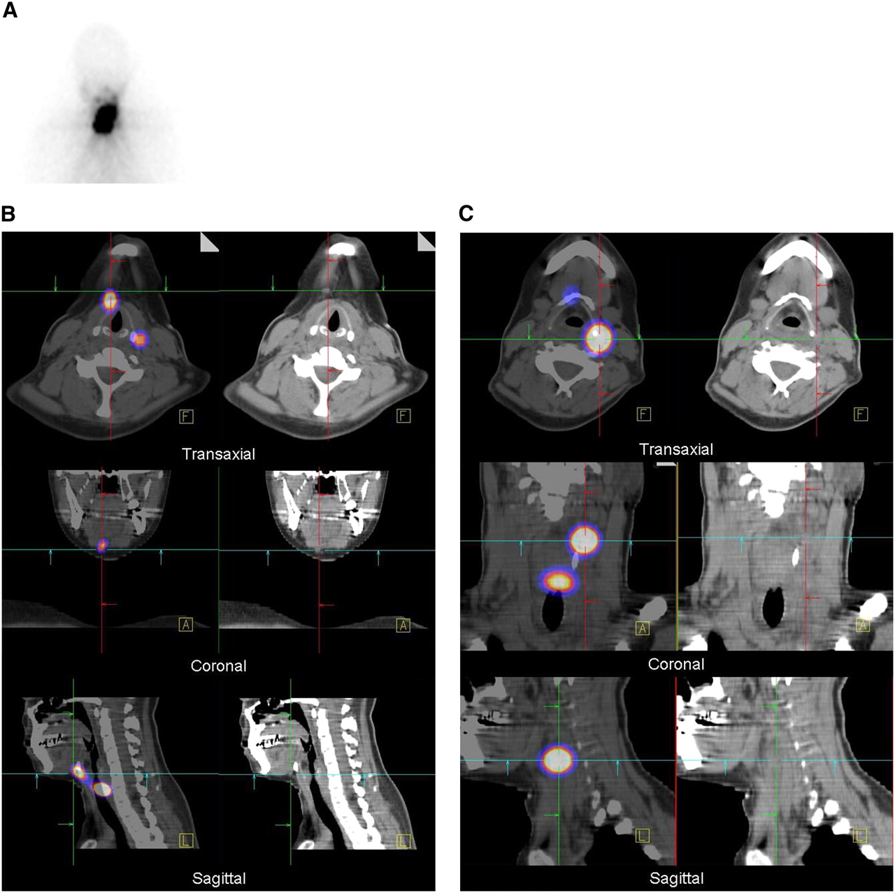
- FIGURE 1.
A 54-y-old woman with differentiated papillary thyroid carcinoma (pT2 N1a [6/21] Mx) after total thyroidectomy and lymph node dissection of centrocervical and left lateral compartment. (A) Planar scintigraphy shows two 131I-avid foci interpreted as thyroid remnant. (B) SPECT/CT (left column) and CT (right column) demonstrate that these foci correspond to LNMs in superior mediastinum (level VII)—shown here for right focus.
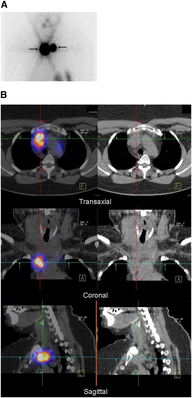
- FIGURE 2.
A 30-y-old woman with differentiated papillary thyroid carcinoma (pT2 N0 [0/10] Mx) after total thyroidectomy and lymph node dissection of centrocervical compartment. (A) Planar scintigraphy shows large 131I-avid foci interpreted as thyroid remnant and indeterminate. (B) SPECT/CT fusion images (left column) and CT (right column) demonstrate 1 focus corresponding to LNM in level II. (C) SPECT/CT (left column) and CT (right column) demonstrate 1 focus corresponding to LNM in level III.
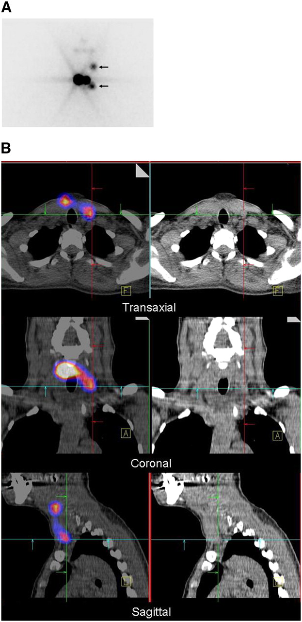
- FIGURE 3.
A 43-y-old woman with differentiated papillary thyroid carcinoma (pT1 N0 [0/7] Mx) after total thyroidectomy and lymph node dissection of centrocervical compartment. (A) Planar scintigraphy shows 131I-avid foci interpreted as LNMs (arrows) and thyroid remnant. (B) SPECT/CT fusion images (left column) and CT (right column) demonstrate that these foci correspond to LNMs in level II and level IV—shown here for lower focus.
- FIGURE 4.
Imaging results in patients classified as N0 (A) or Nx (B) by histopathology. “Nx” denotes classification as indeterminate.


Tables
- TABLE 1
Tumor Stage and TNM Classification as Defined by AJCC/UICC in the Patients Studied
Age (y) Tumor stage TNM classification Stage n Class n <45 I (any T, any N, M0) 23 T1 Nx 8 T1 N0 7 T1 N1 3 T2 NX 1 T2 N0 2 T3 N0 2 II (any T, any N, M1) 1 T3 N1 1 >45 I (T1, N0, M0) 20 T1 Nx 16 T1 N0 4 II (T2, N0, M0) 4 T2 NX 2 T2 N0 2 III (T3, N0, M0 or T1–3, N1a, M0) 7 T3 Nx 2 T3 N0 3 T2 N1a 1 T3 N1a 1 IVA (T4a, N0–1a, M0 or T1–4, N1b, M0) 1 T3 N1b 1 IVB (T4b, any N, M0) 0 IVC (any T, any N, M1) 1 T3a N0 1 - TABLE 2
Planar Findings and SPECT/CT Characterization of Radioiodine-Positive Cervical Foci
Planar findings SPECT/CT characterization No. of lesions Lymph node metastases Lymph node metastases 5 Indeterminate 1 Thyroid remnant 4 Contamination 1 Indeterminate Lymph node metastases 4 Thyroid remnant 10 Contamination 1 Thyroid remnant Lymph node metastases 7 Thyroid remnant 110


Jump to section
Related Articles
Cited By...
- SNMMI Procedure Standard/EANM Practice Guideline for Nuclear Medicine Evaluation and Therapy of Differentiated Thyroid Cancer: Abbreviated Version
- I-131 Postablation SPECT/CT Predicts Relapse of Papillary Thyroid Carcinoma more Accurately than Whole Body Scan
- ENDOCRINE TUMOURS: Imaging in the follow-up of differentiated thyroid cancer: current evidence and future perspectives for a risk-adapted approach
- Radioiodine Scintigraphy with SPECT/CT: An Important Diagnostic Tool for Thyroid Cancer Staging and Risk Stratification
- The SNMMI Practice Guideline for Therapy of Thyroid Disease with 131I 3.0
- Radioiodine Scintigraphy with SPECT/CT: An Important Diagnostic Tool for Thyroid Cancer Staging and Risk Stratification
- Postablation 131I scintigraphy with neck and thorax SPECT-CT and stimulated serum thyroglobulin level predict the outcome of patients with differentiated thyroid cancer
- The Effect of Posttherapy 131I SPECT/CT on Risk Classification and Management of Patients with Differentiated Thyroid Cancer
- The Significance of 99mTc-MAA SPECT/CT Liver Perfusion Imaging in Treatment Planning for 90Y-Microsphere Selective Internal Radiation Treatment
- Single photon emission computed tomography (SPECT)/computed tomography using Iodine-123 in patients with differentiated thyroid cancer: additional value over whole body planar imaging and SPECT
- Reply: Scintigraphic TNM Staging of Tumors: A Proposition
- Scintigraphic TNM Staging of Tumors: A Proposition