


Article Figures & Data
Figures
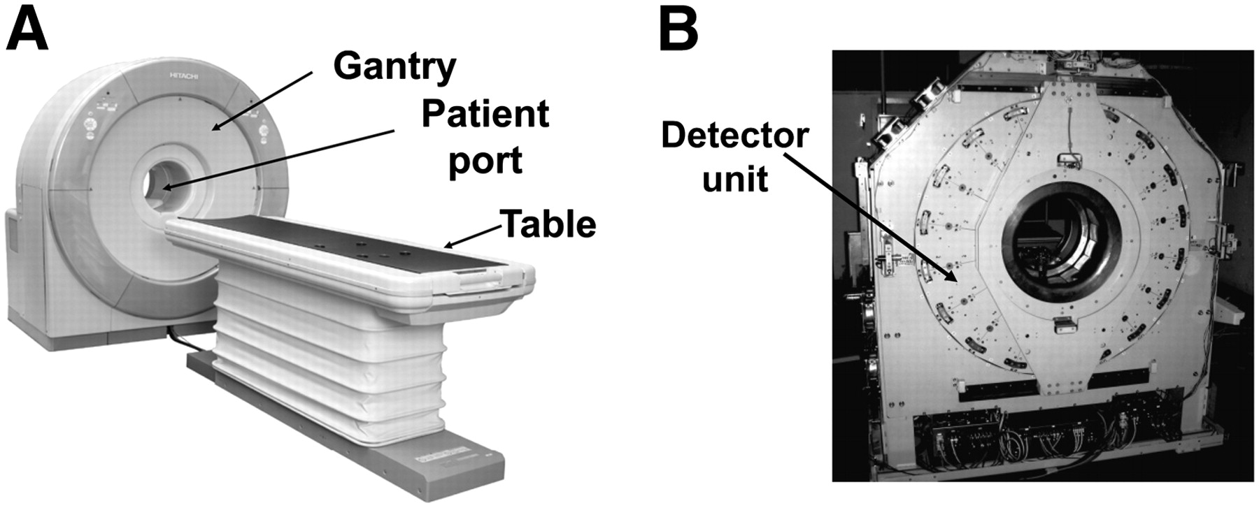
- FIGURE 1.
(A) Prototype 3D PET scanner dedicated to human brain imaging. Diameter of patient port is 350 mm, transaxial FOV is 310 mm, and axial FOV is 250 mm. (B) Eighteen detector units are radially arranged around patient port.
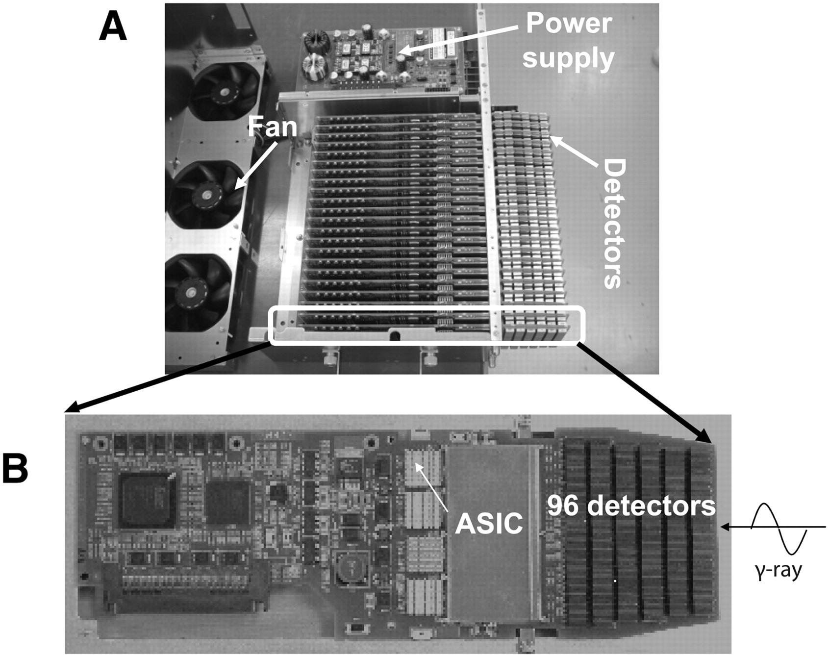
- FIGURE 2.
(A) Structure of detector unit. New detector size is 2 × 4 × 7.5 mm. Dimensions of detector unit are 100 × 400 × 350 mm. Detector boards are arranged in parallel, and detectors are mounted on both sides of each board. (B) Detector board has 96 detectors on each side (192 detectors in total) and signal processors. These processors include application-specific integrated circuits (ASICs) mounted along incident direction of γ-rays. Signals are read by 3-layer DOI system. Each unit has 22 boards and about 4,000 detectors. Entire system is cooled by forced air.
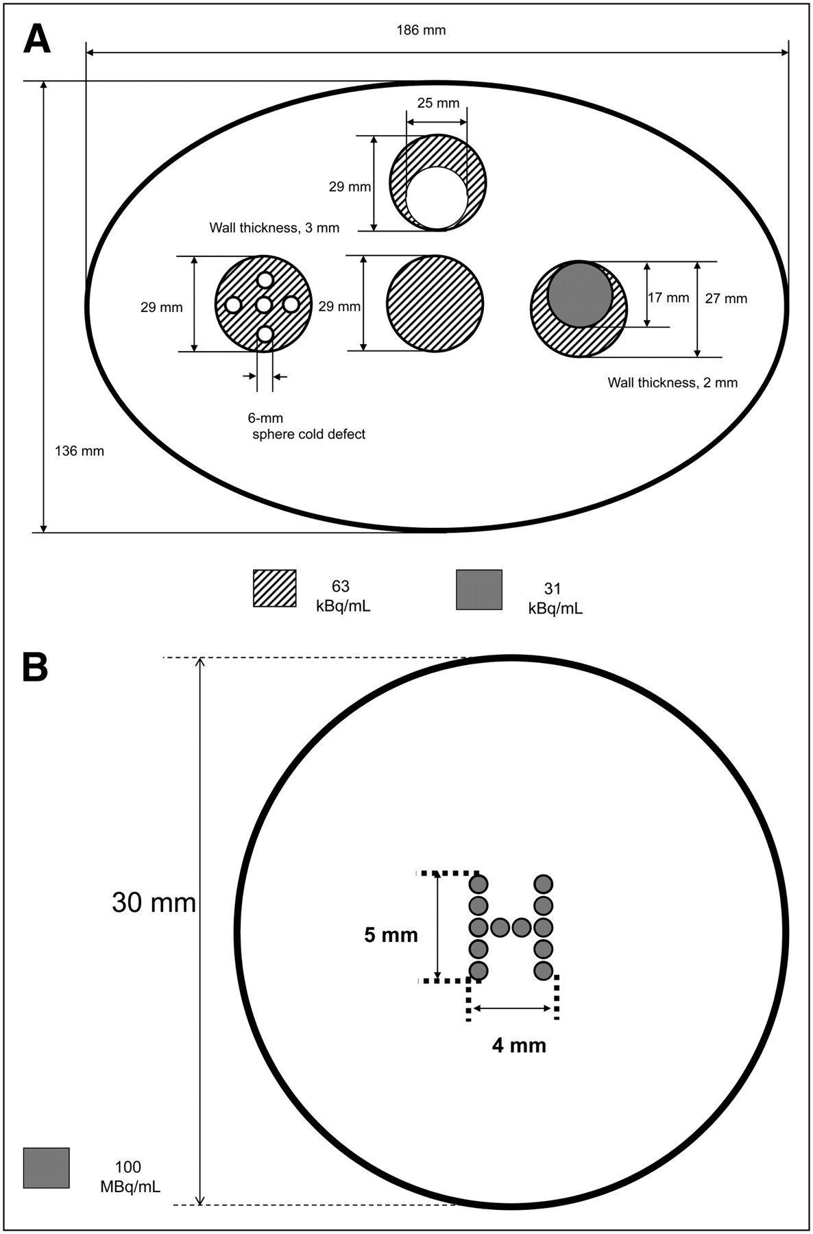
- FIGURE 3.
Scheme of second elliptic phantom (136 × 188 mm) consisting of 4 small compartments (A) and H-shaped hot phantom (B). First compartment had uniform cylinder in middle, second compartment had cylinder with five 6-mm-diameter cold spots, third compartment had cylinder with 25-mm-diameter cold region, and fourth compartment had cylinder filled with half the radioactivity in remaining compartments.
- FIGURE 4.
Time course protocols. Order in time course protocol 1: HR+ PET in whole-body mode, HR+ PET in brain mode, and semiconductor PET; order in time course protocol 2: semiconductor PET, HR+ PET in brain mode, and HR+ PET in whole-body mode. I.V. = intravenously.
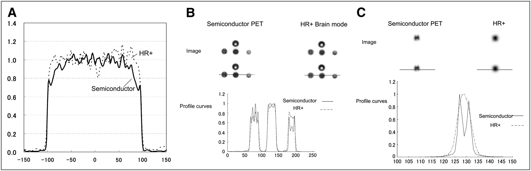
- FIGURE 5.
Phantom images and profile curves obtained by semiconductor PET and HR+ PET. (A) Pool phantom. (B) Tumor phantom. (C) H-shaped hot phantom. Upper parts of B and C show PET images, middle parts show positions of profile curves, and lower parts show profile curves. Profile curves were normalized to maximum counts.
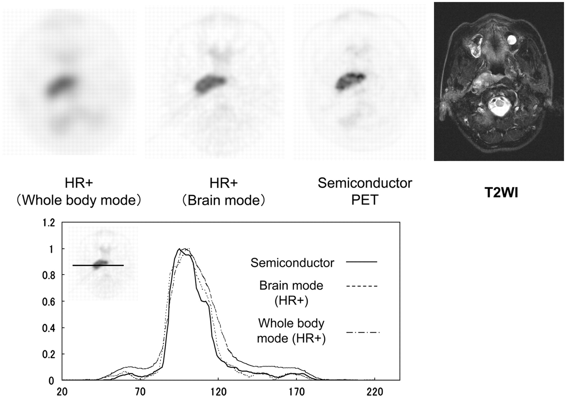
- FIGURE 6.
Whole-body mode (left) and brain mode (middle) scintillator-based PET images and semiconductor PET image (right) of 61-y-old man with nasopharyngeal squamous cell cancer. Semiconductor PET identified intratumoral inhomogeneous glucose metabolism in more detail than HR+ PET. T2-weighted MRI (T2WI) also revealed inhomogeneous intensity in primary lesion (far right). Diagrams show profile curves. Profile curves were normalized to maximum counts. Profile curve for tumor obtained by semiconductor PET was sharpest among 3 images.
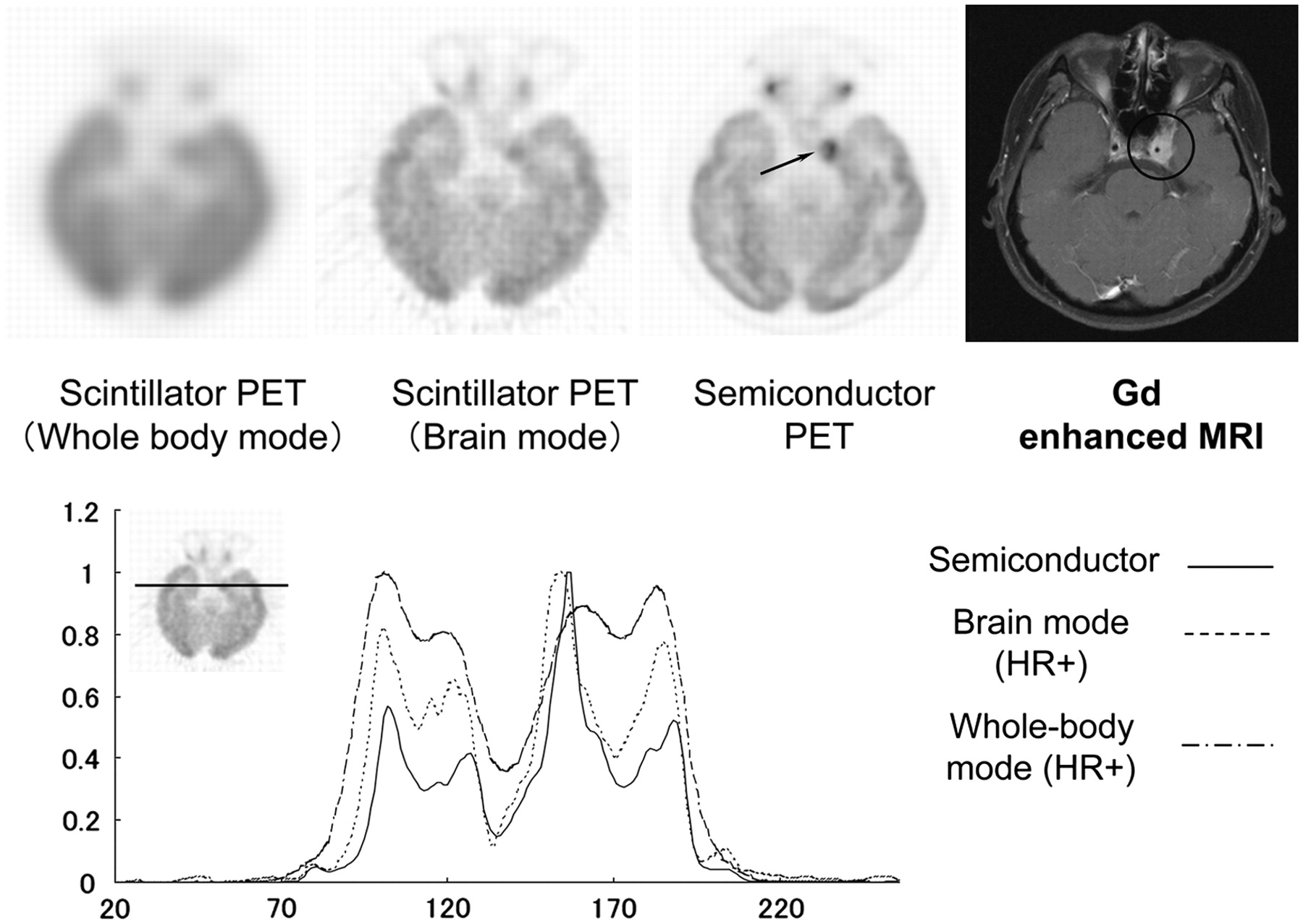
- FIGURE 7.
Whole-body mode (left) and brain mode (middle) scintillator-based PET images and semiconductor PET image (right) of 35-y-old woman with nasopharyngeal squamous cell cancer. Semiconductor PET revealed low uptake in tumor, which corresponded to internal carotid artery (arrow). Gadolinium-enhanced MRI showed that left internal carotid artery penetrated tumor (far right). Diagrams show profile curves. Profile curves were normalized to maximum counts. Profile curve of tumor obtained by semiconductor PET was sharpest among 3 images.


Tables
Performance index Result Standard Spatial resolution (transaxial) 1 cm 2.3 mm NEMA NU 2-2001 10 cm 4.8 mm NEMA NU 2-2001 Spatial resolution (axial) 1 cm 5.1 mm NEMA NU 2-2001 10 cm 5.9 mm NEMA NU 2-2001 Sensitivity (true) at 450–530 keV 17.6 kcps/kBq/mL NEMA NU 2-1994 Scatter fraction (3D) at 450–530 keV 23% NEMA NU 2-1994 NEC-2R (at 450–530 keV) 3.7 kBq/mL 30 kcps NEMA NU 2-1994 7.9 kBq/mL 41 kcps NEMA NU 2-1994 Energy resolution at 511 keV 4.1% Patient Age (y) Sex Pathology TNM classification Comments Findings from primary lesion 1 76 M SCC T2bN2M0 Before treatment Left nasopharyngeal uptake extending to left fossa of Rosenmüller 2 30 M Undifferentiated cancer T3N2M0 Before treatment Bilateral nasopharyngeal–oropharyngeal uptake 3 67 F Undifferentiated cancer T3N2M0 Before treatment Strong uptake in retronasopharyngeal space extending to left fossa of Rosenmüller 4 61 M SCC T3N1M0 Before treatment Strong uptake in retronasopharyngeal space extending to right fossa of Rosenmüller 5 35 F SCC T4N1M0 Before treatment Strong uptake in right nasopharyngeal space extending to right cavernous sinus 6 61 M SCC T1N1M0 Before treatment Uptake along retronasopharyngeal wall and right fossa of Rosenmüller 7 53 M Undifferentiated cancer T1N0M0 Before treatment Uptake along retronasopharyngeal wall and right fossa of Rosenmüller 8 71 F Poorly differentiated SCC T4NxM0 Before treatment Strong uptake in right nasopharyngeal space extending to right cavernous sinus 9 44 M Undifferentiated cancer T4N2M0 Local recurrence in cavernous sinus suggested by MRI after chemoradiation therapy No uptake 10 28 F SCC T3N2M0 Local recurrence in clivus suggested by MRI after chemoradiation therapy No uptake SCC = squamous cell cancer.


