


Article Figures & Data
Figures
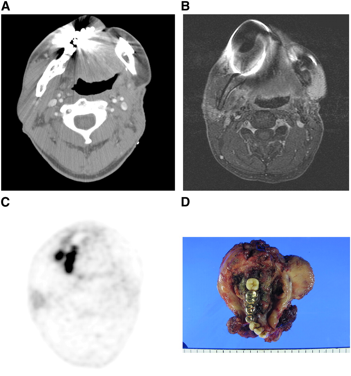
- FIGURE 1.
54-y-old male patient with squamous cell carcinomas of right retromolar area who underwent CT, MRI, and PET/CT scans during initial evaluation of tumor. In contrast-enhanced CT images (A) and in gadolinium-enhanced, fat-suppressed T1-weighted MR images (B), primary tumor was not identified because of metallic artifacts caused by nonremovable dentures. However, in CT-attenuated PET images (C), lesion showed asymmetric glucose uptake at peak SUV of 4.2 in right retromolar trigone and adjacent mandible and then finally was diagnosed as T4 oral cancer. (D) Pathologic examination revealed 5.5-cm-sized squamous cell carcinomas of retromolar trigone that invaded mandible.
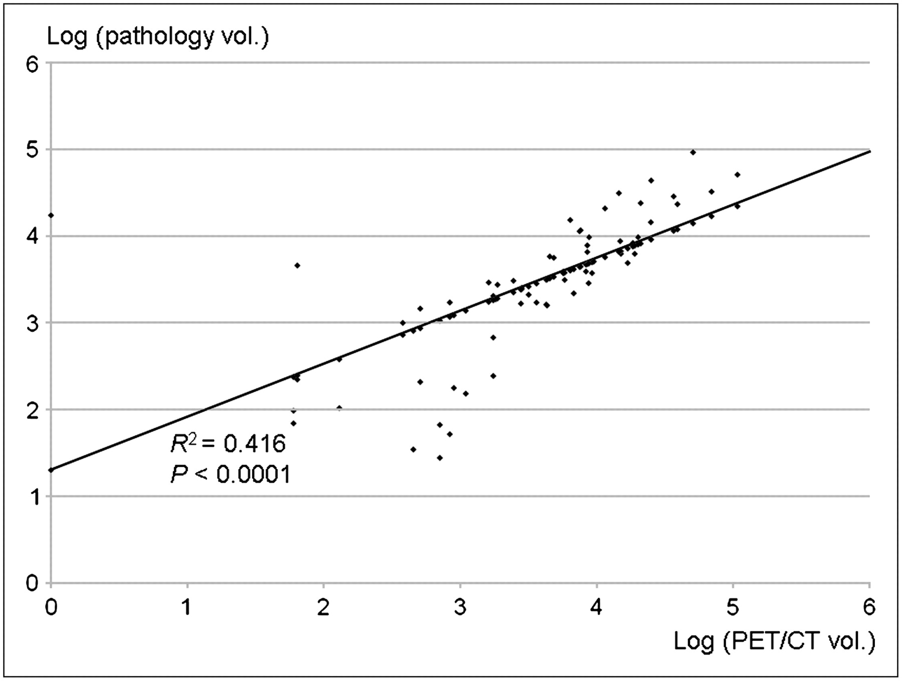
- FIGURE 2.
Regression equation between log (PET/CTSUV 3.5 volume) and log (pathologic volume) was log (pathologic volume) = 0.6041 × log (PET/CTSUV 3.5 volume) + 1.3046. Correlation coefficient (R) and coefficient of determination (R2) were 0.645 and 0.416, suggesting acceptable correlation between 2 variables. vol. = volume.
Tables
No. of Patients with OCC (%) Characteristic With artifacts (n = 64) Without artifacts (n = 40) M:F 41:23 (64.1%:35.9%) 31:9 (77.5%:22.5%) Age (y) Mean ± SD 53.9 ± 10.7 54.5 ± 11.6 Range 22–75 29–75 Primary tumor site Tongue 51 (79.7) 27 (67.5) Floor of mouth 4 (6.3) 5 (12.5) Buccal mucosa 3 (4.7) 5 (12.5) Retromolar trigone 3 (4.7) 1 (2.5) Hard palate 2 (3.1) 1 (2.5) Lip 1 (1.6) 1 (2.5) Pathologic T stage T1 29 (45.3) 14 (35) T2 25 (39.1) 17 (42.5) T3 4 (6.3) 6 (15) T4 6 (9.4) 3 (7.5) Pathologic N stage N0 37 (57.8) 20 (50) N1 9 (14.1) 7 (17.5) N2 17 (26.6) 13 (32.5) N3 1 (1.6) 0 Data for analysis PET/CT 64 (100) 40 (100) CT 64 (100) 27 (67.5) MRI 27 (42.2) 22 (55) Serial pathologic analysis 64 (100) 40 (100) - TABLE 2
Overall Performance of Diagnostic Imaging Modalities for Detection of Primary Tumors in Oral Cavity in Patients with Dental Artifacts
Comparison PET/CT CT MRI P* PET/CT vs. CT (n = 64) 61/64 (95.3%) 48/64 (75.0%) — 0.0016 PET/CT vs. CT vs. MRI (n = 27) 26/27 (96.3%) 21/27 (77.8%) 23/27 (85.2%) PET/CT vs. CT, 0.1764; PET/CT vs. MRI, 0.5391; CT vs. MRI, 1.00 ↵* Statistical analysis by McNemar test with Bonferroni adjustment.
Data are number of identified tumors divided by total number of tumors.
No. of patients Estimated volume (cm3) Pathologic volume (cm3) Difference between estimated and pathologic volumes (P) Correlation Group Diagnostic tool R R2 With artifacts 61 PET/CT (SUV = 2.5) 17.0 ± 24.5 9.2 ± 15.4 0.038 0.76 0.58 PET/CT (SUV = 3.0) 13.3 ± 21.2 0.22 0.75 0.57 PET/CT (SUV = 3.5) 10.8 ± 18.1 0.6 0.76 0.58 48 CT 3.6 ± 6.9 10.7 ± 16.2 0.0063 0.69 0.48 23 MRI 5.1 ± 8.3 12.5 ± 15.5 0.049 0.63 0.39 Without artifacts 40 PET/CT (SUV = 2.5) 13.0 ± 14.3 9.6 ± 14.5 0.29 0.74 0.54 PET/CT (SUV = 3.0) 10.3 ± 12.1 0.82 0.75 0.56 PET/CT (SUV = 3.5) 9.5 ± 11.6 0.97 0.67 0.45 27 CT 3.6 ± 3.3 7.8 ± 8.8 0.024 0.51 0.26 22 MRI 6.2 ± 5.5 11.2 ± 10.1 0.048 0.61 0.37 R = correlation coefficient; R2 = coefficient of determination.
No. of patients PET/CTSUV 3.5 volume (cm3) Pathologic volume (cm3) Log (PET/CTSUV 3.5 volume, mm3) Log (pathologic volume, mm3) Correlation Subgroup R R2 Total 61 10.8 ± 18.1 9.2 ± 15.4 3.47 ± 0.92 3.4 ± 0.86 0.65 0.42 T stage T1 27 3.1 ± 4.9 1.6 ± 2.3 3.02 ± 0.72 2.72 ± 0.76 0.73 0.53 T2 24 8.1 ± 6.5 7.0 ± 5.4 3.73 ± 0.57 3.71 ± 0.32 0.5 0.25 T3–T4 10 38.2 ± 31.3 34.8 ± 24.3 4.44 ± 0.31 4.44 ± 0.31 0.76 0.58 Depth of tumor <1 cm 22 1.7 ± 2.3 0.87 ± 1.1 2.84 ± 0.66 2.52 ± 0.68 0.6 0.37 1–2 cm 29 11.8 ± 14.1 7.2 ± 6.8 3.81 ± 0.58 3.72 ± 0.34 0.65 0.42 >2 cm 10 27.9 ± 32.1 33.2 ± 24.9 4.31 ± 0.43 4.44 ± 0.34 0.85 0.72 Data are mean ± SD. PET/CTSUV 3.5 volume = tumor volume calculated in PET/CT with cutoff point of SUV = 3.5; R = correlation coefficient; R2 = coefficient of determination.


