


Article Figures & Data
Figures
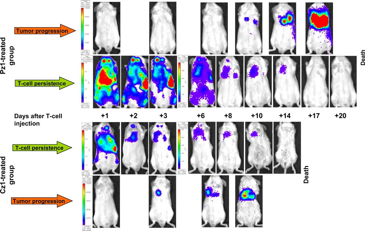
- FIGURE 1.
BLI of adoptively transferred T-cell persistence and tumor progression. T-cell distribution and persistence were imaged with CBRluc and d-luciferin. Tumor progression was imaged with Rluc and coelenterazine. Data for 2 representative animals from Pz1-treated and Cz1-treated groups are shown. Note different scales for assessment of T-cell bioluminescence signal intensities on days +1–3 and +6–20 after T-cell injection. Same scales were used for assessment of tumor BLI data.
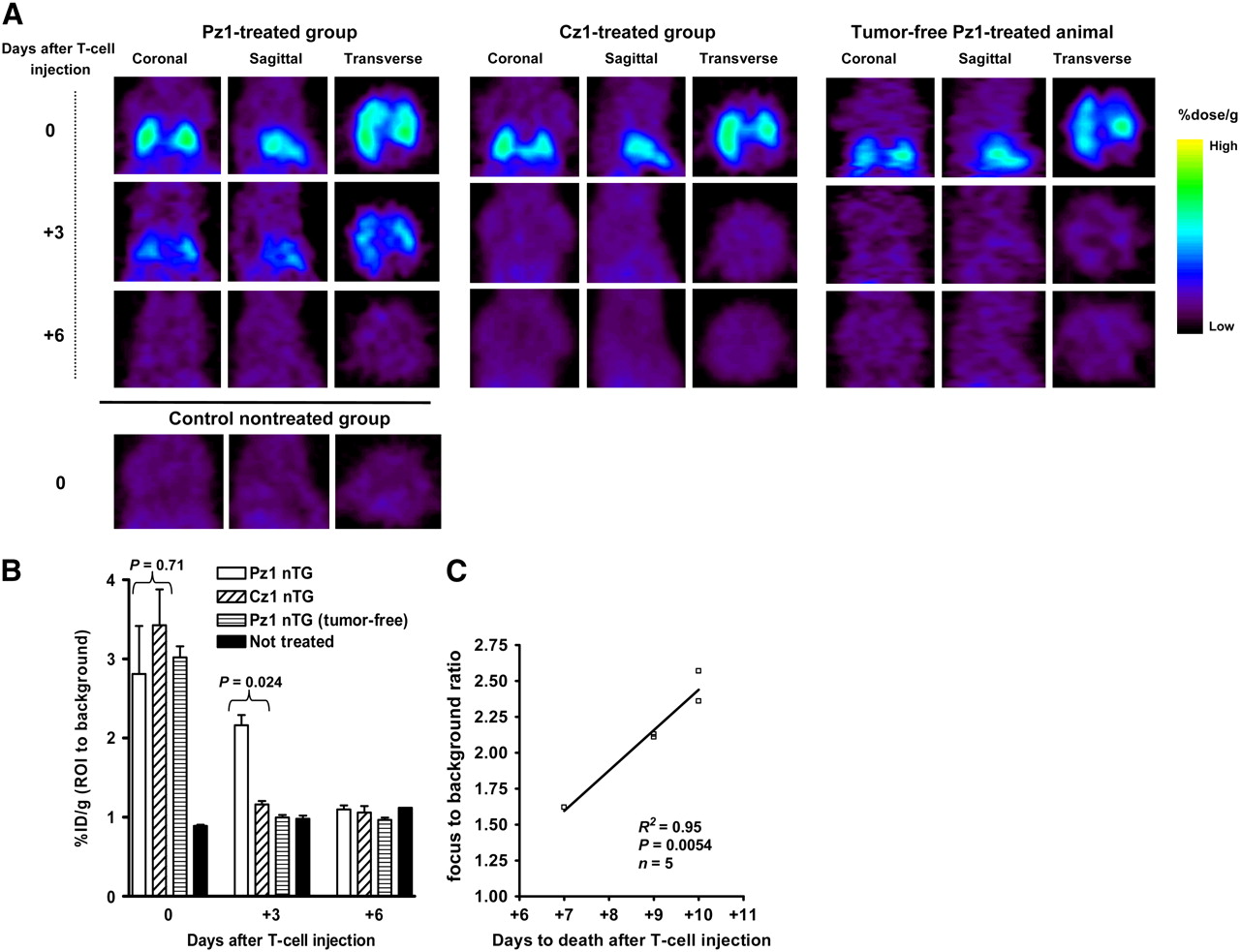
- FIGURE 2.
Quantitative analysis by BLI of adoptively transferred T-cell persistence and tumor progression in individual animals. Solid lines represent bioluminescence signal intensity for T-cell BLI; dotted lines represent that for tumor BLI. (A) All animals in Pz1-treated group showed uniform decrease in bioluminescence signal from T cells with pronounced delay by day +3. Mouse 5 in Pz1-treated group showed early tumor progression, similar to that in Cz1-treated group. (B) In all animals in Cz1-treated group, T-cell bioluminescence signal decreased rapidly without any delay. ph = photons; sr = steradian.
- FIGURE 3.
Small-animal PET imaging of 18F-FEAU accumulation at different time points after adoptive transfer of CAR-positive, HSV1tk/GFP-positive T lymphocytes. (A) Three mutually perpendicular projections for typical Pz1-treated, Cz1-treated, and nontreated tumor-bearing and Pz1-treated tumor-free animals are presented. On day of T-cell injection (day 0), highly intense small-animal PET signal clearly demonstrated presence of pooled T cells in lung areas of treated animals. By day +3, 18F-FEAU accumulation was detected only in Pz1-treated tumor-bearing animals. No radiotracer accumulation was detected on day +6. (B) Small-animal PET signal quantitation performed as described in Materials and Methods. Statistically significant differences in levels of 18F-FEAU accumulation between Pz1- and Cz1-treated tumor-bearing animals were observed on day +3. Columns represent means; bars represent SEMs. (C) Regression analysis of T-cell persistence in and overall survival of Pz1-treated tumor-bearing animals. Ratios of %ID/g in ROI to %ID/g in background for individual animals on day +3 were plotted against time to death. Nearly linear relationship (R2 = 0.95) between small-animal PET signal intensity and survival was revealed.
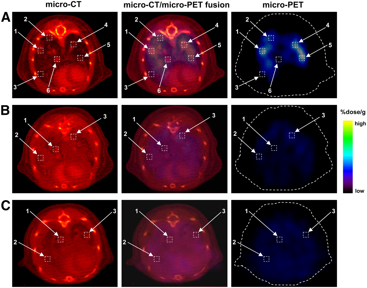
- FIGURE 4.
Small-animal PET/micro-CT fusion imaging and analysis. Analysis of colocalization of 18F-FEAU accumulation on small-animal PET images with tumor regions or thoracic structures (vessels and lung parenchyma) on micro-CT images is shown. One representative tumor-bearing animal each from Pz1-treated (A), Cz1-treated (B), and control nontreated (C) groups is shown. (A) In Pz1-treated animals, highest levels of 18F-FEAU accumulation were detected (boxes 1, 2, 4, and 5) in regions corresponding to tumor foci on micro-CT image. Lower signal intensity was observed in intact lung parenchyma and blood vessel zones (boxes 3 and 6, respectively). (B and C) In contrast, Cz1-treated (B) and nontreated (C) animals had low small-animal PET signal intensities (boxes 1, 2, and 3) in selected anatomic areas on micro-CT images. Heart and blood vessels were enhanced by vasculature contrast agent Fenestra VC. Quantitative analysis of 18F-FEAU accumulation is shown in Table 1.
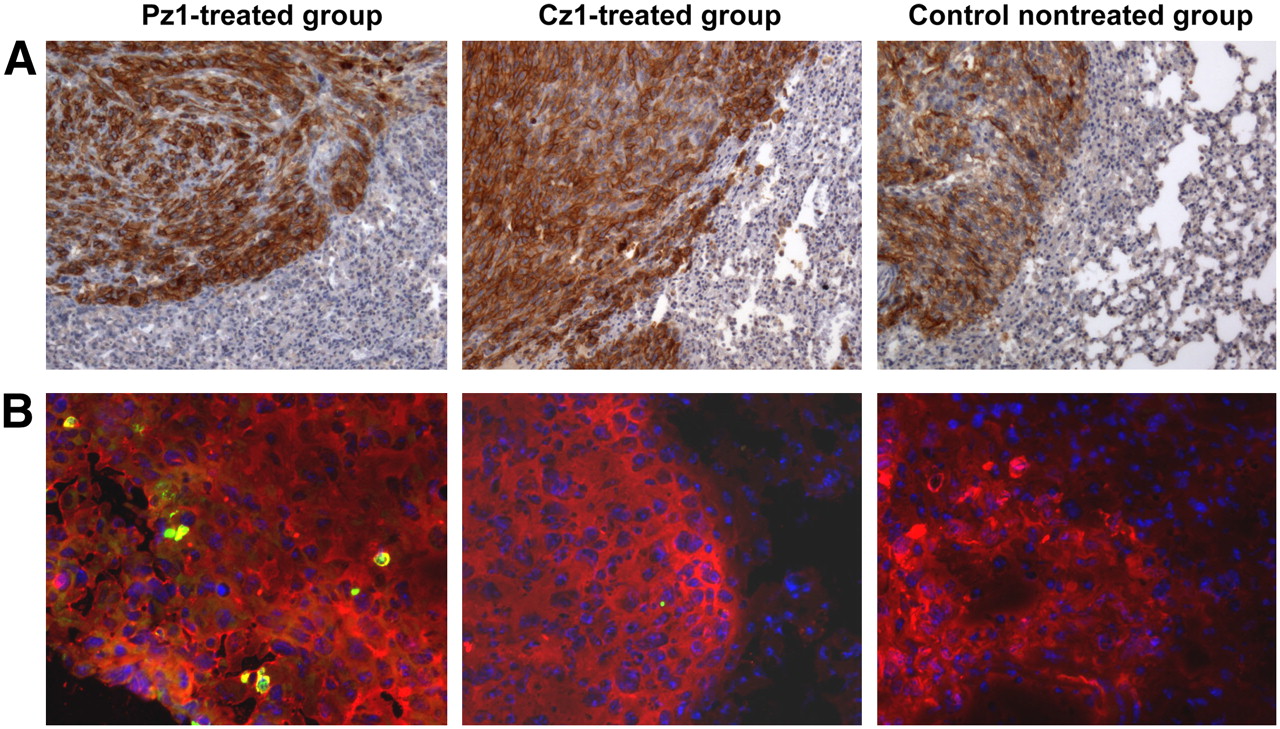
- FIGURE 5.
Histologic analysis of tumor specimens. (A) Anti-hPSMA antibody immunohistochemical staining revealed presence of hPSMA-positive tumor cells in lungs from representative Pz1-treated, Cz1-treated, and nontreated tumor-bearing animals in small-animal PET imaging experiment. Magnification, ×10. (B) Anti-GFP antibody was used for immunofluorescence microscopy of HSV1tk/GFP-expressing T cells (green) infiltrating hPSMA-positive tumors (red; stained with anti-hPSMA antibody). Nuclei were stained with 4,6-diamidino-2-phenylindole (blue). Magnification, ×20. Similar results were obtained in BLI experiment.

Tables
- TABLE 1
Ratios of %ID/g in ROI to %ID/g in Background, as Calculated from Small-Animal PET Images
ROI-to-background ratio for box no.: Group 1 2 3 4 5 6 Pz1 treated 2.25 1.73 1.10 2.11 2.47 1.17 Cz1 treated 1.02 0.98 1.00 Not treated 0.89 0.88 0.94 Box numbers correspond to box numbers in Figure 4.
Supplemental Data
Files in this Data Supplement:


Jump to section
Related Articles
Cited By...
- Dendritic cells accelerate CAR T cells in irradiated tumors through chimeric synapses
- Nanoparticles That Reshape the Tumor Milieu Create a Therapeutic Window for Effective T-cell Therapy in Solid Malignancies
- Immuno-PET Imaging of Engineered Human T Cells in Tumors
- Dynamic imaging for CAR-T-cell therapy
- Comparative Analysis of T Cell Imaging with Human Nuclear Reporter Genes
- Molecular Imaging with Bioluminescence and PET Reveals Viral Oncolysis Kinetics and Tumor Viability
- A New Pyrimidine-Specific Reporter Gene: A Mutated Human Deoxycytidine Kinase Suitable for PET During Treatment with Acycloguanosine-Based Cytotoxic Drugs
- Redirecting T-cell specificity by introducing a tumor-specific chimeric antigen receptor
- Nuclear Imaging of Cancer Cell Therapies