
Main menu

  • Home
  • Content
    • Current
    • Ahead of print
    • Past Issues
    • JNM Supplement
    • SNMMI Annual Meeting Abstracts
    • Continuing Education
    • JNM Podcasts
  • Subscriptions
    • Subscribers
    • Institutional and Non-member
    • Rates
    • Journal Claims
    • Corporate & Special Sales
  • Authors
    • Submit to JNM
    • Information for Authors
    • Assignment of Copyright
    • AQARA requirements
  • Info
    • Reviewers
    • Permissions
    • Advertisers
  • About
    • About Us
    • Editorial Board
    • Contact Information
  • More
    • Alerts
    • Feedback
    • Help
    • SNMMI Journals
  • SNMMI
    • JNM
    • JNMT
    • SNMMI Journals
    • SNMMI

User menu

  • Subscribe
  • My alerts
  • Log in
  • My Cart

Search

  • Advanced search
Journal of Nuclear Medicine
  • SNMMI
    • JNM
    • JNMT
    • SNMMI Journals
    • SNMMI
  • Subscribe
  • My alerts
  • Log in
  • My Cart
Journal of Nuclear Medicine

Advanced Search

  • Home
  • Content
    • Current
    • Ahead of print
    • Past Issues
    • JNM Supplement
    • SNMMI Annual Meeting Abstracts
    • Continuing Education
    • JNM Podcasts
  • Subscriptions
    • Subscribers
    • Institutional and Non-member
    • Rates
    • Journal Claims
    • Corporate & Special Sales
  • Authors
    • Submit to JNM
    • Information for Authors
    • Assignment of Copyright
    • AQARA requirements
  • Info
    • Reviewers
    • Permissions
    • Advertisers
  • About
    • About Us
    • Editorial Board
    • Contact Information
  • More
    • Alerts
    • Feedback
    • Help
    • SNMMI Journals
  • View or Listen to JNM Podcast
  • Visit JNM on Facebook
  • Join JNM on LinkedIn
  • Follow JNM on Twitter
  • Subscribe to our RSS feeds
Research ArticleCLINICAL INVESTIGATIONS

Quantitative Diagnostic Performance of Myocardial Perfusion SPECT with Attenuation Correction in Women

Arik Wolak, Piotr J. Slomka, Mathews B. Fish, Santiago Lorenzo, Daniel S. Berman and Guido Germano
Journal of Nuclear Medicine June 2008, 49 (6) 915-922; DOI: https://doi.org/10.2967/jnumed.107.049387
Arik Wolak
  • Find this author on Google Scholar
  • Find this author on PubMed
  • Search for this author on this site
Piotr J. Slomka
  • Find this author on Google Scholar
  • Find this author on PubMed
  • Search for this author on this site
Mathews B. Fish
  • Find this author on Google Scholar
  • Find this author on PubMed
  • Search for this author on this site
Santiago Lorenzo
  • Find this author on Google Scholar
  • Find this author on PubMed
  • Search for this author on this site
Daniel S. Berman
  • Find this author on Google Scholar
  • Find this author on PubMed
  • Search for this author on this site
Guido Germano
  • Find this author on Google Scholar
  • Find this author on PubMed
  • Search for this author on this site
  • Article
  • Figures & Data
  • Info & Metrics
  • PDF
Loading

Article Figures & Data

Figures

  • Tables
  • FIGURE 1. 
    • Download figure
    • Open in new tab
    • Download powerpoint
    FIGURE 1. 

    Correlations and bias plots (Bland–Altman analysis) for SSS and TPD and for NC and AC.
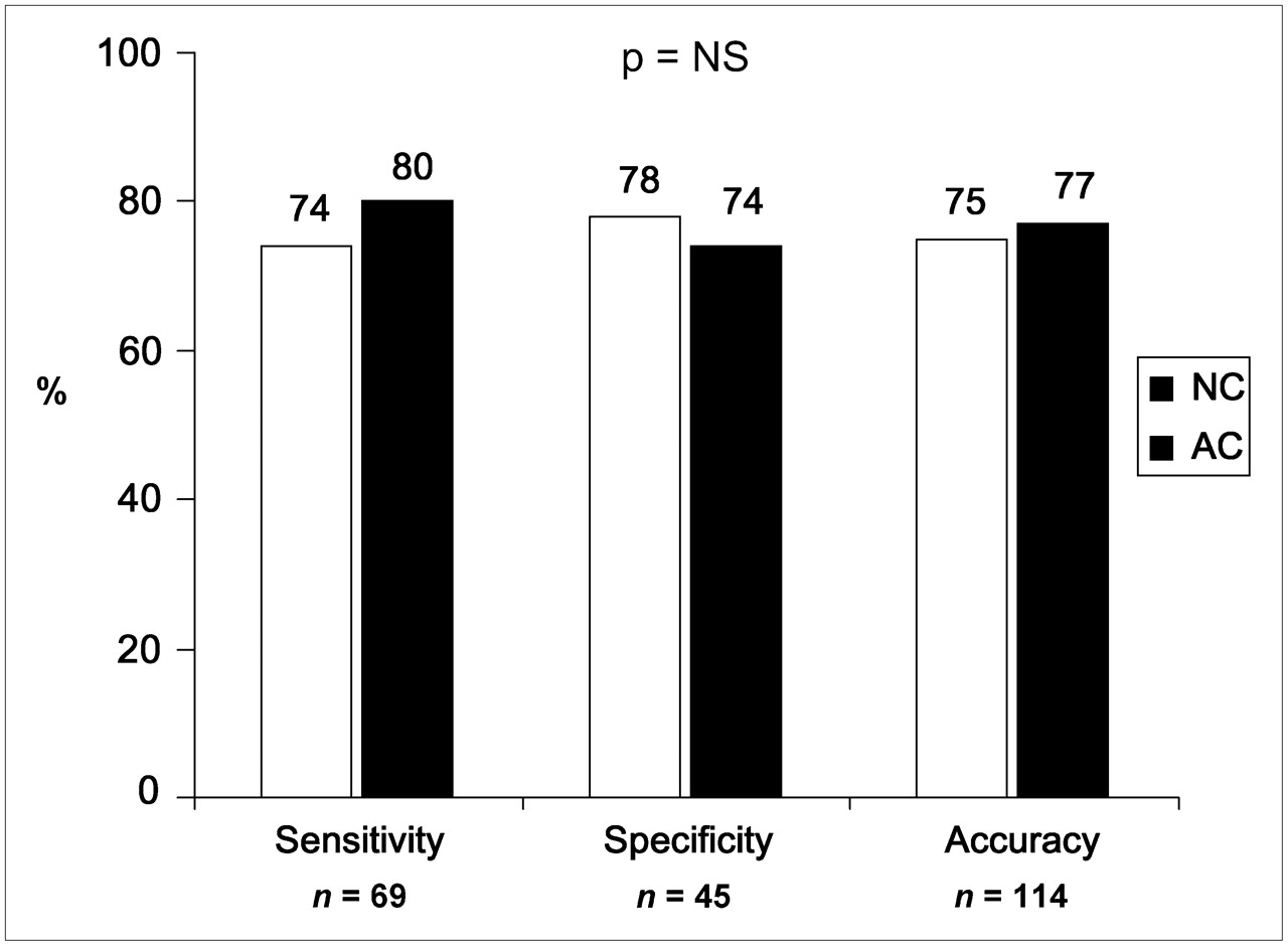
  • FIGURE 2. 
    • Download figure
    • Open in new tab
    • Download powerpoint
    FIGURE 2. 

    Comparison of quantitative results obtained with NC and AC for SSS abnormality threshold of ≥3. NS = not significant.
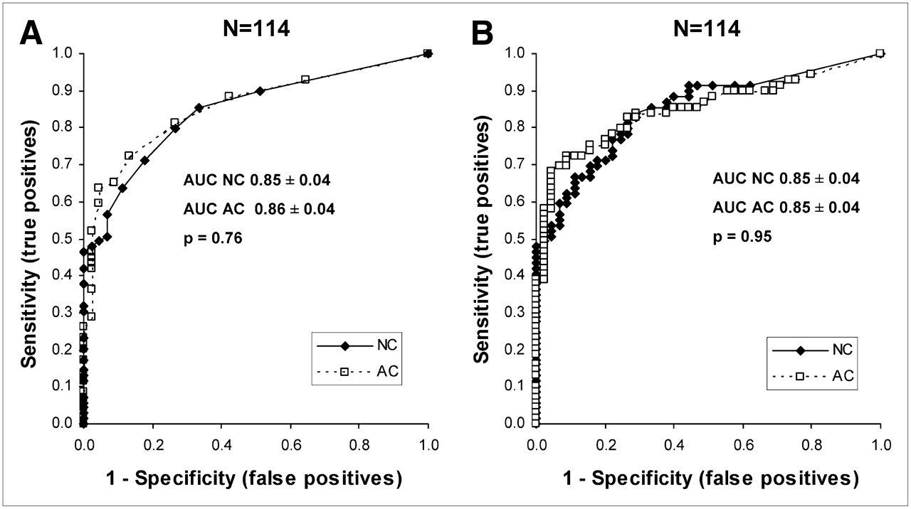
  • FIGURE 3. 
    • Download figure
    • Open in new tab
    • Download powerpoint
    FIGURE 3. 

    ROC curves for detection of ≥70% stenosis with SSS (A) and TPD (B).
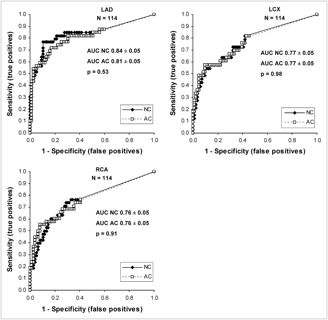
  • FIGURE 4. 
    • Download figure
    • Open in new tab
    • Download powerpoint
    FIGURE 4. 

    ROC curves for detection of ≥70% stenosis by coronary artery territory.
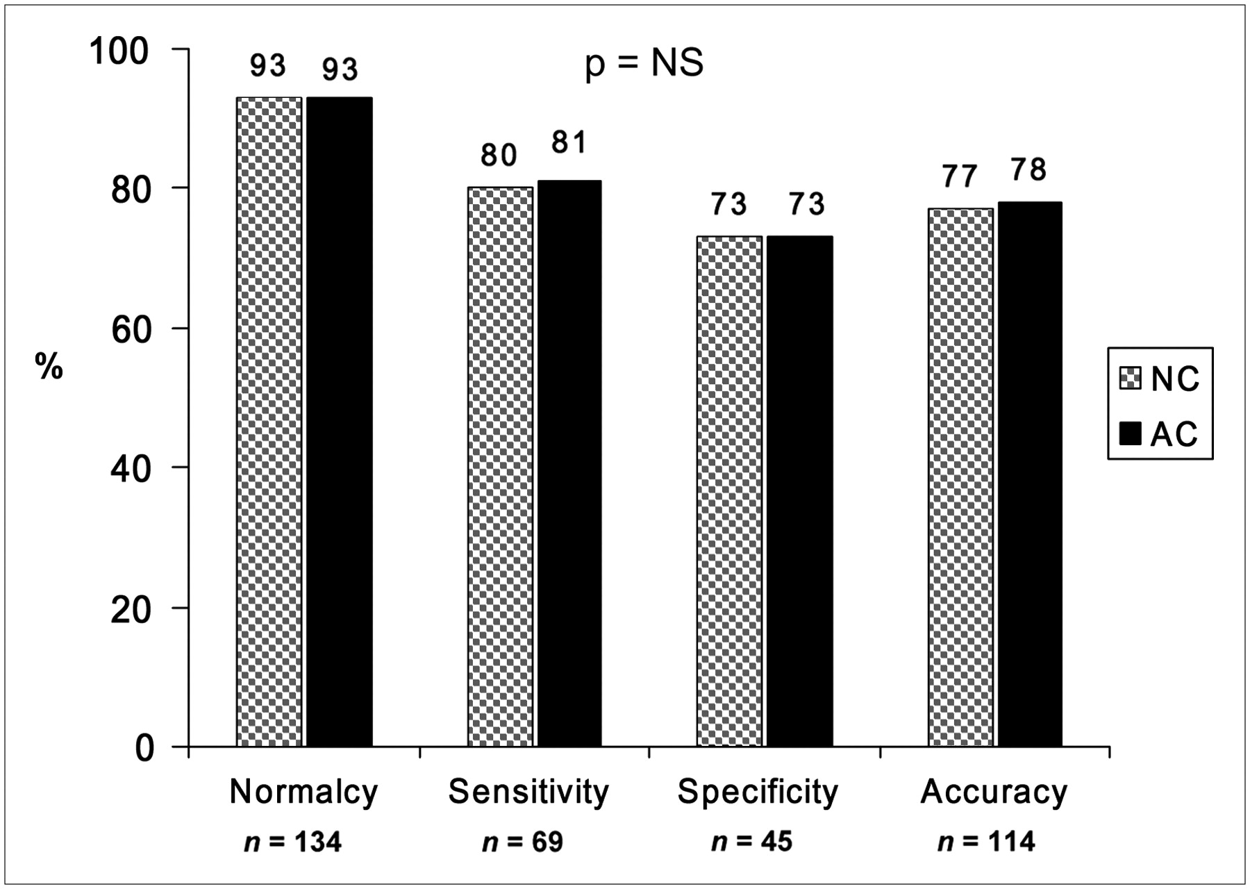
  • FIGURE 5. 
    • Download figure
    • Open in new tab
    • Download powerpoint
    FIGURE 5. 

    Comparison of quantitative results obtained with NC and AC for TPD abnormality threshold of ≥3. NS = not significant.

  • FIGURE 6. 
    • Download figure
    • Open in new tab
    • Download powerpoint
    FIGURE 6. 

    Examples for 3 cases. (A) 55-y-old hypertensive, obese (BMI = 35) patient who had atypical chest pain and who underwent pharmacologic stress MPS. Coronary angiography revealed no evidence of CAD. NC image (top) revealed basal inferior perfusion defect in territory of RCA, whereas AC image (bottom) depicted normal study. (B) 67-y-old overweight (BMI = 26), hypertensive, dyslipidemic, diabetic patient who underwent exercise stress MPS. NC image (top) depicted normal study, whereas AC image (bottom) revealed perfusion defect in territory of LAD. This perfusion defect might have been attributable to apical thinning apparent on AC but not NC studies. (C) 62-y-old hypertensive, diabetic, dyslipidemic, morbidly obese (BMI = 40) patient who had atypical chest pain and who underwent pharmacologic stress MPS. Coronary angiography revealed 90% RCA stenosis. NC image (top) revealed minimal nonsignificant perfusion defect. AC image (bottom), however, correctly revealed basal inferior perfusion defect in territory of RCA.

Tables

  • Figures
    • View popup
    TABLE 1

    Patient Characteristics

    ParameterDatabase group (n = 50)LLk group (n = 134)Angiography group (n = 114)
    Age (y), mean ± SD56.7 ± 14.752.2 ± 5.065.2 ± 21.2
    BMI (kg/m2), mean ± SD30.5 ± 6.228.3 ± 15.230.7 ± 6.0
    BMI of ≤3025 (50)96 (70)65 (47)
    Diabetes8 (16)12 (9)35 (30)
    Hypertension22 (44)57 (42)84 (73)
    Hypercholesterolemia19 (38)47 (34)53 (46)
    Chest pain12 (24)54 (39)85 (74)
    Adenosine0 (0)0 (0)143 (76)
    • Data are reported as number (percentage), unless otherwise indicated.

    • View popup
    TABLE 2

    Angiographic Characteristics for 114 Patients

    ParameterNo. (%) of patients
    No stenosis*45 (40)
    Single-vessel disease31 (27)
    Double-vessel disease23 (20)
    Triple-vessel disease15 (13)
    LAD disease55 (48)
    LCX disease46 (40)
    RCA disease51 (45)
    • ↵* Stenosis of ≥70% luminal diameter narrowing was considered significant.

    • View popup
    TABLE 3

    Quantitative Parameters Obtained for AC and NC Data

    Mean ± SD for:
    Parameter (no. of patients)NCACP
    SSS–all (114)6.7 ± 8.26.9 ± 8.00.5
    SSS–no CAD (45)1.6 ± 2.31.8 ± 2.50.6
    SSS–CAD (69)10.0 ± 9.010.2 ± 8.50.6
    SSS–LLk (134)0.5 ± 10.6 ± 1.20.23
    TPD–all (114)8.5 ± 11.59.4 ± 11.70.07
    TPD–no CAD (45)1.6 ± 2.42.1 ± 2.60.2
    TPD–CAD (69)12.9 ± 12.914.1 ± 12.10.1
    TPD–LLk (134)0. 4 ± 1.00.7 ± 1.10.18
PreviousNext
Back to top

In this issue

Journal of Nuclear Medicine: 49 (6)
Journal of Nuclear Medicine
Vol. 49, Issue 6
June 2008
  • Table of Contents
  • Table of Contents (PDF)
  • About the Cover
  • Index by author
Print
Download PDF
Article Alerts
Sign In to Email Alerts with your Email Address
Email Article

Thank you for your interest in spreading the word on Journal of Nuclear Medicine.

NOTE: We only request your email address so that the person you are recommending the page to knows that you wanted them to see it, and that it is not junk mail. We do not capture any email address.

Enter multiple addresses on separate lines or separate them with commas.
Quantitative Diagnostic Performance of Myocardial Perfusion SPECT with Attenuation Correction in Women
(Your Name) has sent you a message from Journal of Nuclear Medicine
(Your Name) thought you would like to see the Journal of Nuclear Medicine web site.
Citation Tools
Quantitative Diagnostic Performance of Myocardial Perfusion SPECT with Attenuation Correction in Women
Arik Wolak, Piotr J. Slomka, Mathews B. Fish, Santiago Lorenzo, Daniel S. Berman, Guido Germano
Journal of Nuclear Medicine Jun 2008, 49 (6) 915-922; DOI: 10.2967/jnumed.107.049387

Citation Manager Formats

  • BibTeX
  • Bookends
  • EasyBib
  • EndNote (tagged)
  • EndNote 8 (xml)
  • Medlars
  • Mendeley
  • Papers
  • RefWorks Tagged
  • Ref Manager
  • RIS
  • Zotero
Share
Quantitative Diagnostic Performance of Myocardial Perfusion SPECT with Attenuation Correction in Women
Arik Wolak, Piotr J. Slomka, Mathews B. Fish, Santiago Lorenzo, Daniel S. Berman, Guido Germano
Journal of Nuclear Medicine Jun 2008, 49 (6) 915-922; DOI: 10.2967/jnumed.107.049387
Twitter logo Facebook logo LinkedIn logo Mendeley logo
  • Tweet Widget
  • Facebook Like
  • Google Plus One
Bookmark this article

Jump to section

  • Article
    • Abstract
    • MATERIALS AND METHODS
    • RESULTS
    • DISCUSSION
    • CONCLUSION
    • Acknowledgments
    • Footnotes
    • References
  • Figures & Data
  • Info & Metrics
  • PDF

Related Articles

  • This Month in JNM
  • PubMed
  • Google Scholar

Cited By...

  • Diagnostic Performance of Attenuation-Corrected Myocardial Perfusion Imaging for Coronary Artery Disease: A Systematic Review and Meta-Analysis
  • Role of Noninvasive Testing in the Clinical Evaluation of Women With Suspected Ischemic Heart Disease: A Consensus Statement From the American Heart Association
  • Improved Quantification and Normal Limits for Myocardial Perfusion Stress-Rest Change
  • Directions and Magnitudes of Misregistration of CT Attenuation-Corrected Myocardial Perfusion Studies: Incidence, Impact on Image Quality, and Guidance for Reregistration
  • Google Scholar

More in this TOC Section

  • Feasibility of Ultra-Low-Activity 18F-FDG PET/CT Imaging Using a Long–Axial-Field-of-View PET/CT System
  • Cardiac Presynaptic Sympathetic Nervous Function Evaluated by Cardiac PET in Patients with Chronotropic Incompetence Without Heart Failure
  • Validation and Evaluation of a Vendor-Provided Head Motion Correction Algorithm on the uMI Panorama PET/CT System
Show more Clinical Investigations

Similar Articles

SNMMI

© 2025 SNMMI

Powered by HighWire