


Article Figures & Data
Figures
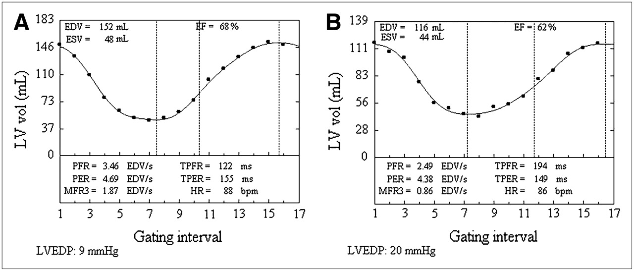
- FIGURE 1.
Left ventricular diastolic filling curves derived from 4D-MSPECT. (A) Curve with normal diastolic filling parameters. (B) Curve with abnormal diastolic filling parameters. LVEDPs determined at subsequent cardiac catheterization are shown. bpm = beats per minute; EF = ejection fraction; ESV = end-systolic volume; HR = heart rate; LV = left ventricular; MFR3 = filling rate during first third of diastole; PER = peak emptying rate; TPER = time to peak emptying rate; vol = volume.
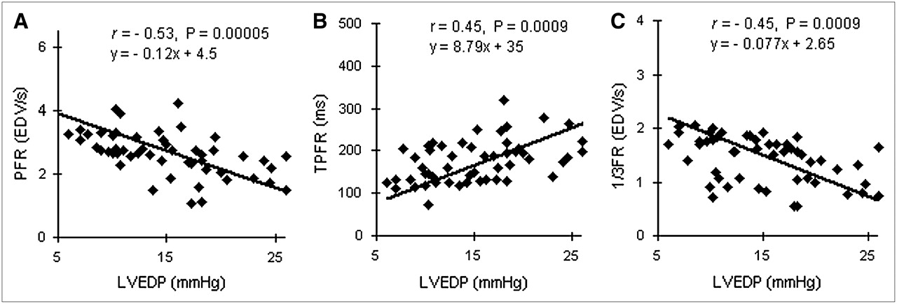
- FIGURE 2.
Correlations between 4D-MSPECT diastolic parameters and LVEDP.
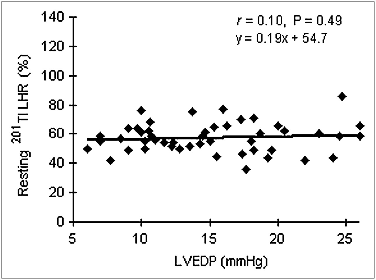
- FIGURE 3.
Correlation between LVEDP and resting 201Tl LHR.
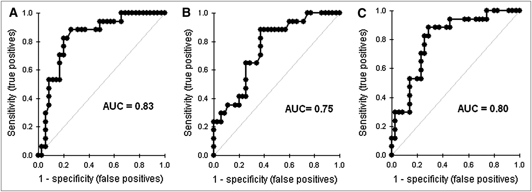
- FIGURE 4.
ROC curve analysis for ability of PFR (A), TPFR (B), and 1/3FR (C) to detect LVEDPs of ≥18 mm Hg or <18 mm Hg.

Tables
Characteristic Mean ± SD or mean (%) Age (y) 58 ± 11 Men* 30 (58) CAD* 38 (73) Diabetes mellitus* 15 (29) Hypertension* 42 (81) Interval between MPI and cardiac catheterization (d) 8 ± 3.50 Heart rate (beats/min) 72 ± 12 Ejection fraction (%) 54.2 ± 4.66 Mean LVEDP (mm Hg) 15.07 ± 5.30 Resting 201Tl LHR 0.57 ± 0.10 PFR (EDV/s) 2.63 ± 0.68 TPFR (ms) 175.77 ± 49.99 1/3FR (EDV/s) 1.43 ± 0.43 ↵* Reported as number of patients.
Diastolic filling variable(s) AUC 95% Confidence interval for AUC Optimal cutoff value(s) for detecting LVEDPs of ≥18 mm Hg Sensitivity (%) Specificity (%) PPV (%) NPV (%) PFR 0.83 0.72–0.95 ≤2.57 EDV/s 82 80 67 90 TPFR 0.75 0.62–0.89 ≥161 ms 88 63 54 92 1/3FR 0.80 0.68–0.93 ≤1.52 EDV/s 82 74 61 90 PFR + 1/3FR ≤2.57 EDV/s + ≤1.52 EDV/s 67 94 86 84


Jump to section
Related Articles
Cited By...
- No citing articles found.