


Article Figures & Data
Figures
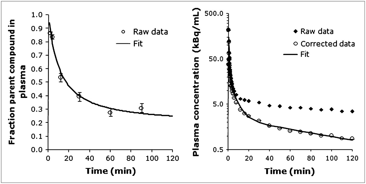
- FIGURE 1.
(A) Fraction of plasma activity associated with unmetabolized 11C-CUMI-101 in baboon plasma during a single scan. Fitted line is estimated from the Hill model (1 − AtB/[tB + C]). (B) Total and metabolite-corrected plasma radioactivity. Fitted line represents the 3-exponential function fit to the data. Error bars represent weights calculated by the delta method.
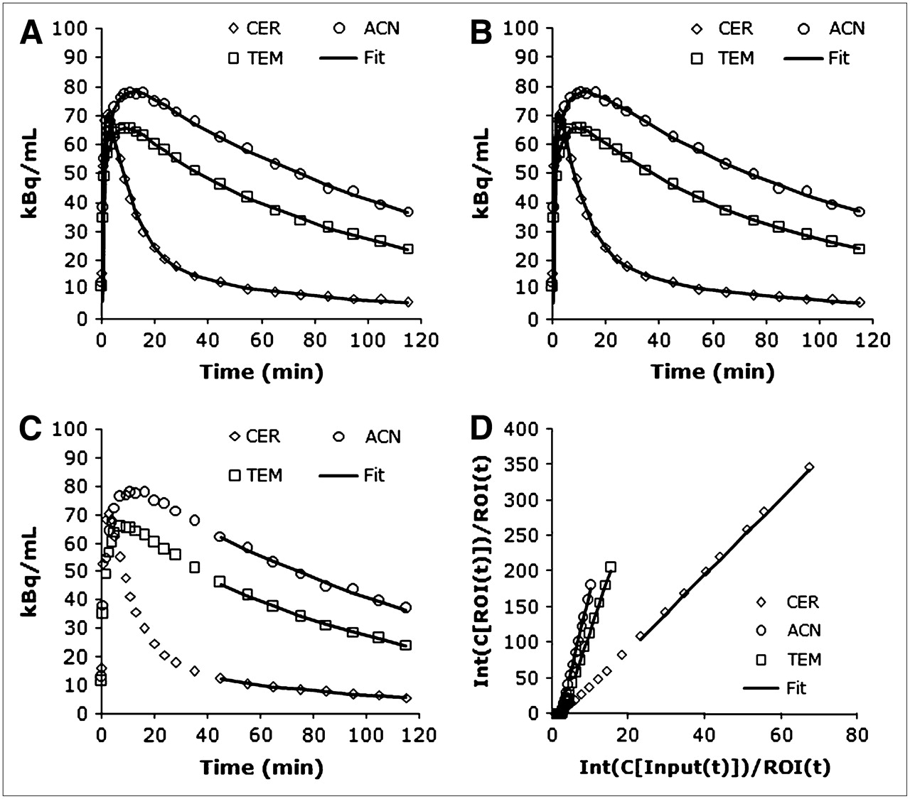
- FIGURE 2.
Regional time–activity curves and corresponding least-squares minimized fits to 3 different models. (A) 2TCNI. (B) Basis pursuit. (C and D) LEGA (native and transformed space, respectively). CER = cerebellum; ACN = anterior cingulate; TEM = temporal cortex.
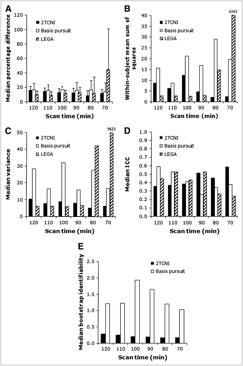
- FIGURE 3.
Modeling metrics comparing 2TCNI, basis pursuit, and LEGA models at 6 different scan durations. (A) PD. (B) WSMSS. (C) Variance. (D) ICC. (E) Identifiability. For all measures, medians are taken across all scans and all ROIs, for both subjects. (A) Error bars represent average deviation from the median. Asterisk in A indicates that the modeling of dorsal raphe did not converge for basis pursuit for the 100-min scan time (for a single study) and thus was left out of the analysis.
- FIGURE 4.
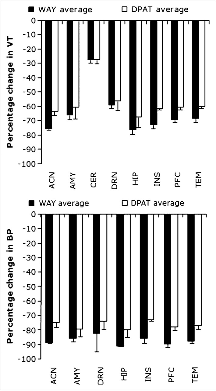
Mean percentage change (PD) in VT (A) and BPF (B) of 11C-CUMI-101 after blockade with 8-OH-DPAT and WAY-100635. Values were derived from the LEGA model with a 100-min scan duration. ACN = anterior cingulate; AMY = amygdala; CER = cerebellum; DRN = dorsal raphe; HIP = hippocampus; INS = insula; PFC = prefrontal cortex; TEM = temporal cortex.
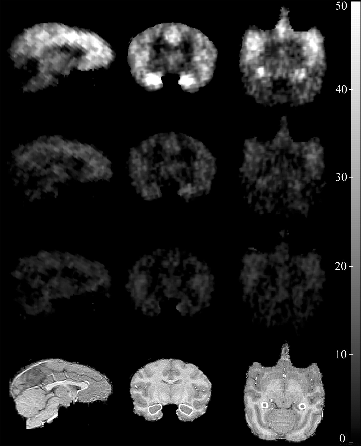
- FIGURE 5.
(First row) Sagittal, coronal, and axial parametric PET images of 11C-CUMI-101 BPF values in baboon brain. (Second and third rows): PET images after blockade by 8-OH-DPAT and WAY-100635, respectively. (Fourth row) Corresponding coregistered MR images. On the MR images, the hippocampus has been highlighted.
- FIGURE 6.
Correlation plots of VT values across various modeling approaches including ROI-analysis vs. voxel-based analyses. (A) 2TCNI voxel vs. 2TCNI ROI (R2 = 0.97). (B) 2TCNI ROI vs. LEGA ROI (R2 = 0.97). (C) Basis pursuit ROI vs. LEGA ROI (R2 = 0.97). (D) 2TCNI voxel vs. LEGA ROI (R2 of 1.00). Line of identity has been added for reference. Data reflect all ROIs in all studies.


Tables
2TCNI Basis pursuit LEGA ROI VT BP VT BP VT BP ACN 90 90 110 100 100 100 AMY 80 80 120 100 90 90 DRN 110 110 120 120 100 90 HIP 110 110 110 110 90 90 INS 80 70 120 100 90 90 PFC 110 100 90 90 80 80 TEM 70 70 90 90 90 80 ACN = anterior cingulate; AMY = amygdala; DRN = dorsal raphe; HIP = hippocampus; INS = insula; PFC = prefrontal cortex; TEM = temporal cortex.
Numbers indicate the shortest scan duration for which the average VT and BPF values from all test–retest experiments for each ROI were between 95% and 105% of the average value, when using 120-min scan duration and also when SD of these percentages is <10%.
Metric 2TCNI LEGA Basis PD 2 1 3 WSMSS 2 1 3 VAR 2 1 3 ICC 2 2 2 ID 1 N/A 2 ↵* With 1 being the best of each criterion.
VAR = variance; ID = identifiability; N/A = not applicable.


Jump to section
Related Articles
Cited By...
- 11C-CUMI-101, a PET Radioligand, Behaves as a Serotonin 1A Receptor Antagonist and Also Binds to {alpha}1 Adrenoceptors in Brain
- Radiosynthesis and Preclinical Evaluation of 18F-F13714 as a Fluorinated 5-HT1A Receptor Agonist Radioligand for PET Neuroimaging
- In Vivo Quantification of Human Serotonin 1A Receptor Using 11C-CUMI-101, an Agonist PET Radiotracer