
Main menu

  • Home
  • Content
    • Current
    • Ahead of print
    • Past Issues
    • JNM Supplement
    • SNMMI Annual Meeting Abstracts
    • Continuing Education
    • JNM Podcasts
  • Subscriptions
    • Subscribers
    • Institutional and Non-member
    • Rates
    • Journal Claims
    • Corporate & Special Sales
  • Authors
    • Submit to JNM
    • Information for Authors
    • Assignment of Copyright
    • AQARA requirements
  • Info
    • Reviewers
    • Permissions
    • Advertisers
  • About
    • About Us
    • Editorial Board
    • Contact Information
  • More
    • Alerts
    • Feedback
    • Help
    • SNMMI Journals
  • SNMMI
    • JNM
    • JNMT
    • SNMMI Journals
    • SNMMI

User menu

  • Subscribe
  • My alerts
  • Log in
  • Log out
  • My Cart

Search

  • Advanced search
Journal of Nuclear Medicine
  • SNMMI
    • JNM
    • JNMT
    • SNMMI Journals
    • SNMMI
  • Subscribe
  • My alerts
  • Log in
  • Log out
  • My Cart
Journal of Nuclear Medicine

Advanced Search

  • Home
  • Content
    • Current
    • Ahead of print
    • Past Issues
    • JNM Supplement
    • SNMMI Annual Meeting Abstracts
    • Continuing Education
    • JNM Podcasts
  • Subscriptions
    • Subscribers
    • Institutional and Non-member
    • Rates
    • Journal Claims
    • Corporate & Special Sales
  • Authors
    • Submit to JNM
    • Information for Authors
    • Assignment of Copyright
    • AQARA requirements
  • Info
    • Reviewers
    • Permissions
    • Advertisers
  • About
    • About Us
    • Editorial Board
    • Contact Information
  • More
    • Alerts
    • Feedback
    • Help
    • SNMMI Journals
  • View or Listen to JNM Podcast
  • Visit JNM on Facebook
  • Join JNM on LinkedIn
  • Follow JNM on Twitter
  • Subscribe to our RSS feeds
OtherCONTINUING EDUCATION

Small-Animal SPECT and SPECT/CT: Important Tools for Preclinical Investigation

Benjamin L. Franc, Paul D. Acton, Carina Mari and Bruce H. Hasegawa
Journal of Nuclear Medicine October 2008, 49 (10) 1651-1663; DOI: https://doi.org/10.2967/jnumed.108.055442
Benjamin L. Franc
  • Find this author on Google Scholar
  • Find this author on PubMed
  • Search for this author on this site
Paul D. Acton
  • Find this author on Google Scholar
  • Find this author on PubMed
  • Search for this author on this site
Carina Mari
  • Find this author on Google Scholar
  • Find this author on PubMed
  • Search for this author on this site
Bruce H. Hasegawa
  • Find this author on Google Scholar
  • Find this author on PubMed
  • Search for this author on this site
  • Article
  • Figures & Data
  • Info & Metrics
  • PDF
Loading

Article Figures & Data

Figures

  • FIGURE 1. 
    • Download figure
    • Open in new tab
    • Download powerpoint
    FIGURE 1. 

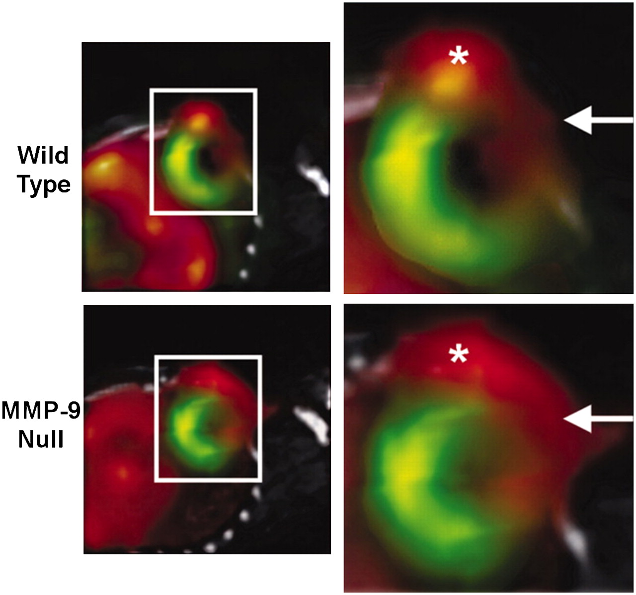
    Coregistered in vivo micro-SPECT/CT images of 201Tl to assess myocardial perfusion and 99mTc-NC100692 targeted at αvβ3 integrin to identify angiogenesis in wild-type and matrix metalloproteinase 9 (MMP-9) null mice. 99mTc-NC100692 micro-SPECT images (red) are fused with 201Tl (green) and CT (gray) images to define uptake of αvβ3-targeted radiotracer relative to 201Tl perfusion defect and anatomic structures within chest (CT performed with both 99mTc and 201Tl data provides template for fusion). Boxed areas on left are magnified on right. Increased 99mTc-NC100692 uptake is seen in anterior-lateral infarct territory (arrows) and 99mTc-NC100692 activity associated with angiogenesis at thoracotomy site (*), demonstrating importance of simply knowing location of sternum as indicated on CT. 99mTc-NC100692 uptake also was noted in liver. (Reprinted with permission of (117).)

  • FIGURE 2. 
    • Download figure
    • Open in new tab
    • Download powerpoint
    FIGURE 2. 

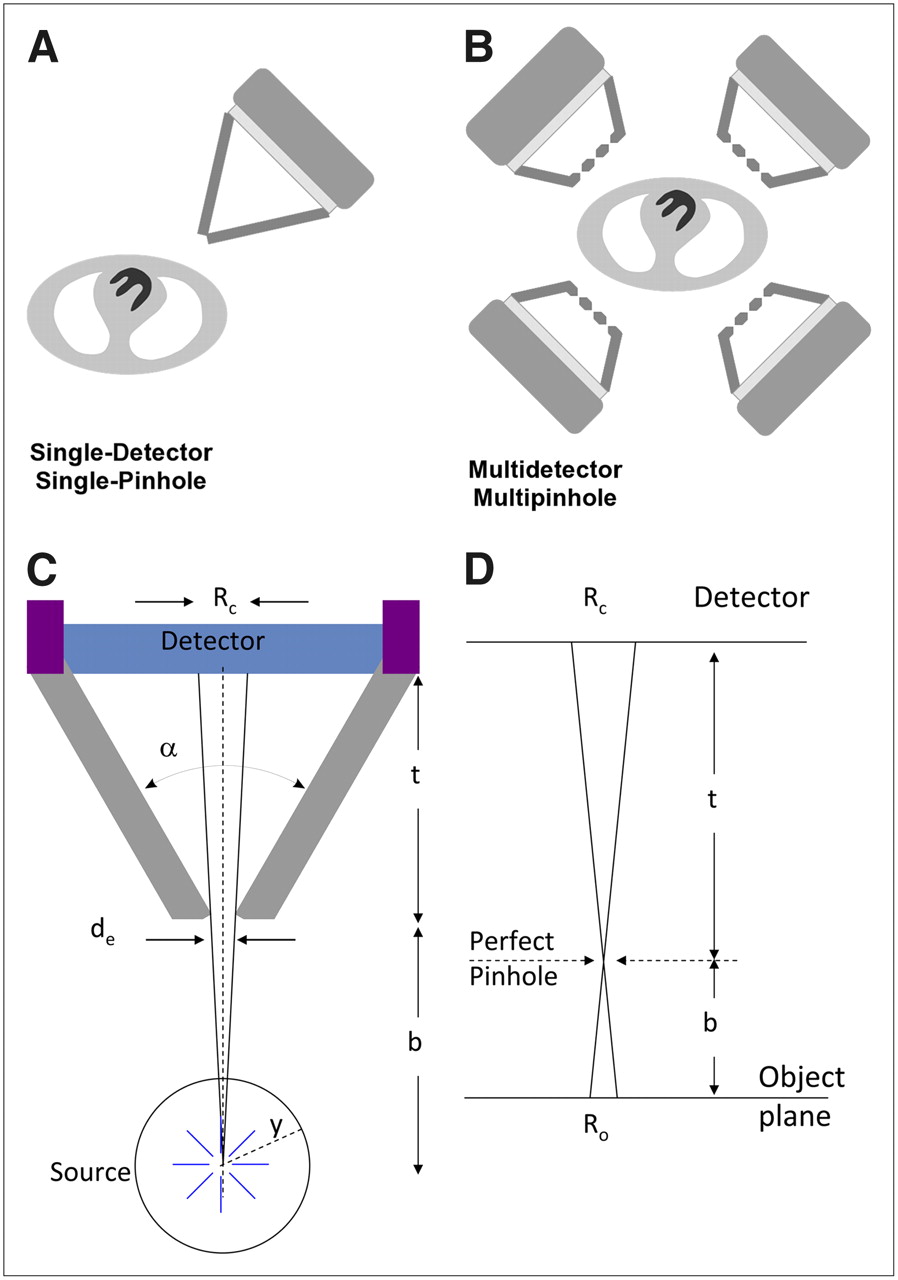
    (A) Early small-animal SPECT systems were developed using single scintillation camera with single-pinhole collimator. (B) Modern small-animal SPECT systems use multiple detectors, each with multipinhole collimators. (C) Schematic of pinhole collimator geometry and relationships between object, its FOV, and its image on plane of detector. Angle between collimator walls (α), linear attenuation coefficient of collimator material (y), and pinhole diameter must be known to derive effective pinhole diameter (de). Geometric resolution measured on detector plane (Rc) is function of de, pinhole aperture-to-object distance (b), and pinhole aperture-to-detector distance (t). In figure, y equals radius of reconstruction circle and Ro equals detector point-spread function projected to object plane (spatial resolution in object plane at distance b from pinhole aperture). More detailed discussion of these relationships may be found in the references authored by Cherry et al., Metzler et al., and Metzler et al. (118–120). (D) Object is magnified by factor b/t onto plane of detector Embedded Image effective resolution can be improved by pinhole magnification.

  • FIGURE 3. 
    • Download figure
    • Open in new tab
    • Download powerpoint
    FIGURE 3. 

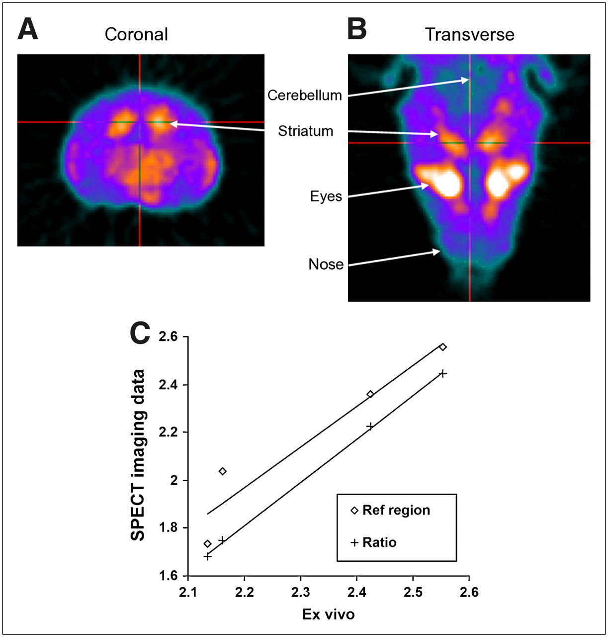
    Coronal (A) and transverse (B) SPECT images showing uptake of DAT imaging agent 99mTc-[2-[[2-[[[3-(4-chlorophenyl)-8-methyl-8-azabicyclo[3,2,1]oct-2-yl]methyl](2-mercaptoethyl)amino]ethyl]amino]ethanethiolato(3-)-N2,N2′,S2,S2′]oxo-[1R-(exo-exo)] (TRODAT-1) in mouse brain 1 h after injection. Generally, greater localization of TRODAT-1 is seen in striatum than in cortical structures. (C) In vivo measurement of striatum-to-cerebellum ratio with SPECT, using kinetic modeling with extraction of input function from cerebellum (Ref region) and from relative striatum-to-cerebellum concentrations at equilibrium (Ratio), showed correlations of R2 = 0.92 (P = 0.04) and R2 = 0.999 (P < 0.01), vs. ex vivo measurements. (Reprinted with permission of (48).)

  • FIGURE 4. 
    • Download figure
    • Open in new tab
    • Download powerpoint
    FIGURE 4. 

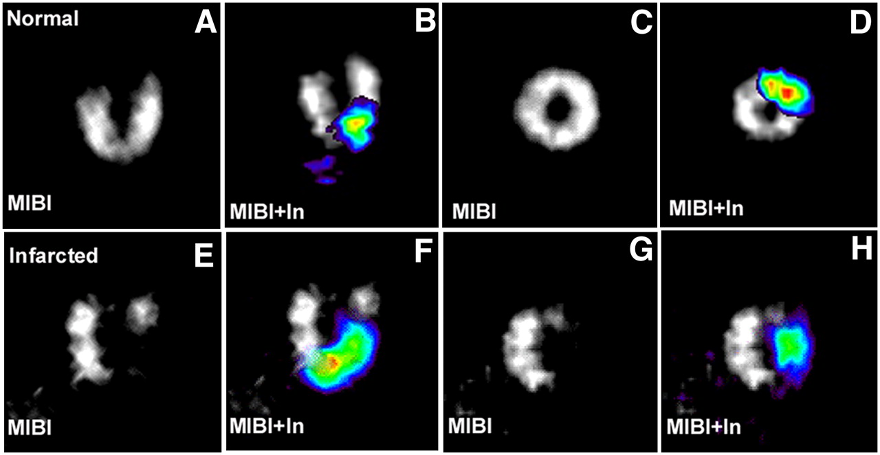
    Cardiac long- and short-axis SPECT images of normal (A and C) and infarcted (E and G) heart using perfusion tracer 99mTc-sestamibi (MIBI). Signal from 111In-labeled stem cells (In, color) was overlaid on gray-scale MIBI images for normal (B and D) and infarcted (F and H) heart.

  • FIGURE 5. 
    • Download figure
    • Open in new tab
    • Download powerpoint
    FIGURE 5. 

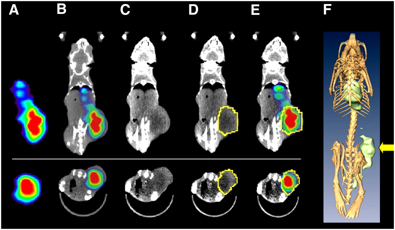
    Small-animal SPECT/CT in oncology research. Coronal (A–E [top], F) and transaxial (A–E [bottom]) images from SPECT/CT study of LNCaP prostate cancer xenograft model 72 h after administration of antibody to PSMA labeled with 177Lu. Tumor uptake is difficult to localize on SPECT images (A) but is easily localized anatomically on SPECT/CT overlay images (B). To quantify uptake of radiopharmaceutical, borders of tumor may be visualized on CT (C). Region of interest is selected on basis of CT (D) and applied to SPECT data (E). Three-dimensional renderings (F; arrow indicates tumor) may be helpful in better understanding anatomic relationships but rarely play role in quantification.

  • FIGURE 6. 
    • Download figure
    • Open in new tab
    • Download powerpoint
    FIGURE 6. 

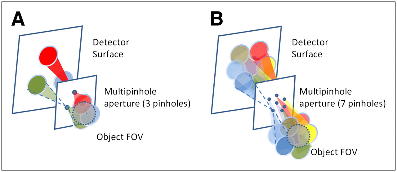
    (A) Schematic of nonoverlapping multipinhole system. Object FOV is projected on detector surface through (in this case) 3 pinholes without overlap. Multipinhole design increases system sensitivity roughly by factor equal to number of pinholes. (B) Schematic of overlapping (multiplexed) multipinhole system. Object FOV is projected on detector surface through (in this case) 7 pinholes. In contrast to system in A, there is significant overlap of projections on detector surface in B.

PreviousNext
Back to top

In this issue

Journal of Nuclear Medicine: 49 (10)
Journal of Nuclear Medicine
Vol. 49, Issue 10
October 2008
  • Table of Contents
  • Table of Contents (PDF)
  • About the Cover
  • Index by author
Print
Download PDF
Article Alerts
Sign In to Email Alerts with your Email Address
Email Article

Thank you for your interest in spreading the word on Journal of Nuclear Medicine.

NOTE: We only request your email address so that the person you are recommending the page to knows that you wanted them to see it, and that it is not junk mail. We do not capture any email address.

Enter multiple addresses on separate lines or separate them with commas.
Small-Animal SPECT and SPECT/CT: Important Tools for Preclinical Investigation
(Your Name) has sent you a message from Journal of Nuclear Medicine
(Your Name) thought you would like to see the Journal of Nuclear Medicine web site.
Citation Tools
Small-Animal SPECT and SPECT/CT: Important Tools for Preclinical Investigation
Benjamin L. Franc, Paul D. Acton, Carina Mari, Bruce H. Hasegawa
Journal of Nuclear Medicine Oct 2008, 49 (10) 1651-1663; DOI: 10.2967/jnumed.108.055442

Citation Manager Formats

  • BibTeX
  • Bookends
  • EasyBib
  • EndNote (tagged)
  • EndNote 8 (xml)
  • Medlars
  • Mendeley
  • Papers
  • RefWorks Tagged
  • Ref Manager
  • RIS
  • Zotero
Share
Small-Animal SPECT and SPECT/CT: Important Tools for Preclinical Investigation
Benjamin L. Franc, Paul D. Acton, Carina Mari, Bruce H. Hasegawa
Journal of Nuclear Medicine Oct 2008, 49 (10) 1651-1663; DOI: 10.2967/jnumed.108.055442
Twitter logo Facebook logo LinkedIn logo Mendeley logo
  • Tweet Widget
  • Facebook Like
  • Google Plus One
Bookmark this article

Jump to section

  • Article
    • Abstract
    • BACKGROUND
    • IMAGE QUANTIFICATION
    • APPLICATION-BASED DESIGN REQUIREMENTS FOR SMALL-ANIMAL SPECT AND SPECT/CT
    • ADVANCES IN SYSTEM DESIGN AND INSTRUMENTATION
    • CONCLUSION
    • Acknowledgments
    • Footnotes
    • References
  • Figures & Data
  • Info & Metrics
  • PDF

Related Articles

  • This Month in JNM
  • PubMed
  • Google Scholar

Cited By...

  • Imaging Capabilities of the Inveon SPECT System Using Single-and Multipinhole Collimators
  • Reproducibility of Serial Left Ventricle Perfusion, Volume, and Ejection Fraction Measurements Using Multiplexed Multipinhole SPECT in Healthy Rats and Rats After Myocardial Infarction
  • Dynamic and Static Small-Animal SPECT in Rats for Monitoring Renal Function After 177Lu-Labeled Tyr3-Octreotate Radionuclide Therapy
  • Google Scholar

More in this TOC Section

  • Approaches to Imaging Immune Activation Using PET
  • Large Language Models and Large Multimodal Models in Medical Imaging: A Primer for Physicians
  • Precision Oncology in Melanoma: Changing Practices
Show more Continuing Education

Similar Articles

SNMMI

© 2025 SNMMI

Powered by HighWire