


Article Figures & Data
Figures
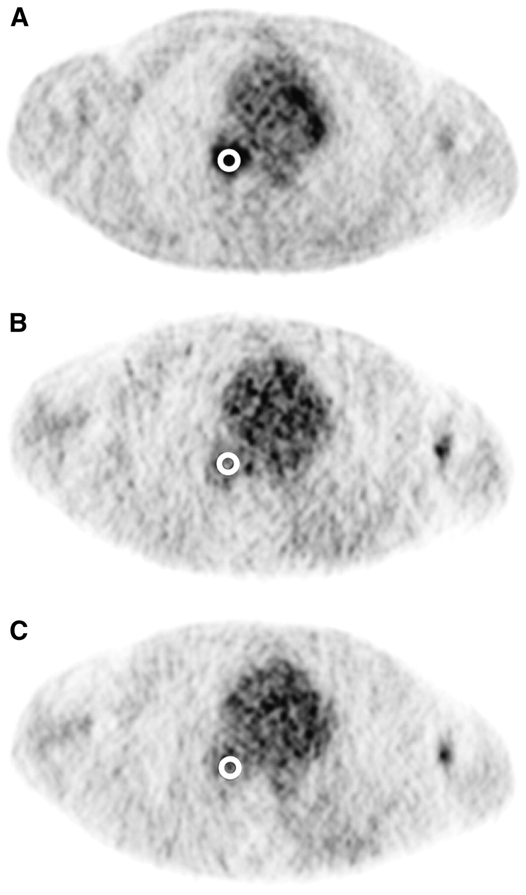
- FIGURE 1.
Circular ROIs (diameter, 15 mm) were manually placed over the most metabolically active part of tumor, as seen in last frame of dynamic 18F-FDG images (A). ROIs were defined in 3 adjacent image slices (not shown). This volume of interest was subsequently applied to 2 15O-water image series (B and C) under assumption that patient did not move between 15O-water and 18F-FDG studies.
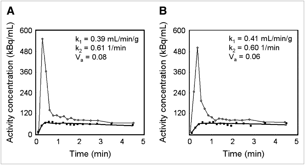
- FIGURE 2.
Example time–activity curves for replicate 15O-water studies performed on same patient. Data for first (A) and second (B) administrations are shown. Circles represent (image-derived) input function and squares denote tumor data. Solid line near tumor data is model fit that produced parameter estimates shown in each figure.
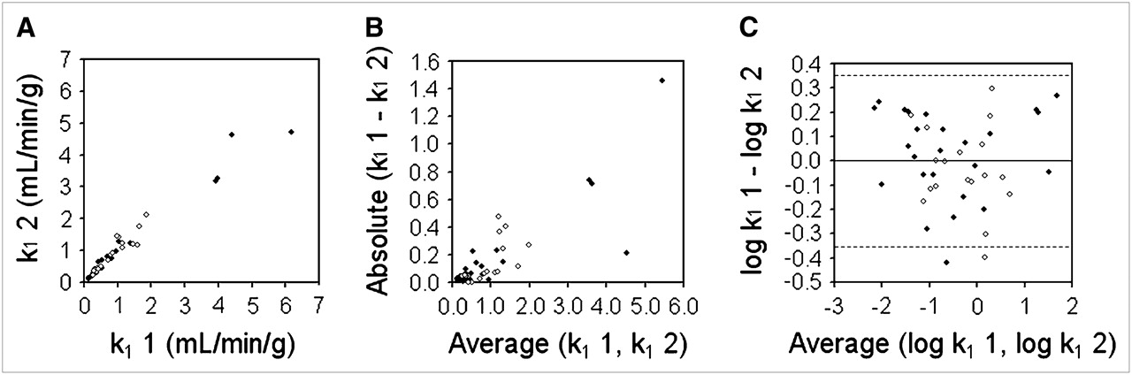
- FIGURE 3.
Bland–Altman analysis of reproducibility of k1 for tumors (TBF). (A) Replicate measures of k1 are plotted against each other. Solid symbols represent data acquired on Advance scanner; open symbols represent data acquired on Discovery RX scanner. (B) Absolute difference between replicate k1 measurements are plotted as function of their mean and show clear proportionality. (C) After log transformation, dsd for k1 was calculated as 0.178. Dashed lines denote 95% CIs on either side of mean (1.96 × dsd).
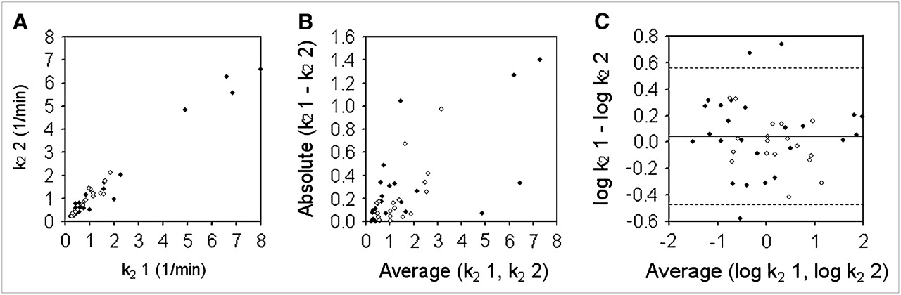
- FIGURE 4.
Bland–Altman analysis of reproducibility of k2 for tumors (efflux rate constant). (A) Replicate measures of k2 are plotted against each other. Solid symbols represent data acquired on Advance scanner; open symbols represent data acquired on Discovery RX scanner. (B) Absolute differences between replicate k2 measurements are plotted as function of their mean and show clear proportionality. (C) After log transformation, dsd for k2 was calculated as 0.259. Dashed lines denote 95% CIs on either side of mean (1.96 × dsd).
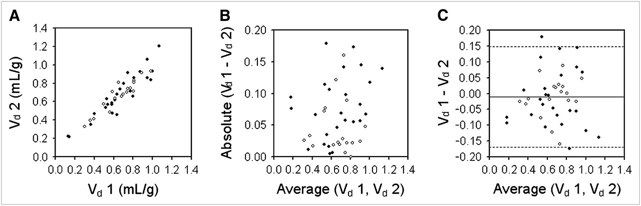
- FIGURE 5.
Bland–Altman analysis of reproducibility of Vd for tumors. (A) Replicate measures of Vd are plotted against each other. Solid symbols represent data acquired on Advance scanner; open symbols represent data acquired on Discovery RX scanner. (B) Absolute differences between replicate Vd measurements are plotted as function of their mean and show no clear proportionality. (C) dsd for Vd was calculated as 0.082 mL/g. Dashed lines denote 95% CIs on either side of mean (1.96 × dsd).


Tables
Parameter Advance Discovery RX Detector material Bismuth germanate Lutetium yttrium orthosilicate Detector size (mm) Tangential 4.0 4.2 Axial 8.1 6.3 Detector thickness (mm) 30 30 Detectors per block 6 (tangential) × 6 (axial) 9 (tangential) × 6 (axial) Blocks (axial direction) 3 4 Detector rings (axial direction) 18 24 Image slices 35 47 Slice thickness (mm) 4.25 3.27 Crystals per ring 672 630 Detectors in whole gantry 12,096 15,120 Septa (mm) Length 120 54 Thickness 1 0.8 Discriminator (keV) Lower level 300 425 Upper level 650 650 Coincidence timing window (ns) 12.5 6.5 Attenuation correction 68Ge pin source 64-slice CT Randoms correction Delayed channel Singles Abbreviation Parameter Calculation dsd SD of the difference  where d is the difference data and n is the number of pairs
wSD Within-subject SD wCV Within-subject coefficient of variation (%) For data in original units: , where is the mean.For log-transformed data: Repeatability (expressed as a percentage of the mean) Injection 1 Injection 2 Parameter Mean Median SD Mean Median SD k1 (mL/min/g) 1.03 0.52 1.28 0.99 0.65 1.09 k2 (1/min) 1.58 1.00 1.79 1.52 1.01 1.60 Va (dimensionless) 0.13 0.08 0.13 0.12 0.08 0.14 Vd (mL/g) 0.66 0.67 0.22 0.67 0.68 0.22


Jump to section
Related Articles
Cited By...
- Whole-Body Parametric Imaging of 18F-FDG PET Using uEXPLORER with Reduced Scanning Time
- Repeatability of swept-source optical coherence tomography retinal and choroidal thickness measurements in neovascular age-related macular degeneration
- 15O-Water PET Study of the Effect of Imatinib, a Selective Platelet-Derived Growth Factor Receptor Inhibitor, Versus Anakinra, an IL-1R Antagonist, on Water-Perfusable Tissue Fraction in Colorectal Cancer Metastases
- Novel Positron Emission Tomography Tracer Distinguishes Normal from Cancerous Cells
- Decreased Blood Flow with Increased Metabolic Activity: A Novel Sign of Pancreatic Tumor Aggressiveness