


Article Figures & Data
Figures
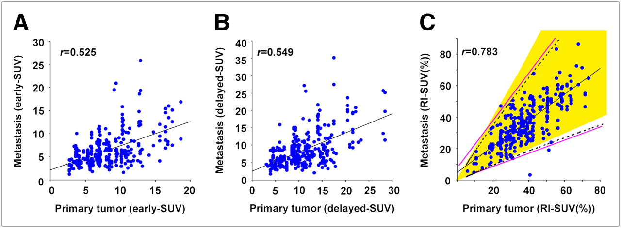
- FIGURE 1.
Correlation between SUV levels of all metastases and primary tumors in PET of lung cancer patients: early imaging (y = 0.523x + 2.123; r = 0.525) (A); delayed imaging (y = 0.551x + 2.542; r = 0.549) (B); and RI SUV (y = 0.829x + 4.667; r = 0.783) (C). Using RI SUV results of 95% prediction interval (broken line), upper linear approximation becomes y = 1.534x + 6.417 and lower linear approximation becomes y = 0.426x + 0.886 (straight pink lines). We indicated yellow area (0.5–2 times RI SUV of primary tumors) for deciding on metastatic lesions.
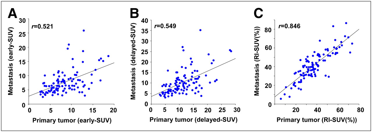
- FIGURE 2.
Correlation between highest SUV levels of all metastases and primary tumors in PET of lung cancer patients: early imaging (y = 0.59x + 2.751; r = 0.521) (A); delayed imaging (y = 0.605x + 3.569; r = 0.549) (B); and RI SUV (y = 0.904x + 7.619; r = 0.846) (C).
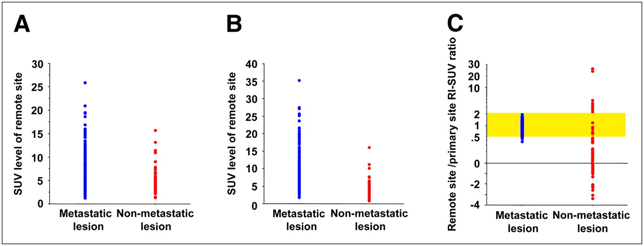
- FIGURE 3.
Comparison of SUV level (early and delayed) and RI SUV ratio of primary lesion and remote site (blue = metastatic uptake; red = nonmetastatic uptake): early imaging (A), delayed imaging (B), and RI SUV (C). In cases in which PET-positive findings are defined as yellow area (0.5–2 times RI SUV of primary tumors), uptakes of metastatic and nonmetastatic lesions are distinguishable.
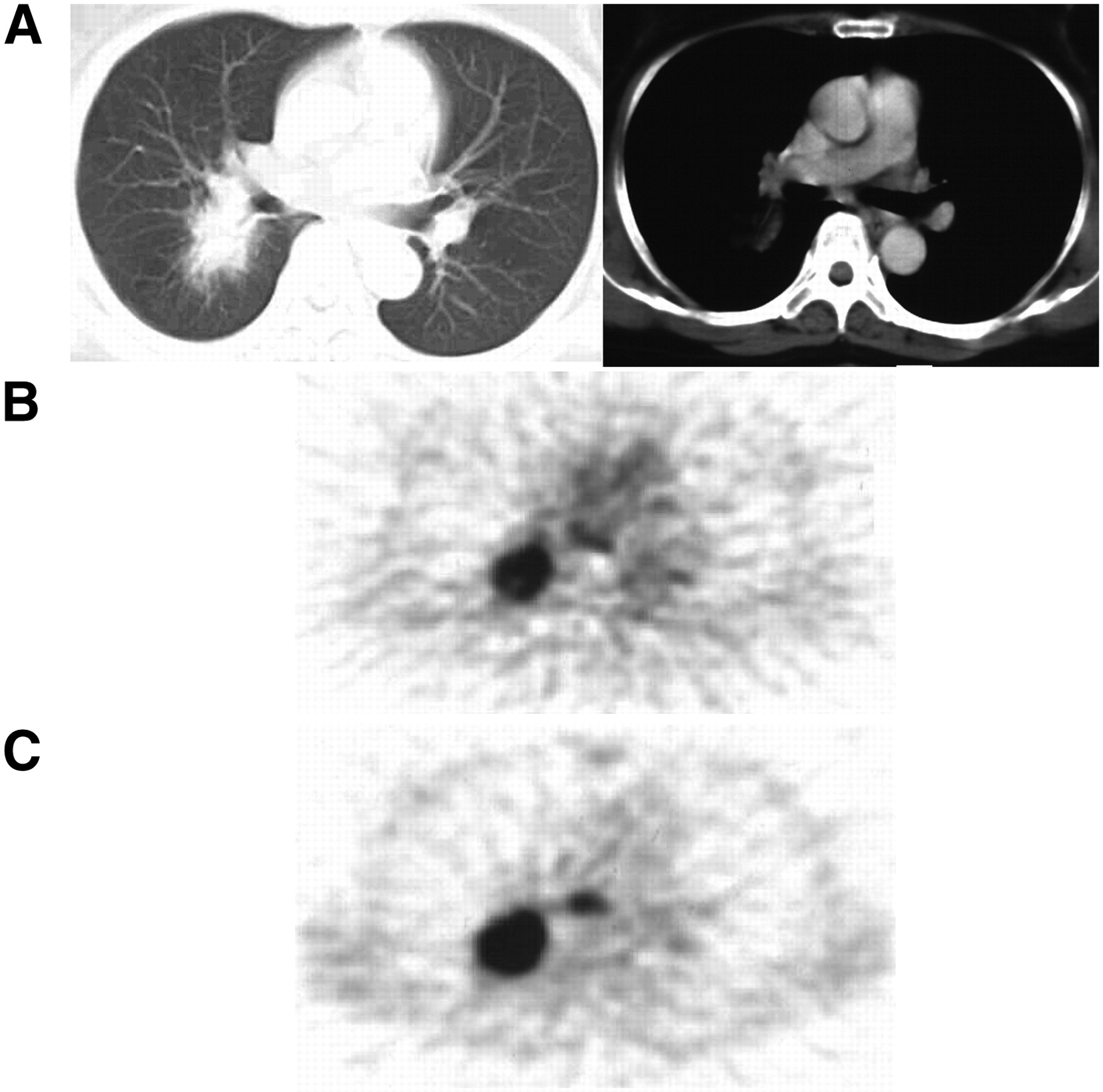
- FIGURE 4.
Representative case of adenocarcinoma and mediastinal lymph node metastasis (lymph node 7) in subcarinal area: chest CT (A), early imaging (B), and delayed imaging (C). CT images show nodule in right lung with no significant mediastinal lymph node swelling. Early imaging shows strong accumulation in nodule and faint accumulation in lymph node 7. PET shows increased uptake in lung nodule (early SUV = 6.85, delayed SUV = 10.01, RI SUV = 46.1%) and uptake in lymph node 7 (early SUV = 3.49, delayed SUV = 5.08, RI SUV = 45.6%).
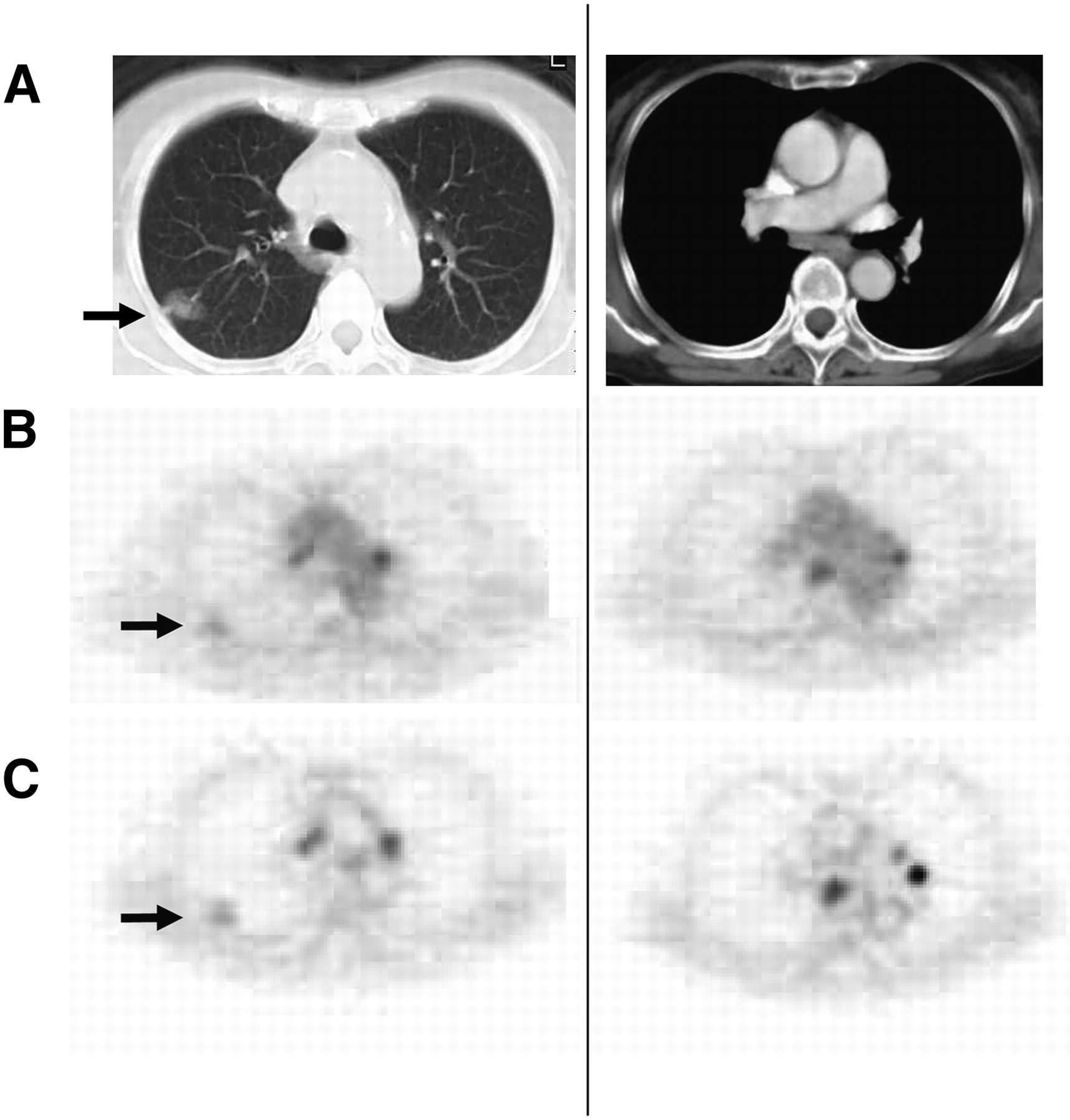
- FIGURE 5.
Representative case of adenocarcinoma and sarcoid reaction of mediastinal lymph node swelling: chest CT (A), early imaging (B), and delayed imaging (C). PET showed 18F-FDG uptake in primary tumor (early SUV = 1.81, delayed SUV = 2.02) (arrows) and focal uptake in mediastinal lymph nodes (lymph node 3: early SUV = 2.91, delayed SUV = 4.2; lymph node 7: early SUV = 3.89, delayed SUV = 5.086). RI SUV in primary tumor was 11.6%; however, RI SUVs in these lymph nodes were much higher (30.8%−44.3%). These nodal uptakes were confirmed at surgery as sarcoid reaction.


Tables
Characteristic Value Total patients (n) 155 M/F (n) 115/40 Mean age (y) 69 ± 9 Age range (y) 45–89 Histopathologic type (n) Adenocarcinoma 83 Squamous cell carcinoma 54 Small cell carcinoma 13 Large cell carcinoma 3 Atypical carcinoid 1 Unclassified 1 Stage (n) IA 30 IB 15 IIA 2 IIB 4 IIIA 27 IIIB 10 IV 67 Malignant lesion Number of lesions Surgery Biopsy Autopsy Clinical follow-up Primary lesion 155 76 77 2 Mediastinal lymph node 85 24 59 2 Hilar lymph node 15 15 Supraclavicular lymph node 6 6 Cervical lymph node 35 35 Intraperitoneal lymph node 8 1 7 Bone 76 4 6 66 Liver 26 1 3 22 Pleura 24 5 7 12 Lung 23 3 4 4 12 Adrenal grand 6 1 1 1 3 Kidney 5 4 1 Peritoneum 3 3 Muscle, skin 2 2 Invasion of atrium 1 1 Total 470 119 194 30 127 Nonmetastatic uptake Number of lesions Surgery Biopsy Clinical follow-up Lymph node (anthracosis) 20 20 Lymph node (follicular hyperplasia) 9 9 Lymph node (anthracosis and follicular hyperplasia) 3 3 Lymph node (granulomatous inflammation) 12 12 Pneumonia 8 5 3 Arthritis 4 4 Urinary tract, hydronephrosis 4 4 Malignancy of another organ (thyroid, larynx) 6 3 3 Parotid tumor (Warthin tumor) 3 3 Incidental colonic 18F-FDG uptake 7 7 Inflammatory disease of abdomen (e.g., gastric ulcer, diverticulitis, or cholecystitis) 12 1 11 Inflammatory disease of head and neck (e.g., sinusitis, parotiditis, or chronic thyroiditis) 21 4 17 Extravasation of 18F-FDG 1 1 Total 110 56 18 36 - TABLE 4
Comparison of Single- and Dual–Time-Point 18F-FDG PET Results for Staging of Nodal Metastasis Based on Patient-by-Patient Analysis (Surgical Cases and Definitive Pathologic N3 cases)
Number of patients Sensitivity Specificity Accuracy Parameter N0 (TN) N1 N2 N3 % 95% CI (%) % 95% CI (%) % 95% CI (%) Early imaging 29/45 4/6 21/21 2/2 93 77–99 64* 49–78 76* 64–85 Delayed imaging 33/45 5/6 21/21 2/2 97 82–100 73* 58–85 82* 72–90 RI SUV 44/45 6/6 21/21 2/2 100 98 88–99 99 93–100 ↵* P < 0.001 vs. RI SUV using McNemar test.
- TABLE 5
Comparison of Single- and Dual–Time-Point 18F-FDG PET Results for Staging of Distant Metastasis Based on Patient-by-Patient Analysis
Number of patients Sensitivity Specificity Accuracy Parameter TP TN FP FN % 95% CI (%) % 95% CI (%) % 95% CI (%) Early imaging 67 52 36 0 100 59* 48–70 77* 69–83 Delayed imaging 67 58 30 0 100 66* 55–76 81* 74–87 RI SUV 67 86 2 0 100 98 92–100 99 95–100 ↵* P < 0.001 vs. RI SUV using McNemar test.
TP = true-positive; TN = true-negative; FP = false-positive; FN = false-negative.
Number of patients overstaged (false-positive) Number of patients understaged (false-negative) Stage No. of patients Early imaging Delayed imaging RI SUV Early imaging Delayed imaging RI SUV N0 45 16 12 1 — — — N1 6 2 1 0 0 0 0 N2 21 0 0 0 0 0 0 N3 2 0 0 0 0 0 0 N staging overall 74 18 (24%) 13 (18%) 1 (1%) 0 0 0 M0 88 36 30 2 — — — M1 67 — — 0 0 0 M staging overall 155 36 (23%) 30 (19%) 2 (1%) 0 0 0


