


Article Figures & Data
Figures
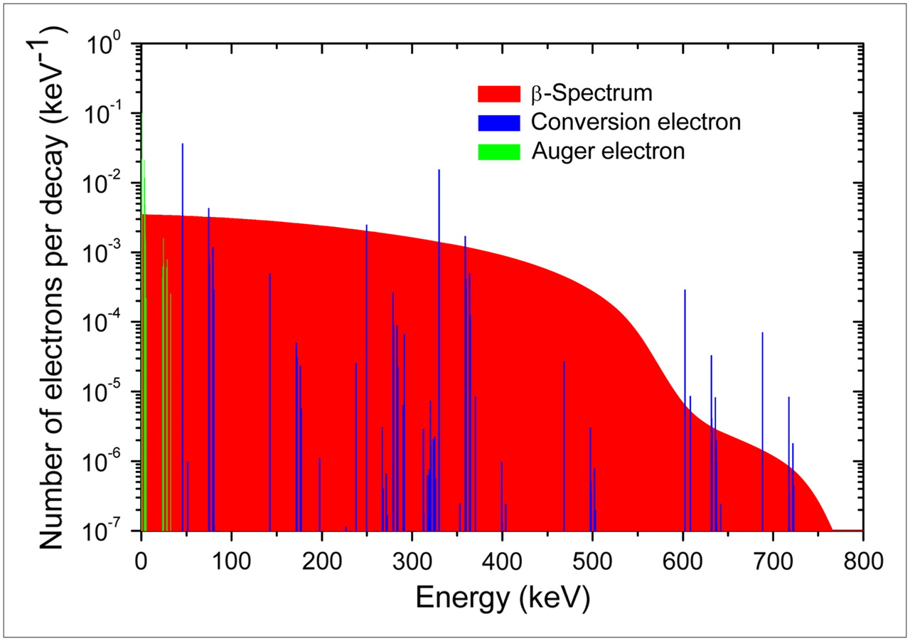
- FIGURE 1.
Mean primary electron spectrum of 131I decays.
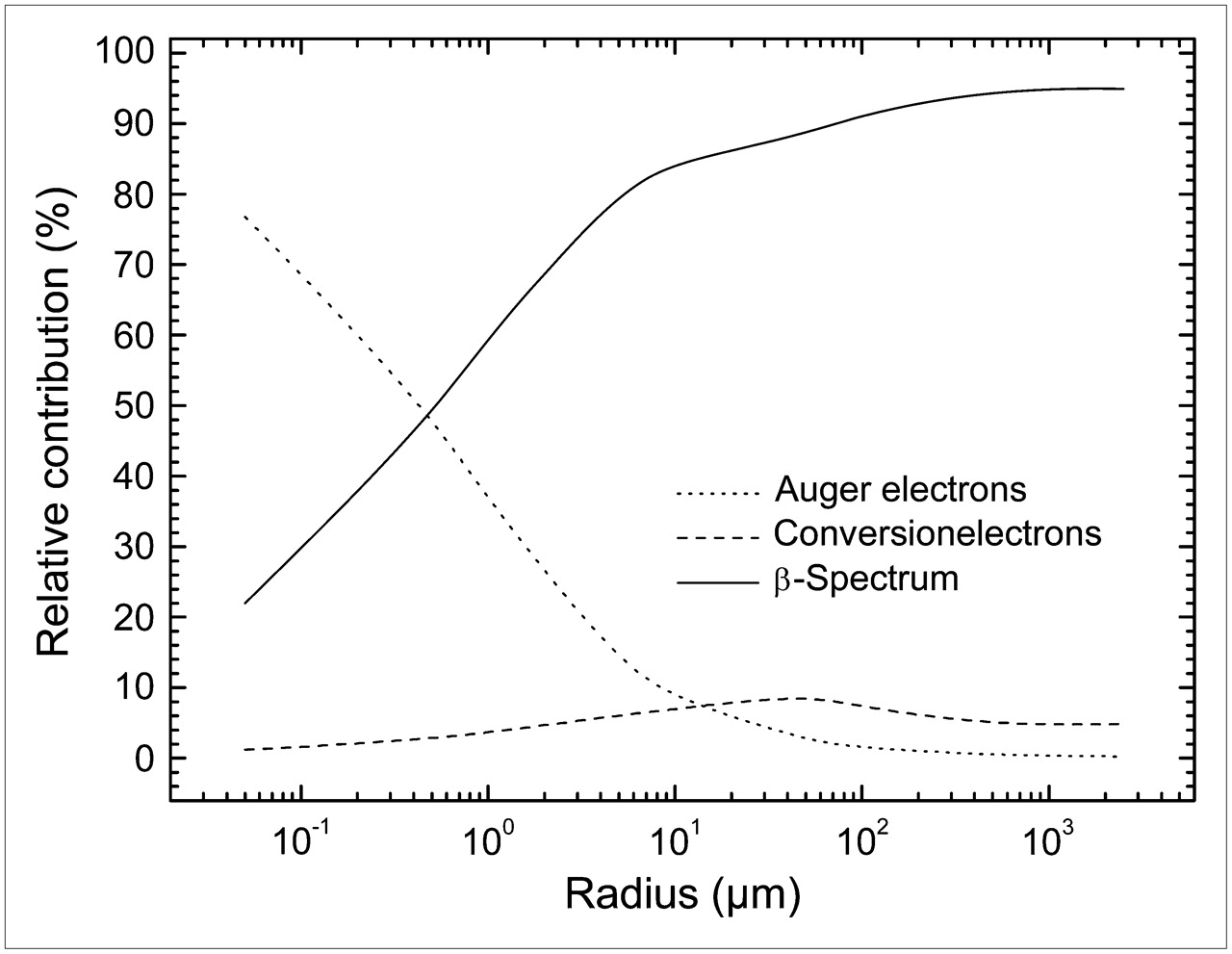
- FIGURE 2.
Two-dimensional plot of a 5-keV electron track obtained with Monte Carlo code CELLDOSE. •, Inelastic interactions of primary electron. ○, Inelastic interactions of secondary electrons.
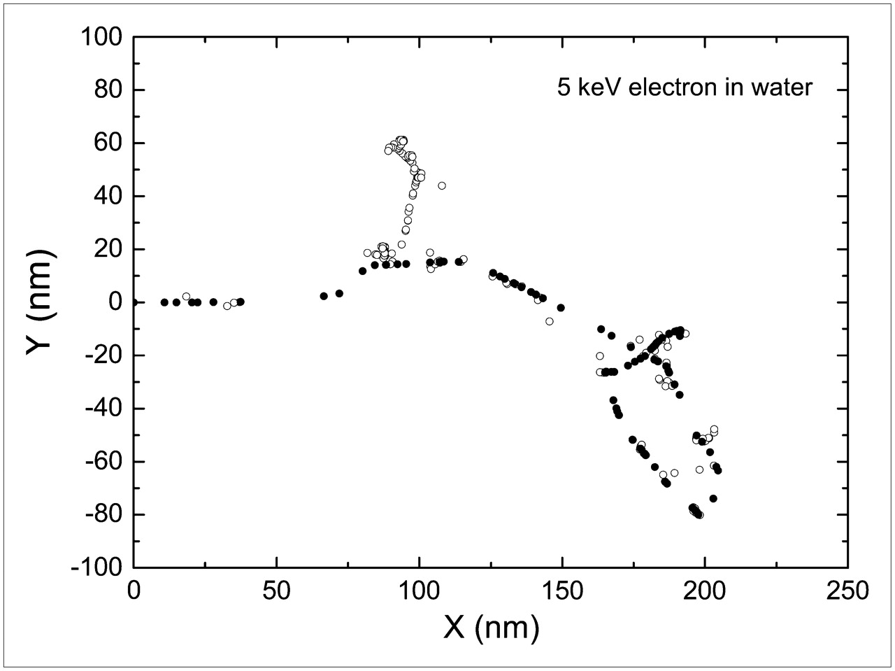
- FIGURE 3.
Fraction of energy retained vs. sphere radius.
- FIGURE 4.
Dose distribution across a sphere of 500 μm radius. Dose was assessed in concentric spheric shells at equidistant intervals of 10 μm starting from the center (first shell, 0–10 μm). Vertical dashed line indicates border of sphere. Profile of dose distribution is normalized using as reference dose at the center (A) or average sphere dose (B).
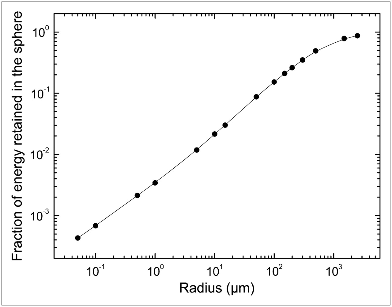
- FIGURE 5.
Plots of relative contribution from β−-particles, Auger electrons, and internal conversion electrons vs. sphere radius.


Tables
- TABLE 1
Fraction of Electron Energy Retained in Each Sphere and S Values: Comparison with S Values Reported by Goddu et al. and by Bardiès and Chatal
Sphere radius (μm) Sphere diameter (μm) Fraction of energy retained in sphere S value (Gy Bq−1s−1): this work S value: Goddu et al. (12,13)* Comparison with present work† S value: Bardiès and Chatal (14)‡ Comparison with present work§ 0.05 0.1 4.3 × 10⁻⁴ 2.51 × 101 0.1 0.2 6.81 × 10⁻⁴ 4.82 0.5 1 2.13 × 10⁻³ 1.22 × 10⁻¹ 1 2 3.42 × 10⁻³ 2.42 × 10⁻² 2.53 × 10⁻² +4.5% 5 10 1.18 × 10⁻² 6.70 × 10⁻⁴ 6.5 × 10⁻⁴ −3% 10 20 2.15 × 10⁻² 1.55 × 10⁻⁴ 1.45 × 10⁻⁴ −6.5% 1.61 × 10⁻⁴ +3.9% 15 30 3.02 × 10⁻² 6.53 × 10⁻⁵ 6.93 × 10⁻⁵ +6.1% 50 100 8.73 × 10⁻² 5.08 × 10⁻⁶ 5.38 × 10⁻⁶ +5.9% 100 200 1.53 × 10⁻¹ 1.12 × 10⁻⁶ 1.03 × 10⁻⁶ −8% 1.2 × 10⁻⁶ +7.1% 150 300 2.11 × 10⁻¹ 4.55 × 10⁻⁷ 4.91 × 10⁻⁷ +7.9% 200 400 2.62 × 10⁻¹ 2.39 × 10⁻⁷ 2.16 × 10⁻⁷ −9.6% 2.59 × 10⁻⁷ +8.4% 300 600 3.5 × 10⁻¹ 9.47 × 10⁻⁸ 1.03 × 10⁻⁷ +8.8% 500 1,000 4.92 × 10⁻¹ 2.86 × 10⁻⁸ 2.8 × 10⁻⁸ −2.1% 3.11 × 10⁻⁸ +8.7% 1,500 3,000 7.84 × 10⁻¹ 1.69 × 10⁻⁹ 1.77 × 10⁻⁹ +4.7% 2,500 5,000 8.71 × 10⁻¹ 4.07 × 10⁻¹⁰ 4.01 × 10⁻¹⁰ −1.5% 4.17 × 10⁻¹⁰ +2.5% Sphere radius (μm) Sphere diameter (μm) S value (Gy Bq−1s−1): present work S value: Li et al. (7)* Comparison with present work† 0.05 0.1 2.51 × 101 0.1 0.2 4.82 0.5 1 1.22 × 10⁻¹ 1 2 2.42 × 10⁻² 5 10 6.70 × 10⁻⁴ 6.8 × 10⁻⁴ +1.5% 15 30 6.53 × 10⁻⁵ 7.2 × 10⁻⁵ +10.3% 50 100 5.08 × 10⁻⁶ 5.5 × 10⁻⁶ +8.3% 150 300 4.55 × 10⁻⁷ 4.9 × 10⁻⁷ +7.7% 500 1,000 2.86 × 10⁻⁸ 3.1 × 10⁻⁸ +8.4% 1,500 3,000 1.69 × 10⁻⁹ 1.8 × 10⁻⁹ +6.5% - TABLE 3
Dose Distribution Inside a Sphere of 500 μm Radius Containing a Homogeneous Distribution of 131I
Distance from center (spheric shells of 10 μm) Relative dose (average sphere dose as reference) Percentage of dose compared with dose at center (%) Center (0; 10 μm) 1.32 100% 90; 100 μm 1.3 98.5% 190; 200 μm 1.26 95.5 290; 300 μm 1.17 88.6 390; 400 μm 1.02 77.3 400; 410 μm 1 75.8 410; 420 μm 0.977 74 420; 430 μm 0.952 72.1 430; 440 μm 0.925 70 440; 450 μm 0.895 67.8 450; 460 μm 0.862 65.3 460; 470 μm 0.825 62.5 470; 480 μm 0.782 59.2 480; 490 μm 0.729 55.2 490; 500 μm 0.657 49.8 - TABLE 4
Relative Contribution to S Values from β−-Particles, Auger Electrons, and Internal Conversion Electrons
Sphere radius (μm) Sphere diameter (μm) Fraction of energy retained in sphere Auger contribution (%) CE contribution (%) β− contribution (%) 0.05 0.1 4.3 × 10⁻⁴ 76.8 1.2 22 0.1 0.2 6.81 × 10⁻⁴ 68.7 1.6 29.7 0.5 1 2.13 × 10⁻³ 48.6 2.8 48.6 1 2 3.42 × 10⁻³ 36.5 3.6 59.9 5 10 1.18 × 10⁻² 13.4 6.1 80.6 10 20 2.15 × 10⁻² 8.5 7 84.5 50 100 8.73 × 10⁻² 2.5 9.1 88.4 100 200 1.53 × 10⁻¹ 1.4 7.2 91.4 500 1,000 4.94 × 10⁻¹ 0.45 4.7 94.9 2,500 5,000 8.71 × 10⁻¹ 0.25 4.8 94.9 CE = conversion electron.


Jump to section
Related Articles
Cited By...
- Membrane and Nuclear Absorbed Doses from 177Lu and 161Tb in Tumor Clusters: Effect of Cellular Heterogeneity and Potential Benefit of Dual Targeting--A Monte Carlo Study
- Membrane and Nuclear Absorbed Doses from 177Lu and 161Tb in Tumor Clusters: Effect of Cellular Heterogeneity and Potential Benefit of Dual Targeting--A Monte Carlo Study
- Multimodal Imaging of 2-Cycle PRRT with 177Lu-DOTA-JR11 and 177Lu-DOTATOC in an Orthotopic Neuroendocrine Xenograft Tumor Mouse Model
- Dose Deposits from 90Y, 177Lu, 111In, and 161Tb in Micrometastases of Various Sizes: Implications for Radiopharmaceutical Therapy
- Cellular Dosimetry of 111In Using Monte Carlo N-Particle Computer Code: Comparison with Analytic Methods and Correlation with In Vitro Cytotoxicity
- 131I Radiation Dose Distribution in Metastases of Thyroid Carcinoma
- Reply: 131I Radiation Dose Distribution in Metastases of Thyroid Carcinoma