


Article Figures & Data
Figures
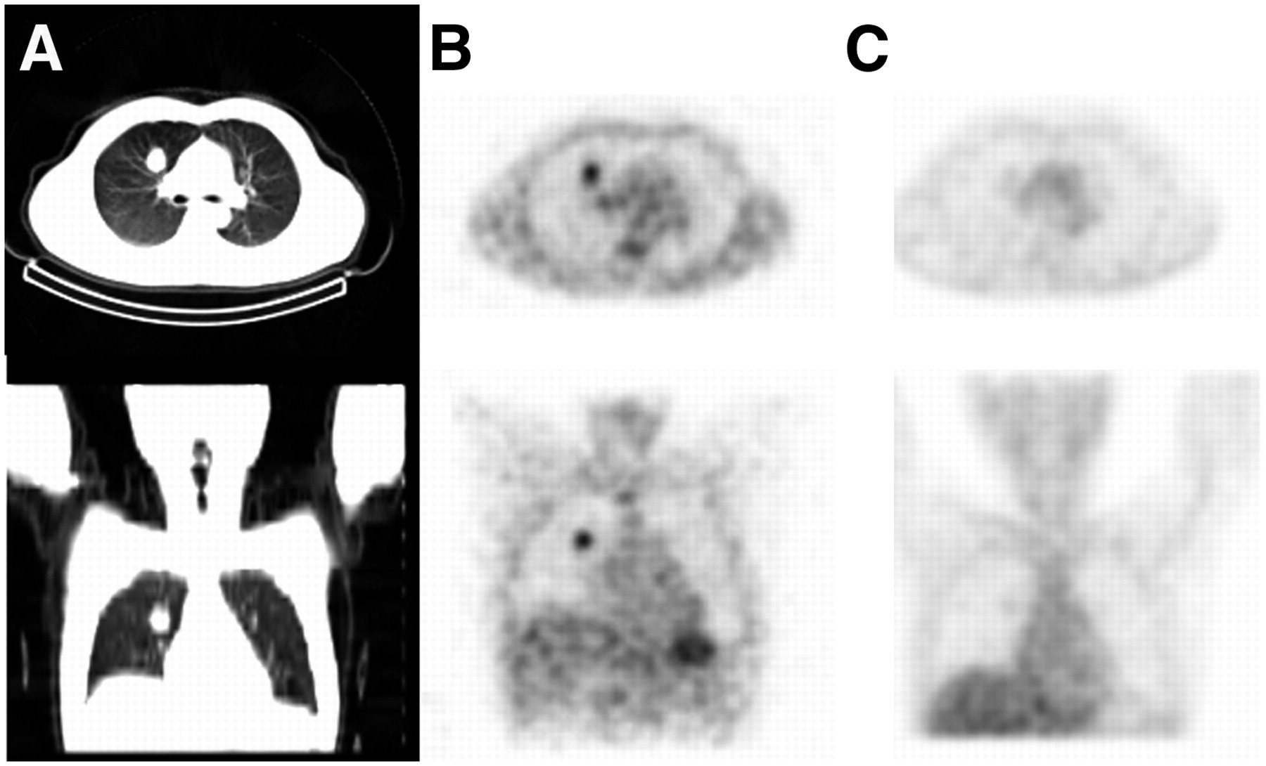
- FIGURE 1.
A 46-y-old male patient with benign clear cell tumor. (A) CT scans showed abnormality in right upper lobe. (B) 18F-FDG coincidence images had intense uptake in lesion. (C) 99mTc-Octreotide images were negative in lesion.
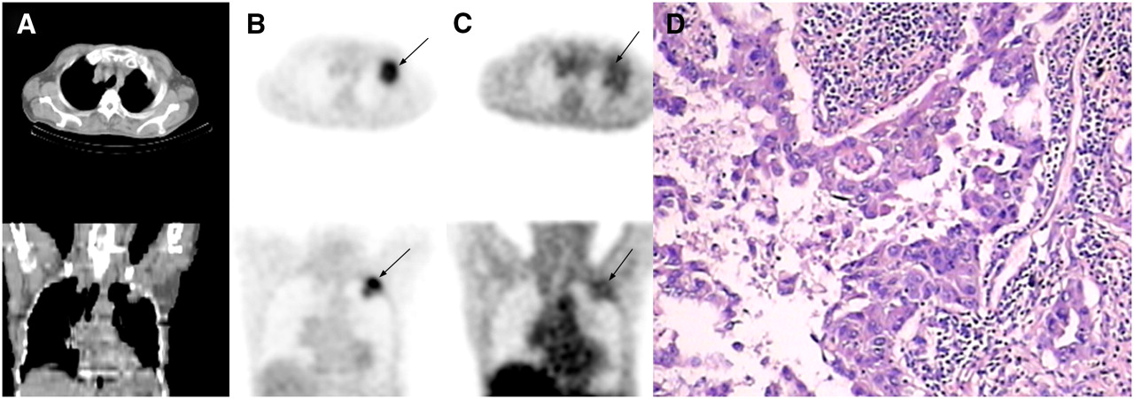
- FIGURE 2.
A 75-y-old male patient with adenosquamous lung cancer in left upper lobe. (A) CT scans showed abnormality in left upper lobe. (B) 18F-FDG coincidence images had intense uptake in lesion (arrows). (C) 99mTc-Octreotide images showed intense uptake in primary tumor and pleura (arrows). (D) Adenosquamous lung cancer and pleural invasion were verified on histology.
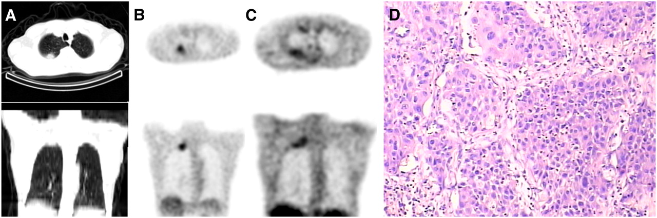
- FIGURE 3.
A 57-y-old male patient with squamous lung cancer. (A) CT scans showed neoplasm high in upper lobe of right lung. (B) 18F-FDG coincidence images had focal uptake in lesion. (C) 99mTc-Octreotide images had focal high uptake in lesion. (D) Squamous lung cancer and pleura and rib invasion were verified on histology.
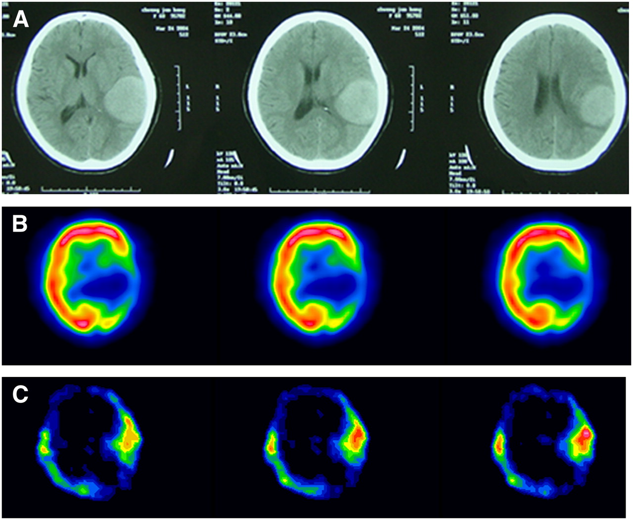
- FIGURE 4.
A 65-y-old woman with lung adenocarcinoma. (A) CT scans revealed brain metastasis and peripheral edema in left temporal lobe. (B) 18F-FDG images showed low uptake in brain lesion. (C) 99mTc-Octreotide images showed focal uptake in brain lesion.
Tables
Patient no. Sex Age (y) Histology Primary lesion (cm) Staging T/Nr T/Nm 1 F 52 Adenocarcinoma 2.0 T1 N3 2.54 5.56 2 M 51 Squamous cell carcinoma 4.2 T2 N3 2.24 6.62 3 M 57 Squamous cell carcinoma 3.0 T2 N0 4.1 9.86 4 M 75 Adenosquamous carcinoma 4.8 T4 N0 3.91 7.34 5 F 55 Adenocarcinoma 3.6 T2 N3 M1 3.29 5.46 6 M 59 Adenocarcinoma 4.2 T3 N3 2.82 6.35 7 M 76 Undifferentiated lung cancer 11.0 T4 N3 5.58 10.56 8 M 72 Adenocarcinoma 3.8 T2 N1 2.30 5.32 9 F 65 Adenocarcinoma 5.2 T4 N3 M1 3.78 8.23 10 F 61 Squamous cell carcinoma 3.2 T2 N0 M1 4.06 7.16 11 M 42 Squamous cell carcinoma 6.2 T4 N3 M1 4.61 5.35 12 M 70 Squamous cell carcinoma 7.8 T4 N3 3.36 10.23 13 F 60 Squamous cell carcinoma 5.3 T2 N3 2.98 9.18 14 M 71 Squamous cell carcinoma 3.5 T2 N2 3.63 6.45 15 M 75 Adenocarcinoma 4.8 T4 N3 M1 2.57 4.12 16 M 70 Squamous cell carcinoma 8.5 T4 N3 3.59 5.36 17 F 63 Adenocarcinoma 3.5 T2 3.87 4.96 18 M 64 Adenocarcinoma 4.5 T2 N3 M1 2.68 5.09 19 M 64 Small-cell lung carcinoma 6.5 T3 N2 3.42 4.49 20 F 64 Adenocarcinoma 3.8 T2 N3 M1 2.62 5.54 21 M 60 Adenocarcinoma 7.1 T3 N2 2.68 5.31 22 M 71 Adenocarcinoma 4.5 T2 N3 M1 4.13 6.23 23 F 44 Small-cell lung carcinoma 5.0 T3 N3 M1 3.32 5.16 24 M 70 Squamous cell carcinoma 8.5 T4 N3 3.36 7.16 25 M 73 Adenocarcinoma 5.1 T2 N0 2.89 4.86 26 M 54 Squamous cell carcinoma 7.5 T4 N3 M1 3.46 8.19 27 M 76 Adenocarcinoma 3.2 T4 N0 3.78 4.61 28 M 51 Small-cell lung carcinoma 7.1 T4 N3 M1 3.63 5.34 29 M 75 Small-cell lung carcinoma 2.8, 5.6 T4 N0 M1 3.98 4.55 30 F 56 Small-cell lung carcinoma 2.0, 5.0 T2 N0 M1 3.68 3.99 31 F 39 Small-cell lung carcinoma 3.2 T4 N0 4.09 5.32 32 F 51 Fibroma 1.2 — 1.29 1.42 33 F 66 Tuberculosis 3.3 — 1.13 2.10 34 F 70 Granuloma 2.3 — 1.56 3.92 35 F 62 Hamartoma 1.6 — 1.49 2.13 36 M 62 Tuberculosis 1.2 — 1.36 3.88 37 M 83 Pneumonia 2.5 — 1.78 1.89 38 M 75 Interstitial pneumonitis 1.0 — 1.39 1.78 39 M 46 Clear cell tumor 2.5 — 1.24 4.12 40 M 75 Tuberculosis 4.0 — 1.35 3.89 41 M 30 Tuberculosis 1.5 — 4.69 6.93 42 M 71 Granuloma 2.5 — 1.59 1.89 43 M 68 Tuberculosis 2.6 — 3.68 4.96 44 M 50 Tuberculosis 3.0 — 3.89 5.69 TNM staging was determined by histopathology or other imaging modalities. Tumor-to-normal tissue uptake ratios of 18F-FDG DHC are expressed as T/Nm. Tumor-to-normal tissue uptake ratios of 99mTc-octreotide imaging are expressed as T/Nr.
- TABLE 2
Efficacy of 99mTc-Octreotide for Detection of Primary Lesion and Lymph Node Involvement Compared with 18F-FDG
18F-FDG DHC 99mTc-Octreotide Lesion location Sensitivity (%) Specificity (%) PPV (%) NPV (%) Sensitivity (%) Specificity (%) PPV (%) NPV (%) Primary lesion 100 (31–31) 46.1 (6–13) 83.8 (31–38) 100 (6–6) 100 (31–31) 75.7 (9–13) 90.1 (31–35) 100 (9–9) Metastatic hilar or mediastinal LN 100 (20–20) 100 (11–11) 100 (20–20) 100 (11–11) 35 (7–20) 100 (7–7) 100 (7–7) 46 (11–24) LN = lymph node.
Data are expressed as percentage, with range in parentheses.
Parameter Value No. of patients 44 Age range (y) 39–83 Women/men 12/32 Benign/malignant lesions 13/31 Surgery/biopsy/bronchoscopy/clinicoradiology 18/12/8/6 Lymph node adenopathy/no lymph node adenopathy 20/11 Clinical T factor for malignant lesions (1/2/3/4) 1/10/3/17 Clinical N status (0/1/2/3) 11/1/3/16 Distant metastasis/no distant metastasis 13/18 Contralateral lung intrametastasis/no lung intrametastasis 2/29 Pleural invasion/no pleural invasion 8/23 Bone metastasis/no bone metastasis 9/22 Brain metastasis/no brain metastasis 2/29


Jump to section
Related Articles
Cited By...
- No citing articles found.